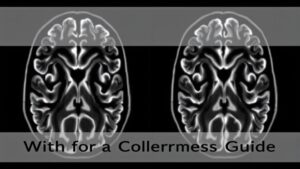
Brain MRI scans create detailed pictures of brain tissue and structures. Magnetic resonance imaging (MRI) uses a powerful magnet and magnetic field to create images of the brain and soft tissues. These images come in two main types: with contrast dye and without contrast dye. According to the American College of Radiology, over 40 million MRI procedures take place annually in the United States, with brain scans being one of the most common uses.
A cranial MRI is a common diagnostic imaging test used to assess neurological conditions. An MRI technologist operates the MRI scanner and ensures patient safety throughout the procedure. MRI scanners use powerful magnets to create high-resolution images of soft tissues, which helps diagnose a wide range of conditions. Both scanning methods help doctors spot problems like tumors, inflammation, and blood vessel issues, giving them clear information to make accurate treatment decisions.
Contrast-Enhanced Magnetic Resonance Imaging (MRI) Scans
Contrast-enhanced MRI gives doctors a clearer view inside your body through special magnetic dyes known as contrast agents, which are administered via IV injection. This process, called a contrast injection, delivers the contrast agent directly into your bloodstream.
These agents light up specific areas on the scan, making it easier to spot health issues that regular MRIs can’t show clearly. Some patients may experience a temporary metallic taste after the IV injection, which is a mild and short-lived side effect.
Your radiologist uses these enhanced images to examine blood vessels, tumors, and inflamed tissues in detail. The contrast agent travels through your blood vessels, creating bright signals that stand out against darker surrounding tissues. This helps doctors locate and measure any abnormal growths or damaged areas with greater accuracy. The contrast agent works by altering the magnetic properties of tissues, and when combined with radio waves, this process helps produce high quality images.
The process works by changing how tissues respond to the magnetic fields in the MRI machine. Small amounts of contrast material make certain body parts appear brighter or darker on the final images. As a result, doctors can see the exact size, shape, and location of problems that need treatment.
These detailed scans guide medical teams in creating precise treatment plans tailored to each patient’s needs. The enhanced images reveal subtle differences between healthy and unhealthy tissues, which proves especially helpful for brain scans, spine problems, and cancer monitoring.
The contrast dye makes blood flow patterns visible too, showing how well different parts of your body receive blood supply. This information helps doctors check for blocked vessels or areas with poor circulation that regular MRIs would miss.
Benefits of Non-Contrast MRI Imaging
Non-contrast MRI offers several clear advantages for both patients and medical teams. Your body creates natural images during these scans without needing any additional substances.
Safety comes as a top benefit – you’ll avoid any risks associated with contrast dyes. Unlike CT scans (computed tomography), X-rays, or ray imaging, MRI does not use ionizing radiation, making it safer for frequent imaging and repeated imaging tests. This makes non-contrast MRI particularly valuable for patients with kidney problems or those who react to contrast materials.
The process moves faster too. Your scan starts right away since there’s no need to inject contrast or wait for it to circulate through your system. Medical teams capture detailed pictures of bones, tissues, and organs in their natural state.
These scans excel at showing structural details in your brain, spine, and joints. While CT scanning and X-ray imaging are useful for certain conditions, MRI provides superior soft tissue detail for brain and spinal cord assessments. Doctors regularly use them to check for:
- Head injuries after accidents
- Birth-related brain conditions
- Basic brain and spine anatomy
- Joint problems and injuries
Of course, the cost stays lower without contrast materials involved. Plus, the simpler procedure means less stress for everyone – both patients and medical staff appreciate the straightforward approach.
Your doctor receives reliable diagnostic information while keeping you comfortable and safe. The natural images help spot problems early, allowing for faster treatment planning. Thus, non-contrast MRI continues serving as a practical choice for many medical situations.
Most patients feel at ease during these scans because they know no additional substances enter their bodies. The medical team focuses entirely on capturing clear images to guide your care effectively.
When Contrast Agents Are Recommended
Your doctor wants to see clearer pictures of what’s happening inside your body? That’s where MRI contrast agents come in handy. These special dyes help doctors spot health issues that regular MRI scans can’t show as clearly. Brain magnetic resonance imaging (MRI) and head MRI are types of MRI exams used to diagnose conditions affecting the brain and spinal cord.
Doctors often recommend contrast agents to check for several medical conditions:
- Brain or spine tumors
- Blood vessel problems
- Swelling or inflammation
- Cancer spread to other areas
- Multiple sclerosis
- Breast cancer
The contrast dye, called gadolinium, makes abnormal tissues stand out from healthy ones on the scan. This helps your medical team spot tiny changes they need to see. Think of it like turning up the brightness on a dim photo – suddenly everything becomes much clearer.
Your radiologist carefully injects just the right amount of contrast to highlight problem areas. This gives your doctors the detailed view they need to make accurate diagnoses and create better treatment plans for you.
Magnetic resonance imaging is a non-invasive technique that helps diagnose conditions by providing detailed images of the brain, spinal cord, and blood vessels.
The whole process works together to give your medical team a precise roadmap of what’s happening in your body. By seeing these enhanced images, they can spot even small issues early and help you get the care you need faster.
Potential Risks and Considerations
Gadolinium contrast agents provide excellent diagnostic images, though they come with specific risks doctors need to consider for each patient. Your doctor will check your medical history and kidney function before scheduling any contrast MRI. Patients with kidney disease are at risk for nephrogenic systemic fibrosis when exposed to certain contrast agents, so this is carefully evaluated.
Some people experience mild reactions like headaches or nausea after receiving gadolinium. Allergic reactions, including mild allergic reaction such as hives or itchiness, are a very slight risk but should be monitored by healthcare staff. More serious reactions remain rare but can include allergic responses or complications in patients with poor kidney function. Recent studies have shown that tiny amounts of gadolinium can stay in brain tissue, though researchers continue studying the long-term effects.
Your healthcare team follows strict safety protocols and appropriate safety guidelines to protect you during contrast-enhanced brain imaging. They screen carefully for risk factors, especially for patients with implanted medical devices or artificial joints, and monitor you throughout the procedure. Patients are typically asked to remove metal and electronic items, including metal objects, and change into a hospital gown before entering the exam room to ensure safety and image quality. The radiologist reviews your complete scan results, noting any unexpected findings that need additional evaluation.
A thorough discussion with your doctor helps address specific concerns about contrast agents. Pregnant women should inform their healthcare provider before undergoing an MRI to ensure all safety considerations are addressed. They’ll explain how the benefits of detailed diagnostic information balance against potential risks for your situation. Regular communication between you and your medical team remains essential throughout the imaging process.
Your doctor creates a personalized plan considering your medical needs and safety factors, including any medical devices or implants. They monitor your response during and after the procedure while staying ready to address any reactions quickly. This careful approach helps deliver valuable diagnostic information while protecting your wellbeing.
Choosing the Right MRI Scanning Approach
Your MRI scanning approach matters—both for accurate diagnosis and your comfort during the procedure. Medical teams look at several key factors to choose the right scanning method for you.
There are different types of MRI machines, including traditional MRI units with a closed cylindrical design, open MRI systems with open sides for greater comfort, and large-diameter MRI scanners that accommodate a wider range of patients. Each MRI unit and scanner offers unique features, such as bore size and magnet strength, which can impact patient experience and the quality of the images produced.
Most MRI exams are performed using a brain MRI coil, a helmet-like device placed around your head, to obtain high quality images of the brain. These images are then reviewed and analyzed on a computer screen by the radiologist.
Your doctor needs to know about your medical background and current symptoms. This helps them pick the most suitable scanning technique for your specific situation. The scanning team considers how different approaches can best show what’s happening in your body.
Each MRI machine offers unique capabilities and settings. The radiologist matches these technical features to your needs. They compare regular scans versus those using a contrast agent, which may be injected during your MRI exam to enhance image clarity and specificity, depending on the type of exam required.
Your comfort stays a top priority throughout this process. The scanning team works to get detailed images while keeping you at ease. They carefully weigh the benefits of each scanning option against potential risks or discomfort.
Many MRI facilities follow guidelines set by professional organizations such as the Radiological Society of North America to ensure safety and quality standards.
The radiologist partners with your doctor to select the most effective scanning approach. They focus on getting precise images that reveal the information needed for your care. This teamwork helps create a scanning plan tailored just for you.
Conclusion
Brain MRI scans with and without contrast help doctors see detailed pictures of brain tissue. These imaging methods catch problems that regular scans can miss. Research from the American College of Radiology shows contrast-enhanced MRIs detect up to 30% more brain abnormalities compared to non-contrast scans. This advanced imaging gives medical teams clear information to create precise treatment plans tailored to each patient’s needs.


