A cuboid fracture is a break in one of the bones in the midfoot. It’s a rare injury caused by trauma, twisting or stress.
For this reason, we are here to explain the cuboid fracture and its causes, symptoms and other natural treatment options.
What is a Cuboid Fracture?
Cuboid fractures are rare, occurring at a rate of 1.8 per 100,000 fractures per year. They can occur alone or with other midfoot fractures making them a part of complex midfoot injuries. Common causes of cuboid fractures are direct impacts or severe crush injuries which can affect the arch, lateral column and overall forefoot function.
Bruising, swelling, pain on the outer foot and difficulty walking are common signs of a cuboid fracture. These symptoms can be debilitating and affect daily activities and mobility so prompt diagnosis and treatment is needed. Whether it’s a isolated cuboid fracture or part of a more complex injury understanding these fractures is key to management.
Cuboid fractures can also occur from indirect trauma such as twisting injuries or stress from repetitive activities. This wide range of causes means you need to have a high index of suspicion in cases of midfoot pain after trauma.
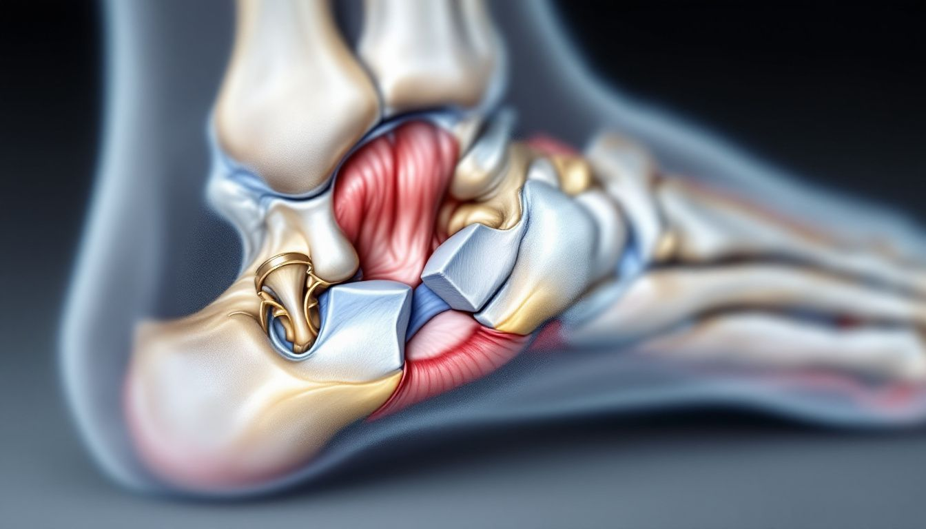
Cuboid Bone Anatomy
The cuboid bone is a structure on the lateral side of the foot which contributes to foot stability and arch formation. It plays a key role in foot mechanics by providing stability and allowing movement during walking and running. Its shape and position makes it important for midfoot structure and provides multiple surfaces for articulation with other foot bones.
The bone’s role in the lateral longitudinal arch of the foot makes it important for foot stability during movement.
The next sections will cover the cuboid’s role in lateral column, articulations and blood supply.
Lateral Column
The cuboid bone supports the lateral column of the foot and affects overall balance and lateral movement. The cuboid is the static lateral component of the foot and is good for maintaining the lateral longitudinal arch during movements such as dorsiflexion and plantar flexion.
This support is important for foot mobility and balance especially during activities that require lateral stability such as walking on uneven surfaces or doing athletic movements.
The cuboid acts as a rigid support for the lateral and medial column and allows the foot to withstand daily activities and high impact sports.
Any damage to this bone such as a fracture can compromise lateral column stability and lead to difficulty in weight bearing and increase risk of further injuries.
Articulations and Blood Supply
The cuboid bone connects to the calcaneus, navicular and the 4th and 5th metatarsals and allows for various foot movements. These articulations are part of the foot’s intricate structure and allows for the range of motion needed for walking, running and other activities. The cuboid’s multiple articular surfaces allows it to support and transmit forces across the foot.
Blood supply to the cuboid is from branches of the lateral plantar artery so the bone is well nourished.
This blood supply is important for the bone’s health and function especially after an injury. Good blood flow helps in healing and prevents osteonecrosis.
Mechanisms of Cuboid Fractures
Cuboid fractures can occur from:
- Direct trauma such as those from vehicle or industrial accidents which can damage the cuboid bone badly.
- Repetitive stress especially in athletes which can cause a cuboid stress fracture due to overuse during running.
- Twisting injuries where the foot suddenly rotates which can also cause a fracture of the cuboid.
Mainly common causes of cuboid fractures are:
- Direct trauma: Impact from accidents or heavy objects.
- Repetitive stress: Overuse during activities like running.
- Twisting injuries: Sudden foot rotation.
- Compression from midfoot injuries.
- Vehicle or industrial accidents.
Cuboid fractures are often associated with other midfoot injuries such as cuneiform fractures and tarsal fractures making diagnosis and treatment more complicated.
Associated Injuries
50–90% of cuboid fractures occur alongside other foot or ankle injuries, such as Lisfranc injuries or fractures of the metatarsals.
Moreover, Cuboid syndrome, a separate but related condition, can develop due to prolonged instability or misalignment.
Nutcracker Fracture
Nutcracker fractures are due to compression mechanism where the cuboid bone is subjected to axial loading. This type of fracture is often seen in high impact sports, falls and accidents where the foot is directly compressed. The compression forces the cuboid between the calcaneus and metatarsals and causes a fracture.
The implications of nutcracker fractures are careful assessment to avoid misdiagnosis and longer recovery period if not treated well. Complications from nutcracker fractures can be chronic pain, instability of the foot and post traumatic arthritis.
Avulsion Fracture
Avulsion fractures of the cuboid occurs when a piece of bone is pulled away by a tendon or ligament. This type of fracture occurs at the site of attachment of ligaments to the cuboid bone. Avulsion fractures can happen during sudden movements or traumatic injuries where the foot is twisted or overstretched.
In some cases, Running, jumping or severe ankle sprains can cause avulsion fractures. These fractures need careful management to ensure proper healing and prevent long term complications.
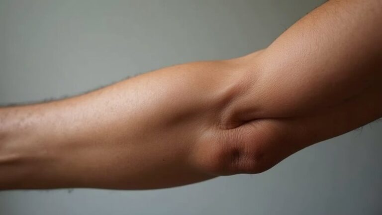
Cuboid Fracture from Rolling Ankle
You’re walking or running when suddenly your ankle rolls inward or outward, causing a sharp, twisting force.
As your foot twists, the bones in your ankle are pushed beyond their normal range of motion. This force can cause a fracture in the cuboid bone, a small but crucial bone on the outer side of your midfoot.
You might feel immediate pain on the outer edge of your foot, and it could be difficult to put weight on it. Swelling and bruising may develop quickly, and the area might feel tender to the touch. You can also notice a sense of instability or weakness in your foot, making it hard to walk normally.
The pain could radiate toward your toes or along the side of your foot, depending on the severity of the injury. If you try to move your foot or push off the ground, the pain might intensify, signaling that something is seriously wrong.
Cuboid Fracture Symptoms
If you have a cuboid fracture, you’ll likely experience a range of symptoms that can affect your ability to walk and move comfortably. Since the cuboid bone is a small, cube-shaped bone on the outer side of your foot, its fracture can cause pain and instability in that area.
1. Pain on the Outer Side of Your Foot
You’ll likely feel a deep, aching pain along the outer edge of your foot, especially near the middle. The pain may get worse when you try to walk or put weight on it.
2. Cuboid fracture Swelling and Bruising
Swelling is likely to develop quickly around the outer foot, and you might see bruising which appears as discoloration (blue, purple, or yellow) around the injured area.
3. Difficulty Walking or Bearing Weight
You may find it painful or even impossible to walk normally. Putting pressure on your foot could cause sharp pain, forcing you to limp or avoid stepping on it altogether.
4. Tenderness to Touch
If you press on the outer side of your foot, it will likely feel sore or sensitive. Certain movements, like rolling your foot inward or flexing it, may make the pain worse.
5. Instability or Weakness in Your Foot
Also, you can feel like your foot is unstable, making it hard to balance. This is because the cuboid bone plays a key role in supporting your arch and stabilizing your foot.
6. Pain that Worsens with Activity
Standing, walking, or pushing off your foot (like when running or jumping) can intensify the pain. If the fracture is minor, you may feel okay at rest but experience pain as soon as you start moving.
Unexpected right ?
7. Stiffness and Limited Range of Motion
If you try to move your foot, you may notice stiffness or a limited range of motion, as the fracture disrupts the normal mechanics of the foot. This stiffness can make everyday movements, like climbing stairs or shifting your weight, more difficult.
How to Diagnose a Cuboid Fracture
Cuboid fractures are diagnosed through physical examination and imaging. Physical examination reveals localized tenderness and swelling in the lateral foot which are the key indicators of a cuboid fracture. But due to the cuboid’s unique anatomy, these fractures can be missed without thorough clinical evaluation and imaging.
Imaging like MRI and X-rays are important for confirmation of diagnosis. X-rays are important for initial assessment but MRI is useful for detecting fractures that are not visible on plain X-rays.
Below are the roles of MRI and X-rays in diagnosing cuboid fractures.
Magnetic Resonance Imaging (MRI)
As you can see, MRI is highly sensitive in detecting cuboid fractures that are not visible on plain X-rays. This is important for confirming doubts in cuboid fracture diagnosis especially when other imaging techniques miss the fracture. MRI is useful in identifying soft tissue injuries associated with cuboid fractures such as ligament tears or bone marrow edema.
In cases where symptoms persist after initial treatment, MRI provides important information for further clinical decisions.
Plain Film Radiography
X-rays is important for initial assessment of cuboid fractures, it provides information on fracture presence and alignment. This is often the first step in diagnosing cuboid fractures and helps to determine the severity of the injury. But X-rays may miss occult fractures due to overlapping bone structures and may require further imaging in some cases.
Despite of these limitations, X-rays is still important in initial evaluation of cuboid fractures. It gives a clear view of the bone and can identify any displacement or misalignment that needs immediate attention.
Classification of Cuboid Fractures
Cuboid fractures can be classified into different types based on its nature and severity, including isolated fracture classification.
These classification helps in treatment decisions and outcome prediction. For example cuboid fractures can be stable or unstable and can be classified as simple, comminuted, intra-articular or extra-articular.
Key classification types:
- Type 1: Simple avulsion injuries attached to the capsule of the calcaneo-cuboid joint.
- Type 2: Isolated extra-articular body of cuboid.
- Type 3: Intra-articular injuries within the cuboid only.
- Type 4: Midfoot injuries also involving tarsometatarsal injuries.
- Type 5: Mid-tarsal joint damage with lateral column crushing.
Treatment of Cuboid Fractures
Treatment of cuboid fractures depends on the type and severity of the fracture. Conservative management is recommended for non-displaced fractures, immobilization and partial weight-bearing.
Surgical management is needed for displaced fractures or when there is significant shortening of the lateral column.
Treatment depends on the type of fracture and overall health of the patient. Both conservative and surgical management aims to restore foot function and prevent long term complications.
Below are the treatment options.
Conservative Management
Conservative management includes immobilization and partial weight-bearing which is effective for non-displaced fractures. This involves use of fracture boot to support the foot and prevent further injury. Pain relief and activity modification is also part of conservative management.
In general conservative management is effective for non-displaced cuboid fractures and patients can recover without surgery.
Surgical Management
Surgical management is indicated when there is significant displacement or when the lateral column of the foot is shortened more than 3mm. Open reduction and internal fixation is the common surgical management, to restore bone length and joint integrity. In severe cases external fixation may be needed to maintain structural integrity.
Surgical management aims to restore the normal anatomy and function of the affected joints especially the calcaneocuboid joint. This is important to prevent long term functional problems and for successful recovery.
Recovery and Rehabilitation
Rehabilitation after cuboid fracture is important to restore function and minimize recovery time. Rehabilitation exercises should start gently within 2 weeks, ankle mobility exercises to promote healing. By 4th week patients can already walk without boot and focus on exercises that strengthens and stretches.
Recovery is gradual weight-bearing and strengthening exercises to get back to normal activities.
Complications
Cuboid fractures can cause pain, foot deformity and decreased function. Delayed diagnosis and treatment can cause persistent pain and decreased foot function. Arthritis and foot fractures are more likely if the cuboid fracture is not properly aligned during healing.
Complications:
- Arthritis: Early degenerative arthritis can occur especially in the calcaneocuboid joint.
- Foot deformities: Loss of length in the lateral column may require surgical lengthening with bone grafts.
- Nonunion: Rare but can occur nonunion of cuboid fracture can cause chronic pain.
Long-Term Outcome
Full recovery from cuboid fracture can take up to 3 months where some swelling and pain may still be present. Patients are generally advised to go back to sports activities after 6 weeks if they feel they can and comfortable. Prognosis depends on the type of fracture and if it’s surgically or conservatively managed.
Long term complications of cuboid fractures are persistent pain, stiffness and instability of the foot. Regular follow up is important to monitor the healing and to prevent long term functional impairment.
Wrap Up
With all of this in mind, Cuboid fractures are relatively rare, accounting for about 5–10% of all foot fractures. They are often underdiagnosed because they can be mistaken for simple ankle sprains or soft tissue injuries.
As mentioned, conservatively or surgically managed, treatment is to restore foot function and prevent long term complications. Follow proper rehabilitation and monitor for complications and you can recover well and back to normal activities.