
You may ponder whether that bump on your head is just a harmless bone growth or something more serious. Exostosis of the skull, a benign overgrowth of bone, often forms due to genetics, repeated irritation, or inflammation, and while it’s usually painless, it can cause discomfort or pressure depending on its size and location. Whether you’re managing a tiny lump or noticeable swelling, comprehension of the causes and next steps can ease your mind-you’re not alone in this.
Definition and Characteristics of Skull Exostosis
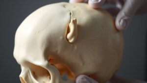
Skull exostosis, a bony outgrowth on the skull, is a rare but usually harmless condition. You could notice it as a small, hard lump on your head, but it’s typically a benign bone growth that doesn’t cause problems.
While some people have a genetic predisposition to developing these growths, they often don’t show symptoms. In the event you do experience discomfort or cosmetic concerns, your doctor might recommend imaging studies like X-rays or CT scans for diagnosis.
Most cases don’t need treatment, but in the event that the growth presses on nerves or causes pain, surgical removal could be an option. The key takeaway? It’s usually nothing to worry about, but getting it checked guarantees peace of mind.
Common Causes and Risk Factors of Skull Exostosis
While many assume a bump on the head comes from a recent bump or bruise, skull exostosis often develops over time due to foundational causes you couldn’t expect. These benign bone growths, like osteomas, aren’t usually painful but can stem from surprising triggers.
Here’s what may lead to them:
- Hereditary factors: Provided your family has a history of bone growths, like hereditary multiple exostoses (HME), you’re more likely to develop them too.
- Chronic irritation: Repeated pressure or minor injuries to your skull-think tight headgear or sports-can spur exostosis.
- Environmental factors: Long-term inflammation or exposure to irritants might encourage bone overgrowth.
- Joint issues: Conditions like osteoarthritis near the skull can trigger bone spurs.
Understanding these causes helps you spot risks promptly, even should exostosis stays harmless.
Symptoms Associated With Skull Exostosis
In case you’ve noticed a firm, slow-growing bump on your head, it couldn’t be something you bumped into-it could be skull exostosis. This bony growth often feels hard and stationary under your skin.
While many cases are asymptomatic and only spotted during imaging tests for other issues, some people experience localized pain or discomfort, especially when the exostosis presses on nerves or tissues. You may also feel stiffness in your neck or occasional headaches when the growth affects nearby muscles.
Rarely, complications like cosmetic changes or pressure on critical structures arise, which can lead to surgical intervention. Most of the time, though, it’s harmless and doesn’t need treatment unless symptoms become bothersome.
Should you be concerned, a doctor can help determine the next steps.
Diagnostic Methods for Skull Exostosis
You’ll likely start with a physical exam where your doctor checks for unusual bumps or tenderness on your skull.
In case they suspect exostosis, they’ll probably order imaging tests like X-rays or CT scans to get a clear depiction of the growth.
These steps help rule out other conditions and confirm whether the bump is harmless or needs further attention.
Physical Examination Findings
During a physical exam for skull exostosis, your doctor will carefully feel along your head to check for unusual bumps or hard spots. They’ll look for signs of a bony growth and ask about your medical history, including any symptoms like pain or discomfort.
Here’s what to expect:
- Palpation: Your doctor will gently press on your skull to locate any firm, irregular lumps that could signal an exostosis.
- Visual Inspection: They’ll check for visible deformities or asymmetry in your head’s shape.
- Symptom Discussion: You’ll be asked about any headaches, tenderness, or changes you’ve noticed.
- Next Steps: Should a bony growth be suspected, imaging tests could be recommended to confirm the diagnosis.
This thorough exam helps rule out other conditions and guides further testing.
Imaging Techniques Used
Several imaging techniques can help confirm whether you have a skull exostosis, giving doctors a clear representation of any bony growths.
X-rays are often the initial step, showing the location and size of the bony outgrowths. Provided more detail is needed, CT scans provide sharper images, revealing the exostosis’s exact shape and how it affects nearby structures.
An MRI could be used should there be concern about soft tissue involvement, like nerves or blood vessels. For cases where multiple growths are suspected, bone scintigraphy can scan your entire skeleton, highlighting areas with abnormal bone activity.
These imaging techniques guarantee an accurate diagnosis, helping your doctor plan the best treatment. Each method has its strengths, so your doctor will choose the right one based on your specific needs.
Differential Diagnosis Steps
Because skull exostosis can sometimes mimic other conditions, doctors take careful steps to rule out similar issues and confirm the diagnosis.
Here’s how they pinpoint it:
- Physical examination: They’ll check for lumps, tenderness, or unusual bone growths on your skull, noting any family history of bone disorders.
- Imaging techniques: X-rays give an initial look, while CT scans offer detailed 3D views to spot the exostosis’s size and shape.
- Biopsy: Should the growth look suspicious, they may remove a small sample to test for cancer or other abnormalities.
- Specialist input: Orthopedic surgeons or radiologists weigh in to interpret results and guide next steps.
This careful differential diagnosis guarantees you get the right answers-and the right care.
Imaging Techniques Used in Diagnosis
In case you’ve been informed you could have exostosis of the skull, imaging tests can help your doctor get a clear visual representation of what’s going on. X-rays are often the initial step, showing abnormal bone growths on your skull.
Should more detail be needed, CT scans provide cross-sectional images to pinpoint the size and location of the exostosis. MRIs help evaluate soft tissue nearby, checking whether nerves or other structures are affected.
Bone scans can detect increased bone activity, which could signal changes in the exostosis.
These imaging techniques guarantee an accurate diagnosis, helping your doctor plan the best next steps. Each test has its strengths, so your medical team will choose the right one based on your specific situation.
Non-Surgical Treatment Options
Should you be handling skull exostosis, managing pain is often your initial step-over-the-counter NSAIDs can help reduce inflammation and discomfort.
Physical therapy may also be recommended to improve movement and strengthen nearby muscles, making daily life easier.
These non-surgical options can keep symptoms under control without needing more invasive treatments.
Pain Management Strategies
Once exostosis of the skull causes discomfort, you don’t always need surgery to find relief. Non-surgical treatment can effectively manage pain and inflammation, helping you stay comfortable. Here’s how you can tackle symptoms without going under the knife:
- NSAIDs like ibuprofen reduce swelling and ease pain, making daily tasks more manageable.
- Ice packs applied for 15–20 minutes soothe the area, numbing sharp discomfort.
- Gentle physical therapy strengthens muscles around the growth, improving mobility and reducing strain.
- Activity adjustments-like avoiding tight headwear-prevent irritation and flare-ups.
Regular check-ins with your doctor guarantee your management plan stays effective.
By combining these strategies, you’ll keep pain under control while avoiding unnecessary procedures.
Listen to your body and tweak habits as needed-small changes make a big difference.
Physical Therapy Benefits
Physical therapy offers a practical way to ease discomfort from skull exostosis without surgery. By focusing on pain relief and mobility improvement, a therapist can guide you through targeted exercises to reduce tension around the affected area.
Strengthening exercises for your neck and shoulders help stabilize your head, easing strain caused by the bony growth. Manual therapy techniques, like gentle massage or joint mobilization, increase blood flow and relax tight muscles, making movement easier.
You could also learn posture adjustments to avoid aggravating the exostosis during daily tasks. Therapists sometimes use tools like ultrasound to soothe pain and promote healing.
With consistent sessions, you’ll likely notice less discomfort and better flexibility, helping you manage skull exostosis more comfortably.
Surgical Interventions for Severe Cases
Whenever exostosis of the skull becomes severe, surgery could be the best option to relieve pressure on nerves, ease pain, or prevent further complications.
Here’s what you should know about surgical interventions:
- Craniotomy or osteotomy: Surgeons might remove the exostosis by carefully cutting into the skull, preserving healthy bone.
- Guided by imaging studies: CT scans or MRIs pinpoint the exact location, guaranteeing precision during the procedure.
- Recovery trajectory: Most patients heal well, with minimal long-term effects in the event the growth is fully removed.
- Monitoring for recurrence: Follow-ups help catch any regrowth promptly, so you stay ahead of potential issues.
Surgery isn’t always needed, but at the time it is, these steps guarantee you’re back on track with less discomfort.
Trust your medical team-they’ll guide you through each phase.
Potential Complications of Skull Exostosis
You may experience nerve compression when the growth presses on nearby nerves, leading to pain or numbness.
Surgical intervention can help, but it’s not without risks like infection or slow healing.
Being aware of these complications helps you make informed choices about treatment.
Nerve Compression Risks
Because skull exostosis involves bony overgrowths, they can sometimes press on nearby nerves, leading to discomfort or even more serious issues like numbness or tingling in your face or scalp.
Should the exostosis grow near critical nerve pathways, you could experience:
- Persistent pain or dull aches in your head or neck.
- Tingling or numbness in your scalp, face, or jaw, signaling nerve compression.
- Headaches that worsen over time, especially should the exostosis irritate sensitive nerves.
- Weakness or muscle twitching in facial areas, hinting at potential neurological deficits.
Prompt diagnosis is key to preventing long-term damage.
Should symptoms persist, your doctor could recommend treatment, including surgical intervention to relieve pressure on affected nerves.
Don’t ignore these signs-getting checked promptly can make a big difference.
Surgical Intervention Outcomes
While surgery can relieve pressure from skull exostosis, it’s essential to know what to expect afterward-including possible complications.
Surgical intervention is often successful, but like any procedure, it carries risks. You may face infection, bleeding, or reactions to anesthesia, though pre-surgery checks help reduce these.
Post-surgery, swelling and bruising are common but usually fade in weeks. Rarely, pain or slow healing can occur.
Most exostosis growths are benign, but in hereditary cases, there’s a small chance they could turn malignant, like chondrosarcoma. That’s why follow-ups matter-they catch recurrence promptly.
Stay alert for new symptoms and keep appointments to guarantee long-term success. Your care team will guide you, so don’t hesitate to ask questions.
Recovery takes patience, but relief is worth it.
Recovery and Post-Treatment Care
After surgery for exostosis of the skull, your recovery will take several weeks, so it’s vital to take it easy and let your body heal.
Follow your doctor’s post-treatment care plan closely to guarantee a smooth healing period.
- Pain management: Use prescribed or over-the-counter medications as directed to stay comfortable, but don’t ignore sudden increases in pain-report them.
- Incision care: Keep the surgical site clean and dry to prevent infection, and follow instructions for dressing changes.
- Follow-up appointments: Attend all scheduled visits so your doctor can monitor progress and catch any issues promptly.
- Physical therapy: Should it be recommended, gently ease back into movement to restore function without straining your body.
Listen to your body and avoid heavy lifting or intense activity until cleared by your healthcare team.
Long-Term Outlook and Prognosis
Though skull exostosis could sound concerning, most people experience a positive long-term outlook. Since these growths are typically benign, they rarely cause serious problems. Should you not have symptoms, you might only need regular monitoring to guarantee nothing changes. For those who do experience headaches or neurological issues, surgical intervention often resolves the problem with a high success rate and low chance of recurrence.
Here’s a quick breakdown of what to expect:
| Factor | Impact | Action |
|---|---|---|
| Benign Nature | Rarely becomes harmful | Monitor unless symptomatic |
| Symptoms | May require treatment | Seek medical advice |
| Surgical Option | High success rate | Consider should symptoms worsen |
| Prognosis | Generally excellent | Regular check-ups recommended |
| Recovery | Quick return to normal activities | Follow post-op care |
Your prognosis is likely good, especially with proper care.


