Facial dermatomes are the areas of the face that are innervated by the trigeminal nerve. Recognizing these areas is essential for diagnosing and managing facial pain and neurological disorders.
In this post, I’ll review the anatomy, mapping, and clinical applications of facial dermatomes.
What are Dermatomes?
Dermatomes refer to the areas of skin supplied by specific spinal nerve roots, including the dorsal root ganglion. Dermatomes are a fundamental concept in neurology and pain medicine.
Each spinal nerve root innervates a specific region of the body, making them an essential building block for understanding sensory exams and diagnosing nerve and spinal cord pathology.
Dermatome maps are more than just a curiosity; they’re a practical clinical tool.
The maps link each skin area to its spinal nerve, making it easy to find the right dermatome based on spinal nerve anatomy.
Getting a handle on dermatomes is crucial when you’re checking someone’s sensory responses during a neurological exam.
By learning which patches of skin connect to each spinal nerve, you can figure out which nerves might be injured or affected by a condition.
This is super helpful for things like shingles, where a virus targets a specific skin area.
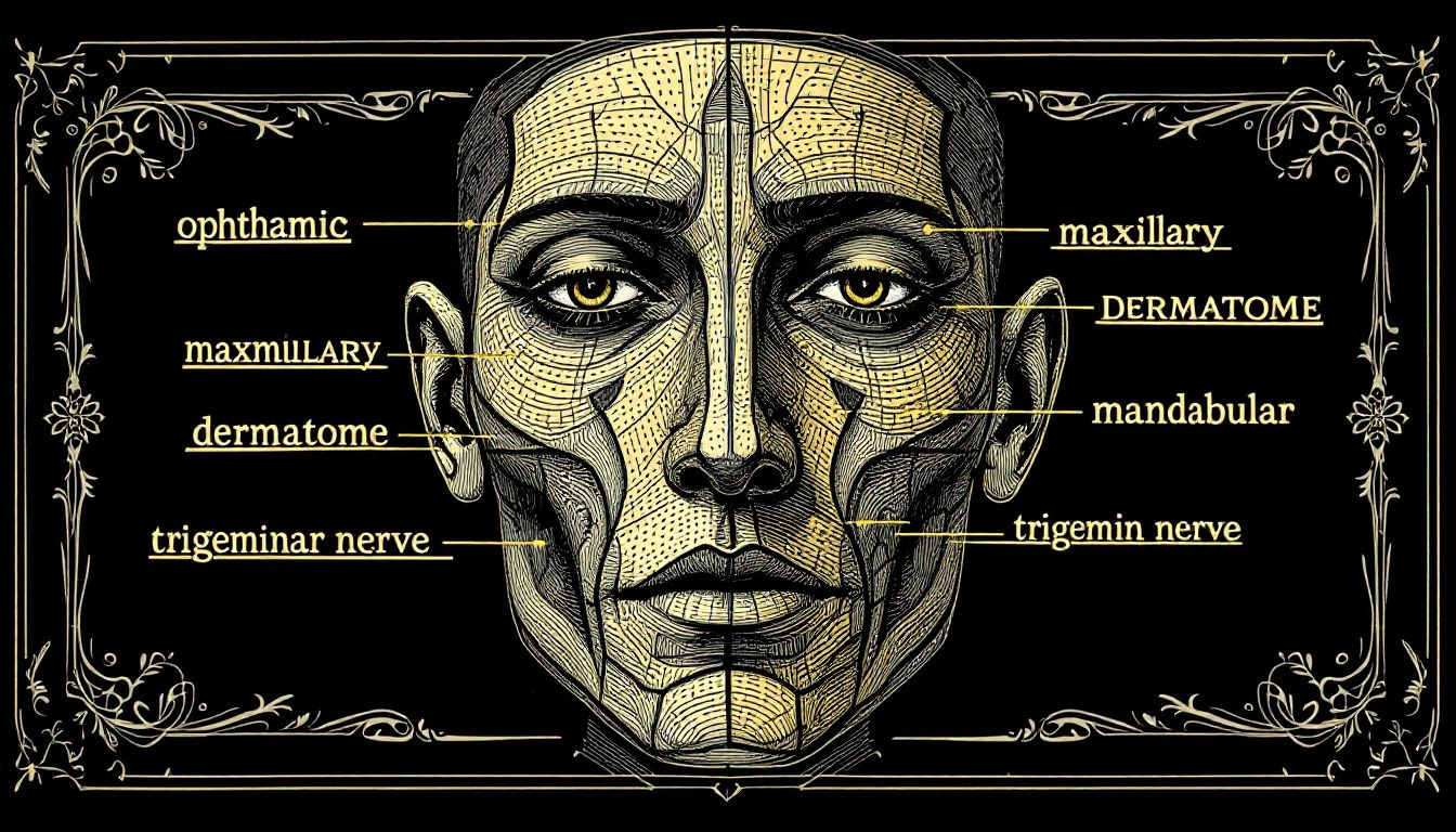
Facial Dermatome Anatomy
Facial dermatomes are supplied by the trigeminal nerve (CN V), also known as the fifth cranial nerve.
The trigeminal nerve carries sensory information from the face to the brain.
It has three main branches: V1 (ophthalmic), V2 (maxillary), and V3 (mandibular). Each branch supplies sensation to a specific part of the face.
Each branch of the trigeminal nerve supplies a specific region of the face:
- V1 (ophthalmic branch): upper eyelid, forehead, and anterior scalp
- V2 (maxillary branch): lower eyelid, upper lip, cheek, and portions of the nasal mucosa
- V3 (mandibular branch): lower lip, chin, jaw, and anterior two-thirds of the tongue. The mandibular branch also has motor fibers that control chewing (mastication) and opening the jaw (tongue deviation to the side).
The trigeminal nerve branches segment the face into areas supplied by different spinal nerves. This segmentation is important for facial pain diagnosis and management.
Facial Dermatome Map
The facial dermatome map divides the face into regions supplied by each branch of the trigeminal nerve.
The map is important for understanding facial sensory anatomy and evaluating facial disorders.
The facial dermatome map is a practical clinical tool, not just a diagram to memorize. It helps you quickly determine which trigeminal nerve branch is affected by a patient’s condition.
The map is a useful reference for visualizing the sensory distribution across the face.
Ophthalmic Branch (V1)
The ophthalmic branch (V1) supplies the upper eyelid, forehead, and anterior scalp.
This narrow region of the facial dermatome map also includes the tip of the nose, nasal mucosa, and dorsum of the nose.
The ophthalmic branch is important clinically because it supplies the cornea, which is critical for eye sensation and protection. V1 is responsible for reflexive eye closure and is essential for protecting the eye.
Conditions like herpes zoster ophthalmicus can cause severe complications, including corneal ulcers and vision loss, if left untreated.
Maxillary Branch (V2)
The maxillary branch (V2) supplies the lower eyelid, upper lip, and cheek. This branch also innervates the mucosa of the maxillary sinus and portions of the nasal mucosa.
It’s also good for diagnosing conditions like trigeminal neuralgia, as well as evaluating pain in the midface region.
Accurate V2 mapping is critical for identifying the correct area of pain and providing appropriate care for your patients.
Mandibular Branch (V3)
The mandibular branch (V3) supplies the:
- lower lip
- chin
- jaw
- external ear
- lower teeth and gums
The mandibular branch includes motor fibers that regulate the muscles involved in chewing and opening the jaw, along with causing the tongue to deviate to the side.
From a clinical perspective, the mandibular branch is noteworthy as it covers regions often affected by dental problems and temporomandibular joint (TMJ) disorders
Clinical Relevance
Facial dermatomes are important to understand and map clinically.
Accurate facial dermatome mapping is essential for diagnosing and managing conditions like trigeminal neuralgia and herpes zoster.
Of course, mapping facial dermatomes is vital for assessing patients with conditions such as trigeminal neuralgia and herpes zoster.
Both conditions typically present with localized pain and sensory symptoms that correspond to specific dermatomes.

Trigeminal Neuralgia
Trigeminal neuralgia is a chronic, debilitating condition characterized by severe, electric shock-like pain in the face.
The pain is typically confined to one side of the face and is often triggered by chewing, brushing teeth, smiling, or applying makeup.
This condition is more common in women and typically affects individuals over the age of 50.
The cause of trigeminal neuralgia is often pressure on the trigeminal nerve by a nearby blood vessel, but it can also be related to multiple sclerosis, tumors, facial trauma, or other lesions compressing the nerve.
Herpes Zoster (Shingles)
Herpes zoster, also known as shingles, is caused by the reactivation of the Varicella–Zoster virus.
The condition is characterized by discoloration, pain, and a band of blisters along an affected dermatome.
Herpes zoster is more common in individuals over the age of 50 and typically lasts three to five weeks.
Antiviral medications and pain medications are common treatments for herpes zoster. A vaccine for chickenpox (Varicella) is also protective against herpes zoster.
Accurate diagnosis and treatment are important, especially when the ophthalmic branch (V1) is involved.
Dermatome Maps Vary
Dermatome maps are not consistent from person to person.
The maps can vary due to differences in segmental spinal nerve anatomy and intrathecal intersegmental anastomoses. These anastomoses are connections between spinal nerve roots that can occur at various levels.
One example of this is the C4 dermatome. On the Keegan and Garrett map, the C4 dermatome is represented in a narrow band at the base of the neck and upper anterior clavicles.
The C5 dermatome is typically more prominent in this region. This is just one example of the variations that can occur between dermatome maps.
Note : It’s essential to consult multiple resources and use clinical judgment when evaluating your patients.
Read More : Hand Dermatomes
Clinical Relevance
It’s not uncommon to find different dermatome maps in various resources. While this can be confusing, it’s also an educational opportunity.
By comparing the maps and understanding the reasons for variations, you can develop a more complete understanding of human dermatomes.
Diagnostic Evaluation
Evaluating conditions that affect dermatomes requires knowledge of dermatome maps and sensory assessment techniques.
Dermatome testing, often performed using a pin and cotton wool, is used to assess light touch and pain.
For more complete sensory evaluation, it’s important to test both light touch and pinprick sensation.
Recommendations
A circumferential pattern is recommended for testing dermatomes to help differentiate between sensory loss across dermatomes.
Light touch is often a less sensitive test than pain for evaluating dermatomes.
It’s important to use both techniques during your sensory exams. Always ask your patients to alert you if they experience any areas of numbness or weakness, as this is critical information for your diagnosis.
As with any anatomical concept, it’s important to remember that dermatome patterns vary from person to person.
During your neurological exams, you must take the time to appreciate the unique patterns on each of your patients.
Apply your knowledge of dermatomes to provide appropriate care, and always consider the sensory level when evaluating your patients.
Accurate diagnosis of spinal cord and peripheral nerve pathology depends on your understanding of dermatomes.
Trustworthy Educational Resources
Trustworthy educational resources are critical for understanding dermatomes.
Kenhub’s dermatome guide is a valuable learning resource that is based on published literature and validated by experts.
These resources are perfect for medical students, physicians, and anyone else interested in learning about dermatomes in depth.
Credible educational resources are important for understanding the educational and clinical implications of dermatome maps.
These resources are essential for both learning about dermatomes and applying that knowledge in the clinical setting.
Final Thoughts
So that’s it. Dermatomes help us understand skin sensations and find nerve issues.
The facial dermatomes, supplied by the trigeminal nerve, are important to understand due to conditions like trigeminal neuralgia and herpes zoster.
Accurate mapping and diagnosis of these dermatomes is critical for providing appropriate care.
As you continue to learn about and apply your knowledge of dermatomes, remember that every touch and sensation on the skin holds a story. The skin is the reveal of the nervous system’s intricate workings.