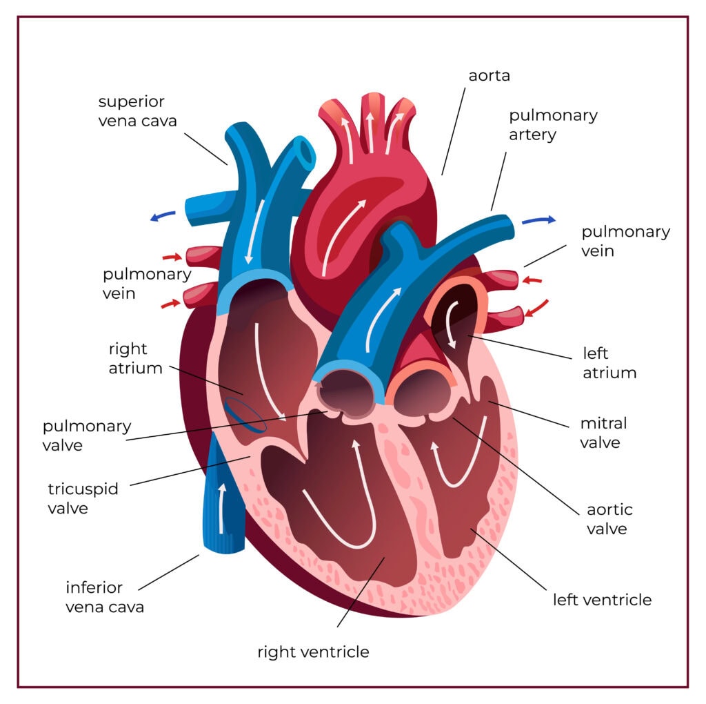
A heart diagram visually explains how the human heart works. It shows the heart’s chambers, valves, and blood flow.
The heart consists of four chambers and four valves that regulate blood flow, ensuring efficient circulation of oxygenated and deoxygenated blood throughout the body. This article will help you understand the heart’s structure and function through detailed diagrams
Heart Diagram
Heart diagrams are powerful visual tools that help us grasp the complex structure and function of the human heart. These diagrams are not just for medical students or professionals; they are invaluable for anyone looking to understand how their heart works. These diagrams simplify understanding of how the heart pumps blood and how its parts interact by illustrating its anatomy.
More than just static images, interactive heart diagrams allow users to label different parts of the heart, enhancing their learning experience. These visual aids are particularly useful in understanding cardiovascular diseases, as they can clearly show how conditions like coronary artery disease affect heart function.
These diagrams make the circulatory system more comprehensible and less of a mystery.
Anatomy of a Healthy Heart
The heart, about the size of a fist and weighing around 10 ounces, is a vital component of our cardiovascular system. Nestled behind and slightly to the left of the sternum, between the right and left lungs, the heart’s intricate structure is crucial for its function.
Exploring the heart’s chambers, valves, and layers of the heart wall reveals their unique roles in maintaining efficient blood flow and overall health.
Chambers of the Heart
The heart’s four chambers are divided into two upper chambers, the atria, and two lower chambers, the ventricles. The right atrium receives deoxygenated blood from the body, which then flows into the right ventricle to be pumped to the lungs for oxygenation. The left atrium, on the other hand, receives oxygenated blood from the lungs and passes it to the left ventricle, the strongest chamber, which then pumps it to the rest of the body.
The septum, a muscular wall, separates the left and right sides of the heart, preventing the mixing of oxygenated and deoxygenated blood. This division is crucial for the efficient functioning of the cardiovascular system, ensuring that oxygen-rich blood is distributed to the body while deoxygenated blood is sent to the lungs for reoxygenation.
Valves of the Heart
The heart’s four valves ensure that blood flows in one direction, preventing any backflow. The atrioventricular valves, which include the tricuspid valve between the right atrium and right ventricle and the mitral valve between the left atrium and left ventricle, play a crucial role in this process. These valves open to allow blood flow from the atria to the ventricles and close to prevent backflow during ventricular contractions.
The semilunar valves, including the pulmonary valve at the exit of the right ventricle and the aortic valve at the exit of the left ventricle, ensure that blood flows smoothly from the ventricles into the arteries. These valves open when the ventricles contract and close to prevent blood from flowing back into the heart once it has been pumped out.
Layers of the Heart Wall
The heart wall consists of three layers, each with a specific function. The outermost layer, the epicardium, provides a protective covering for the heart. Beneath it lies the thick middle layer, the myocardium, composed of cardiac muscle responsible for the heart’s powerful contractions.
The inner layer, the endocardium, lines the heart chambers and covers the heart valves. This smooth lining ensures that blood flows efficiently within the heart, reducing friction and preventing damage to the heart’s interior surfaces.
Together, these layers form a robust structure that supports the heart’s continuous pumping action.
Blood Flow Through the Heart
Blood flow through the heart follows a precise sequence, ensuring that oxygenated and deoxygenated blood are efficiently circulated. Grasping this process is crucial for understanding how the heart functions within the cardiovascular system.
We’ll explore the detailed pathways and the role of heart valves in maintaining this vital flow.
Pathway of Blood
Blood enters the heart through the right atrium from the body via the superior and inferior vena cavae. From the right atrium, blood flows into the right ventricle, which then pumps it to the lungs through the pulmonary arteries for oxygenation. This journey marks the transition from deoxygenated to oxygenated blood.
Once oxygenated, blood returns to the heart through the pulmonary veins into the left atrium. It then moves into the left ventricle, which pumps the oxygen-rich blood to the rest of the body through the aorta. This continuous cycle ensures that oxygen and nutrients are delivered to tissues, while waste products like carbon dioxide are removed.
Role of Valves in Blood Flow
Heart valves play a critical role in maintaining unidirectional blood flow. The atrioventricular valves, located between the atria and ventricles, open to allow blood to flow from the atria to the ventricles and close to prevent backflow during ventricular contractions. This mechanism ensures efficient blood circulation within the heart.
Semilunar valves, located at the exits of the ventricles, open when the ventricles contract to allow blood to flow into the arteries. They then close to prevent blood from flowing back into the ventricles once it has exited the heart. The proper functioning of these valves is crucial for maintaining the heart’s efficiency and preventing complications.
Circulatory System Overview
The circulatory system is a complex network that transports blood, nutrients, gases, and waste products throughout the body. The heart, as a powerful muscular pump, plays a central role in this system, ensuring that blood continuously cycles through the body to maintain homeostasis and overall health.
Let’s delve into the specifics of oxygenated and deoxygenated blood and the major blood vessels involved.
Oxygenated vs Deoxygenated Blood
Oxygenated blood, rich in oxygen, is essential for supplying the body’s tissues with the oxygen they need for cellular respiration. After gas exchange in the lungs, oxygenated blood returns to the left atrium through the pulmonary veins and then moves into the left ventricle to be pumped to the rest of the body.
In contrast, deoxygenated blood passes, which has delivered its oxygen and picked up carbon dioxide and other waste products from the tissues, returns to the heart through the right atrium. It then flows into the right ventricle, which pumps it to the lungs for reoxygenation.
This continuous cycle ensures that oxygen and nutrients are delivered to the body’s cells while waste products are efficiently removed.
Major Blood Vessels
The major blood vessels play crucial roles in the circulatory system. The aorta, the largest artery in the body, carry blood from the left ventricle to the systemic circulation, delivering oxygenated blood and nutrients to tissues and organs.
The superior and inferior vena cava are the primary veins that return deoxygenated blood from the body back to the heart. Additionally, the pulmonary arteries transport deoxygenated blood from the right ventricle to the lungs for oxygenation. These vessels ensure that blood flows efficiently between the heart, lungs, and the rest of the body.
Heart Diagram Labeling and Identification
Heart diagrams are not only educational tools but also interactive aids that enhance understanding of the heart’s complex structure. Labeling exercises help individuals better understand the functions and interconnections of different heart parts.
This section covers how to label key parts of the heart and understand variations in heart diagrams.
Labeling Key Parts of the Heart
Labeling the heart’s major parts involves identifying the four chambers, valves, and key blood vessels. The right atrium collects deoxygenated blood from the body, which then flows into the right ventricle to be pumped to the lungs. The left atrium receives oxygenated blood from the lungs and passes it to the left ventricle, which pumps it out to the body through the aorta.
Heart valves, such as the tricuspid, pulmonary, mitral, and aortic valves, ensure that blood flows in one direction, preventing backflow. The vena cavae and pulmonary veins play crucial roles in returning deoxygenated and oxygenated blood to the heart.
Common Heart Diagram Variations
Heart diagrams can vary widely, from simple anatomical illustrations to complex physiological representations. Anatomical diagrams focus on the physical structure of the heart, detailing its chambers, valves, and vessels. These diagrams are essential for understanding the heart’s layout and spatial relationships.
Physiological diagrams, on the other hand, emphasize the functional aspects of the heart, such as blood flow patterns and electrical impulses. These diagrams are particularly useful for visualizing how the heart works during different phases of the cardiac cycle. Both types of diagrams provide valuable insights, depending on the educational or clinical context.
Common Heart Conditions Visualized
Understanding common heart conditions through visual aids can significantly enhance our comprehension of these diseases. Heart diagrams can illustrate how conditions like coronary artery disease and congenital heart defects affect heart function and structure.
Let’s delve into these conditions in more detail.
Coronary Artery Disease
Coronary artery disease (CAD) is a leading cause of cardiovascular diseases, often referred to as a ‘silent killer’ due to its asymptomatic nature until severe events like heart attacks occur.
This condition occurs when the blood vessels that supply blood to the heart muscle become narrowed or blocked, reducing blood flow and oxygen supply. Visualizing CAD through diagrams can help us understand how these blockages impact heart function and lead to complications such as acute coronary syndrome.
Treatment options for CAD include surgical procedures like angioplasty or bypass surgery, which aim to restore blood flow to the heart muscle. Cardiac rehabilitation programs also offer structured support for patients recovering from heart-related conditions, helping them to improve their heart health and prevent future complications.
Congenital Heart Defects
Congenital heart defects are structural abnormalities present at birth that can affect the heart’s function and blood flow. These defects can range from minor issues, such as small holes in the heart, to more severe problems like leaky valves or malformed heart chambers. Visual representations of these defects help illustrate how they alter the heart’s anatomy and impact its ability to pump blood efficiently.
Some congenital heart defects may require medical intervention, either soon after birth or later in life, to correct the structural issues and improve heart function. Diagrams of these conditions aid in diagnosis and treatment planning, ultimately enhancing the quality of life for those affected.
Bottom Line
The heart’s intricate structure and function are vital to our survival, and understanding how it works can empower us to take better care of it. From the anatomy of a healthy heart to the pathways of blood flow and the roles of various heart components, this blog post has provided a comprehensive guide.