As we age, our muscles tend to lose their elasticity and flexibility. This can lead to discomfort, pain, and even injuries. One area of the body that often suffers from reduced flexibility is the hips.
The hip joint is a ball-and-socket joint that allows for a wide range of motion. It is surrounded by several muscles, tendons, and ligaments that work together to support movement. When these muscles become tight or stiff, it can limit the hip’s range of motion and cause discomfort.
The major hip muscles include the flexors, extensors, abductors, adductors, and rotators. Each group plays a vital role in activities such as walking, running, and maintaining balance. This article will provide a detailed guide on the anatomy and functions of these important muscles.
Key Takeaways
Hip muscles are key players in stability and movement, categorized into flexors, extensors, abductors, adductors, and rotators.
Injury prevention is crucial; common issues include hip flexor strain, gluteal strains, and conditions like piriformis syndrome.
Nerve supply is vital for hip muscle function, with the femoral and obturator nerves facilitating leg movements, and damage can lead to mobility issues.
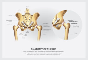
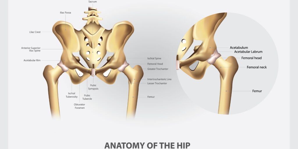
Hip Muscle Anatomy
Hip muscles provide stability and mobility, enabling activities such as walking, running, and maintaining posture. The hip joint connects the lower limbs to the axial skeleton, allowing for various movements and supporting body weight. Movements like sitting, standing, and jumping rely on these muscles, joints, and hip bones.
Hip joints are integral to balance and overall body movement. The hip muscles can be classified based on their functions into categories like flexors, extensors, abductors, adductors, and rotators.
The major groups of hip muscles include the gluteal group, lateral rotator group, adductor group, and iliopsoas group. Each of these groups plays a unique role in facilitating various hip movements and maintaining stability.
Hip Flexors
Hip flexors bring the knee closer to the chest, especially during walking and running. They are crucial for activities involving hip flexion, such as climbing stairs, running, and cycling.
Iliopsoas
The iliopsoas muscle maintains postural stability and is the body’s most significant hip flexor. It consists of three muscles: iliacus, psoas major, and psoas minor, each contributing to hip flexion and stability. The iliopsoas is formed by the iliacus and psoas major, which together facilitate hip flexion and help stabilize the lumbar spine.
The psoas major muscle originates from the lumbar vertebrae and inserts onto the lesser trochanter of the femur. Meanwhile, the iliacus muscle arises from the iliac fossa and also attaches to the lesser trochanter, assisting in hip flexion.
Rectus Femoris
The rectus femoris is one of the quadriceps muscles that serves dual roles in flexing the hip and extending the knee. It has two points of origin: the anterior inferior iliac spine and the superior margin of the acetabulum.
This muscle performs dual functions, aiding in both hip flexion and knee extension. Its dual functionality is vital for movements involving both the hip and knee joints.
Sartorius
Sartorius, the longest muscle in the body, aids in flexing, abducting, and laterally rotating the hip. It starts at the anterior superior iliac spine and inserts at the medial aspect of the tibia. This muscle aids in hip flexion, abduction, and lateral rotation of the thigh.
Its unique positioning and length allow it to assist in various movements of the hip and thigh.
Tensor Fasciae Latae
The tensor fasciae latae stabilizes the hip and assists in its flexion and abduction. It originates from the iliac crest and the anterior superior iliac spine. It contributes to the flexion and abduction of the hip joint, and stabilizes the pelvis during ambulation.
It is crucial for maintaining balance and stability during movement.
Hip Extensors
Hip extensors, including gluteus maximus, biceps femoris, semitendinosus, and semimembranosus, primarily move the leg backward and support posture. They are crucial for activities like standing up, climbing stairs, and pushing heavy objects.
Gluteus Maximus
The gluteus maximus originates from the posterior part of the ilium and the lateral mass of the sacrum. Its main role is to extend and externally rotate the thigh, particularly during activities like rising from a seated position. This muscle is the largest of the gluteal muscles and significantly contributes to the shape of the buttock.
Its strength and size enable powerful movements involving hip extension.
Hamstrings
The hamstring muscles, including the biceps femoris, semitendinosus, and semimembranosus, play key roles in both hip extension and knee flexion. The biceps femoris, one of the hamstrings, is especially important for these functions.
The semitendinosus and semimembranosus muscles also contribute to hip extension and help in the medial rotation of the thigh. They are essential for movements involving the hip and knee joints.
Hip Abductors
Hip abductors move the leg away from the body’s midline and prevent waddling or Trendelenburg gait. These muscles are located on the outer side of the hip and buttock.
Gluteus Medius
The gluteus medius starts from the gluteal surface of the ilium. It is located between the anterior and posterior gluteal line. It stabilizes the pelvis during activities like walking and standing.
It inserts on the greater trochanter of the femur, aiding in thigh abduction and pelvis stabilization during movement. Its function is essential for balance and proper gait.
Gluteus Minimus
The gluteus minimus also arises from the ilium but is located beneath the gluteus medius, contributing to the hip’s internal rotation. Its contraction assists in both hip abduction and the internal rotation of the thigh.
The gluteus minimus originates at the gluteal surface of the ilium and attaches to the anterior aspect of the greater trochanter. It aids in thigh abduction and internal rotation while also stabilizing the pelvis.
Tensor Fasciae Latae
The tensor fasciae latae muscle helps maintain stability in the pelvis while the hip is abducted. It functions to abduct the hip and maintain pelvic stability during movement.
Hip Adductors
Hip adductors are essential for drawing the thigh towards the body’s midline, enabling movements such as crossing legs, swimming, and squeezing legs during exercises like hip thrusts. These muscles play a significant role in various daily activities and sports.
Adductor Longus
The adductor longus originates from the pubis and inserts on the middle third of the linea aspera of the femur. It assists in flexing the thigh when extended and contributes to adduction at the hip joint. It is essential for movements that bring the legs together and stabilize the hip joint.
Adductor Brevis
The adductor brevis starts from the body of the pubis and the inferior pubic ramus. Its insertion is at the posterior surface of the femur, proximal to the adductor longus.
It plays a significant role in drawing the thigh towards the body during movements, aiding in hip adduction. It is crucial for balance and proper alignment during dynamic activities.
Adductor Magnus
The adductor magnus has two parts with distinct origins: one from the ischiopubic ramus and the other from the ischial tuberosity. It is responsible for both adducting and extending the thigh at the hip joint. It assists in thigh adduction and extension, essential for movements requiring strong and stable hip joints.
Gracilis
The gracilis muscle originates from the inferior ramus of the pubis. It functions by adducting the thigh and aiding in flexion of the leg at the knee.
Gracilis inserts onto the medial surface of the proximal tibia, primarily flexing the leg at the knee and aiding in hip adduction. It is essential for movements requiring hip and knee coordination.
Pectineus
The pectineus muscle originates from the superior ramus of the pubis and extends to the upper part of the linea aspera and lesser trochanter of the femur. This positioning allows it to aid in both adduction and flexion of the thigh at the hip joint.
It contributes to flexing and adducting the thigh, essential for movements that bring the legs together and stabilize the hip. Its dual function makes it an essential part of the adductor group.
Hip Rotators
Hip rotators facilitate internal and external rotation of the hip joint, enabling a range of movements. They are essential for activities like walking, running, and any motion involving turning the leg.
External Rotators
The external rotators, including:
the piriformis
quadratus femoris
superior gemellus
inferior gemellus
obturator externus
play a key role in laterally rotating the hip. For example, the piriformis muscle runs from the sacrum to the greater trochanter and is crucial for this function. They also assist in hip abduction and stabilization during movements.
The quadratus femoris, located on the ischial tuberosity, is involved in externally rotating and adducting the hip joint.
Internal Rotators
Muscles involved in internal rotation of the hip include the gluteus minimus and tensor fasciae latae. The gluteus minimus is vital for the internal rotation of the hip and stabilizes the pelvis during movement.
The tensor fasciae latae aids in internal rotation of the hip while also stabilizing the hip and knee joints. Together, these muscles ensure smooth and controlled hip movements.
Hip Stabilizers
Hip stabilizers maintain pelvic alignment and stability during various activities. They ensure pelvic stability, enabling proper posture and movement.
Iliopsoas
The iliopsoas muscle is the main hip flexor and also aids in external rotation, significantly contributing to the stabilization of the hip joint. This muscle stabilizes the pelvis when standing, crucial for various daily activities.
Its role in pelvic stability and hip flexion is vital for maintaining balance and proper alignment during movements. Without the iliopsoas, standing and walking would be greatly impaired.
Gluteus Medius
The gluteus medius plays a vital role in maintaining pelvic stability, especially while walking or running. It is essential for keeping the pelvis level, preventing it from dropping on the opposite side.
During walking, the gluteus medius keeps the pelvis stable, enabling smooth and balanced movements. Its function is crucial for avoiding gait abnormalities.
Adductors
The adductor muscles assist in stabilizing the hip joint by controlling movements and maintaining proper alignment during various activities. These muscles are vital for controlling lateral movements of the hip and maintaining stability during dynamic activities.
Their role in stabilizing the hip joint ensures smooth, coordinated movements, preventing injuries and maintaining balance. The adductors are essential for activities that involve lateral movements.
Nerve Supply to Hip Muscles
Nerves in the hip region facilitate muscle movement and deliver sensory information such as touch and pain. They ensure that hip muscles function correctly and respond to stimuli.
Femoral Nerve
The femoral nerve primarily innervates muscles that are responsible for knee extension and hip flexion. It is the largest nerve in the lumbar plexus, comprising fibers from the L2-L4 spinal nerves. Damage to the femoral nerve can lead to issues like quadriceps weakness, affecting mobility.
It is essential for movements involving the hip and knee joints.
Obturator Nerve
The obturator nerve innervates the adductor muscles of the thigh, which are essential for bringing the legs together. It emerges from the lumbar plexus and primarily innervates the adductor muscles of the thigh.
Injury to the obturator nerve can result in difficulty with leg adduction and impact gait. It is responsible for sensation in the skin of the medial thigh, facilitating movements like adduction.
Sciatic Nerve
The sciatic nerve is responsible for innervating the hamstring muscles and the adductor magnus. It is the largest single nerve in the body, branching into the tibial and common peroneal nerves.
Compression of the sciatic nerve can lead to pain radiating down the leg, known as sciatica. It innervates muscles in the posterior thigh and all muscles in the leg and foot, affecting both movement and sensation.
Superior Gluteal Nerve
The superior gluteal nerve innervates the gluteus medius, gluteus minimus, and tensor fasciae latae muscles. Damage to this nerve can result in a trendelenburg gait, where the pelvis tilts towards the non-affected side. It plays a vital role in hip abduction and stabilizing the pelvis during walking.
Its function is crucial for balance and proper gait.
Inferior Gluteal Nerve
The inferior gluteal nerve primarily innervates the gluteus maximus muscle, which is essential for hip extension. Injury to this nerve can compromise the ability to stand from a seated position or climb stairs. It is crucial for movements involving powerful hip extension.
Its function is vital for activities requiring strong, stable hip joints.
Diagram
Common Disorders and Injuries of Hip Muscles
Hip injuries are prevalent among individuals of all ages, particularly in athletes, seniors, and those who are overweight. Recognizing common disorders and injuries of hip muscles aids in their prevention and treatment.
Hip Flexor Strain
Hip flexor strains often occur due to overuse or sudden movements, leading to pain in the front of the hip. Common symptoms are pain, tightness in the hip, and difficulty walking.
Treatment typically includes rest, ice, and over-the-counter medications to reduce pain and inflammation. Physical therapy may also be recommended to strengthen the hip muscles and prevent future injuries.
Gluteal Muscle Strain
Strains of the gluteal muscles can result in pain while performing lateral movements or rising from a seated position. Gluteal strains can occur during activities that involve sudden movements or excessive force.
Common treatments include physical therapy and targeted stretching exercises to alleviate pain and restore function. Strengthening gluteal muscles can prevent future strains.
Piriformis Syndrome
Piriformis syndrome causes pain in the buttock area, which may radiate down the leg due to sciatic nerve compression. This condition can lead to sciatica symptoms, making daily activities challenging.
Diagnosis typically involves physical examinations and imaging tests like MRI to rule out other conditions. Treatment may include physical therapy, stretching exercises, and medications to reduce pain and inflammation.
Hip Impingement
Hip impingement can lead to discomfort during hip flexion and internal rotation, affecting movement quality. It can cause joint pain and limited range of motion in the hip joint.
Two main types of hip impingement are CAM and Pincer, each affecting the hip differently. Treatment options may include physical therapy, medications, and in severe cases, surgery to relieve symptoms.
Primary Hip Flexors
Primary hip flexors include the iliacus, psoas major, and rectus femoris. The iliopsoas muscle, formed by the iliacus and psoas major, significantly contributes to maintaining postural stability while standing. These hip flexor muscles play crucial roles in activities requiring hip flexion, such as walking and running.
The rectus femoris muscle attaches at the anterior inferior iliac spine and just superior to the acetabulum, flexing the hip and extending the knee.
Secondary Hip Flexors
Secondary hip flexors, such as the pectineus and sartorius muscles, contribute significantly to hip flexion and stabilization during movements. The pectineus functions as both a hip flexor and secondary adductor.
The sartorius, the longest muscle in the body, assists in hip flexion, adduction, and external rotation. Its unique positioning supports various movements of the hip and thigh.
Clinical Points
The femoral triangle is an important anatomical region that contains vessels and nerves supplying the thigh and is relevant in surgeries and injuries. Understanding the anatomy of this region, including the femoral artery, is crucial for medical professionals.
An abnormal Trendelenburg gait is characterized by a lateral trunk tilt towards the affected side during the stance phase. Duchenne–Trendelenburg syndrome demonstrates weakness of the hip abductors, highlighting the importance of these muscles.
Frequently Asked Questions
What are the primary hip flexor muscles?
The main hip flexor muscles you should know are the iliacus, psoas major, and rectus femoris. These guys help you lift your knees and bend at the hip!
What causes hip flexor strains?
Hip flexor strains usually happen from overuse or making sudden movements, so it’s important to warm up properly and avoid pushing too hard. Taking care of your hip flexors can help prevent this pain in the front of your hip.
How can I prevent hip muscle injuries?
To prevent hip muscle injuries, focus on maintaining good posture and incorporating targeted exercises to strengthen your hip muscles. Avoiding repetitive motions is also key to keeping those hips healthy!
What is the role of the gluteus medius in hip stability?
The gluteus medius is key for keeping your pelvis stable while you walk or run, so it stops your hip from dropping on the opposite side. Basically, strong gluteus medius muscles help keep you balanced and moving efficiently!
How is piriformis syndrome diagnosed?
Piriformis syndrome is typically diagnosed with a combination of physical exams and imaging tests like MRI to eliminate other possible issues. It’s all about pinpointing that pesky pain!