The human brain is a complex organ divided into three main regions: the forebrain, midbrain, and hindbrain, each responsible for various cognitive and autonomic functions. And major parts of the brain, including the cerebral cortex, cerebellum, and brainstem, work together to manage tasks ranging from sensory perception to vital life functions.
Want to see a clear human brain diagram?
For this reason, we are here to show you the visual diagram of human brain and explains the main brain regions and their functions.
Overview of the Human Brain
The human brain stands as the most intricate organ in the human body, orchestrating a symphony of intelligence, sensory interpretation, and motor control. It is the epicenter of our thoughts, memories, and speech, governing every aspect of our behavior and bodily functions.
The brain’s ability to control movements, organ functions, and responses to stress underscores its vital role in sustaining life, much like the adult human brain.
The nervous system, which includes the brain and spinal cord, is made up of two primary cell types: neurons and glial cells. Neurons are the signal transmitters, responsible for the rapid communication within the brain and to the rest of the body. Glial cells, on the other hand, provide essential support and maintenance for neurons, ensuring the brain’s optimal functioning.
Sounds good, right?
This intricate network of nerve cells enables the brain to perform a vast array of tasks, from simple reflexes to complex cognitive functions, including the central nervous system.
Various parts of the brain work in concert to control thoughts, memories, movement, sensory perception, and organ functions. The cerebral cortex, often referred to as gray matter, is where most of this information processing occurs.
Major Divisions of the Brain
The human brain is a complex organ divided into three main regions:
- The forebrain, which is the largest part, responsible for higher cognitive functions, sensory interpretation, and voluntary motor activities
- The midbrain, which plays a crucial role in vision, hearing, and motor control
- The hindbrain, which manages vital automatic functions such as heart rate and breathing
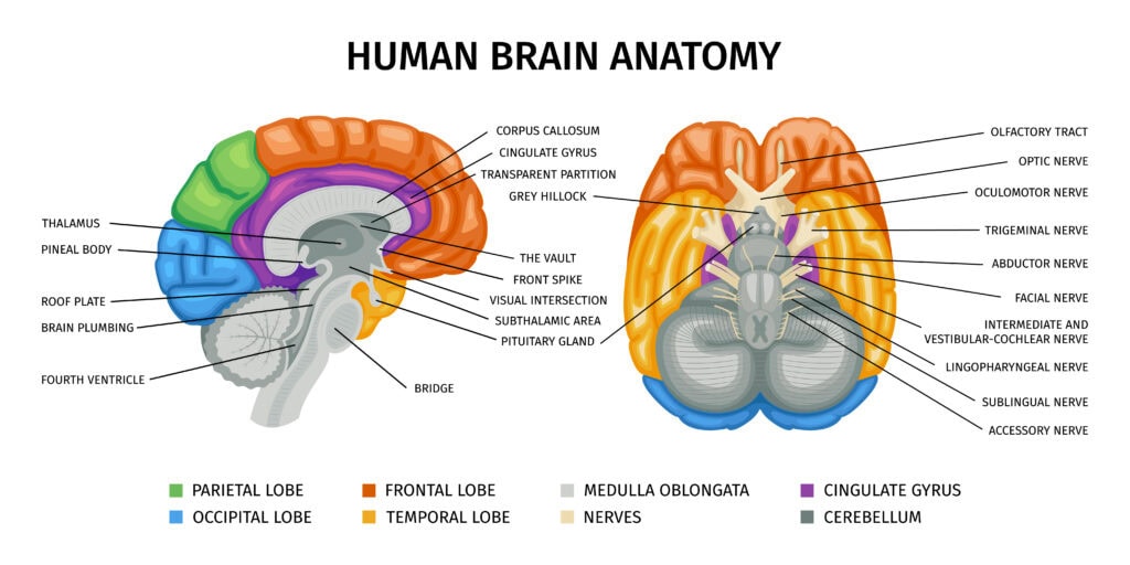
The primary sections of the brain are the cerebrum, cerebellum, and brainstem. The cerebrum, the largest segment, is divided into two hemispheres responsible for higher cognitive functions like reasoning and speech. The cerebellum, located at the back of the brain, coordinates voluntary movements and maintains balance.
The brainstem, connecting the cerebrum and cerebellum to the spinal cord, controls essential autonomic functions like breathing and heart rate. Together, these divisions ensure the seamless operation of the entire brain.
The Cerebrum
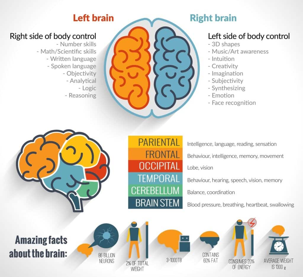
The cerebrum, perched atop the brain, is the largest and most developed part, responsible for conscious thoughts, memories, and voluntary actions. This brain region is divided into two cerebral hemispheres, each controlling the opposite side of the brain and connected by the corpus callosum a thick band of nerve fibers that facilitates communication between the two sides. The cerebral cortex, or gray matter, is where most of the brain’s information processing occurs.
The cerebrum is further divided into four lobes, each with unique functional capabilities. These lobes, the frontal, parietal, temporal, and occipital-work together to interpret sensory information, manage higher cognitive functions such as reasoning and judgment, and control voluntary movements.
Frontal Lobe
The frontal lobes are the brain’s command center, overseeing motor skills, speech, and intellectual functions. The primary motor cortex, located in the frontal lobe, is responsible for producing voluntary movements, while the premotor cortex guides eye and head movements and contributes to our sense of orientation. Broca’s area, also situated in the frontal lobe, is crucial for speech production.
The prefrontal cortex, another critical area within the frontal lobe, plays a vital role in memory, intelligence, concentration, temper, and personality. This region is essential for higher cognitive functions, helping us plan, make decisions, and exhibit self-control. Damage to the frontal lobes can significantly impact these abilities, underscoring their importance in our daily lives.
Parietal Lobe
The parietal lobes play a crucial role in processing sensory information such as touch, temperature, and pain. They help us navigate our environment by integrating sensory input from various parts of the body and interpreting spatial relationships.
This region is essential for understanding and responding to our surroundings, making it a key player in our sensory perception and coordination.
Temporal Lobe
The temporal lobes are essential for processing auditory information and are integral to our ability to perceive sounds. These lobes house regions that are critical for forming and retrieving memories, making them vital for both hearing and memory functions. The temporal lobes also play a role in understanding language and speech, contributing to our communication abilities.
Within the temporal lobes, areas such as the hippocampus are crucial for memory formation and spatial navigation. This region helps us remember where we have been and how to get there, playing an essential role in our daily lives. Damage to the temporal lobes can lead to memory loss and difficulties in processing auditory information, highlighting their importance in our cognitive functions.
Occipital Lobe
The occipital lobes, located at the back of the brain, are primarily responsible for interpreting visual information. These lobes process visual data, including colors and shapes, enabling us to understand and respond to what we see. The right occipital lobe interprets information from the left visual field, while the left lobe processes information from the right visual field.
This region’s role in visual perception is crucial for our ability to navigate the world. It allows us to recognize objects, track movements, and interpret visual cues. Any damage to the occipital lobes can result in visual impairments, underscoring their importance in our sensory processing and overall perception.
The Cerebellum
The cerebellum, located at the back of the brain, is a powerhouse of coordination, balance, and fine motor skills. Despite representing only about 10% of the brain’s total volume, it houses more than half of all the brain’s neurons. This dense collection of neurons allows the cerebellum to make real-time adjustments to motor commands based on sensory feedback, ensuring smooth and coordinated movements.
Although the cerebellum does not initiate movements, it enhances their accuracy and adaptability. It fine-tunes motor activities, helping maintain posture and balance, and plays a significant role in learning new motor skills through a trial-and-error process. Dysfunction in the cerebellum can lead to movement disorders, including uncoordinated voluntary movements and balance difficulties.
The cerebellum receives inputs primarily from the same side of the body, controlling outputs to the same side. This ipsilateral control is crucial for the cerebellum’s role in fine-tuning motor actions and adapting movements to changing conditions.
The Brainstem
The brainstem, a vital structure that connects the cerebrum and cerebellum to the spinal cord, manages essential autonomic functions like breathing and heart rate. It consists of three main parts: the midbrain, pons, and medulla oblongata, each with specific roles in maintaining vital bodily functions. The brainstem’s control over vital automatic functions underscores its importance in sustaining life.
Acting as a conduit for signals between the brain and spinal cord, the brainstem ensures seamless nervous system operation. It is involved in regulating cardiac rhythms, blood pressure, and respiratory rates, making it indispensable for our survival.
The following subsections will delve into the specific functions of the midbrain, pons, and medulla oblongata.
Midbrain
The midbrain, a small yet crucial part of the brainstem, is involved in functions such as vision, hearing, and motor control. It contains structures like the superior colliculus, which processes visual information and coordinates eye movements, and the inferior colliculus, which integrates and relays sound signals. These functions are essential for our ability to perceive and respond to visual and auditory stimuli.
Additionally, the midbrain contributes to motor control through areas like the substantia nigra, which plays a significant role in movement regulation and coordination. Damage to the midbrain can affect these functions, leading to difficulties in visual and auditory processing and motor control. Understanding the midbrain’s role highlights its importance in our sensory perception and movement.
Pons
The pons, situated between the midbrain and medulla oblongata, serves as a critical relay station for signals traveling between different parts of the brain. It facilitates communication between the cerebrum and cerebellum, aiding in both sensory processing and motor control. This relay function ensures that the brain’s various regions can work together seamlessly.
In addition to relaying signals, the pons is involved in regulating facial sensations and movements. It plays a role in controlling activities such as chewing, eye movements, and facial expressions. The pons’ ability to manage these functions underscores its importance in our daily interactions and sensory experiences.
Medulla Oblongata
The medulla oblongata, the lower part of the brainstem, automatically manages critical life-sustaining processes such as heart rate and blood circulation. It is responsible for controlling vital bodily functions, including respiration and cardiovascular stability. The medulla oblongata’s regulation of blood circulation is essential for delivering oxygen and nutrients to tissues throughout the body.
Overall, the medulla oblongata plays a crucial role in maintaining homeostasis by regulating vital functions. Its ability to manage these processes is fundamental to our survival, making it one of the most important structures in the brain.
Human Brain Diagram
Start by drawing a large bean or oval shape for the outline, which can be adjusted in size as desired. Adding squiggly lines across the outline can simulate the wrinkled appearance of the brain’s surface. To maintain anatomical accuracy, sketch curves that separate various parts like the frontal and temporal lobes.
Once the basic shape is drawn, add half-moon shapes along segment lines to illustrate the brain’s texture. Incorporate the brain stem by drawing a narrow tube extending from the base of the brain and adding a curved shape for the cerebellum.
Using colors can enhance the visual appeal of the diagram, helping to differentiate between the various brain parts. Labeling each part, such as the frontal lobe and cerebellum, can be useful for educational purposes.
Protective Structures of the Brain
The human brain is shielded by several protective structures that provide physical protection and support. These structures include the skull, meninges, and cerebrospinal fluid, each playing a crucial role in safeguarding the brain from injury and maintaining its stability.
Skull
The skull, or cranium, is the bony structure that surrounds and protects the brain from external injury. It comprises 22 bones that are fused together, creating a protective cavity for the brain.
The skull’s robust construction ensures that the brain is shielded from impacts and other potential dangers, highlighting its importance in brain protection.
Meninges
The meninges consist of three layers that protect the brain:
- Dura mater – the tough outer layer that provides a durable protective barrier.
- Arachnoid – located beneath the dura mater, this web-like structure cushions the brain.
- Pia mater – the innermost layer that directly covers the brain’s surface and follows its contours, providing additional protection and support.
The space between the dura and arachnoid membranes is called the subdural space, which, along with the subarachnoid space, allows for the circulation of cerebrospinal fluid.
These spaces and layers work together to protect the brain from mechanical damage and help maintain a stable environment for its functions.
Cerebrospinal Fluid
Cerebrospinal fluid (CSF) cushions the brain and spinal cord from injury, acting as a buffer against external trauma.
This fluid circulates around the brain and spinal cord, providing a cushioning effect that reduces the impact of sudden movements and shocks. CSF also helps maintain intracranial pressure and provides a stable chemical environment for the brain.
Produced in the brain’s ventricles, CSF is continually circulated and replaced, ensuring that the brain remains protected and its functions are not disrupted. This constant circulation helps remove waste products and deliver nutrients, highlighting the critical role of cerebrospinal fluid in brain health and protection.
Neurons and Glial Cells
The human brain is composed of two main types of cells: neurons and glial cells. Neurons are the primary signal transmitters, capable of both electrical and chemical communication, allowing for complex signaling within the nervous system.
Glial cells, on the other hand, play an important supportive role, providing nourishment, maintaining homeostasis, and forming the myelin sheath around axons.
Recent research has identified more than 3,000 different human brain cell types, surpassing the known cell types in the human lung. This diversity of cell types underscores the complexity of the brain and the intricate interplay between neurons and glial cells.
Neurons
Neurons are composed of three main parts: the cell body, dendrites, and axon. The cell body contains the nucleus and organelles that support the neuron’s functions. Dendrites receive signals from other neurons, while the axon transmits signals to other neurons or muscles. This structure allows neurons to communicate and process information effectively.
Neurons transmit signals through electrical impulses known as action potentials. These impulses travel along the axon and reach the synapses, where neurotransmitters are released to communicate with other neurons. This complex signaling network is essential for processing and transmitting information throughout the brain and nervous system.
Glial Cells
The human brain contains around 86 billion glial cells. This amount greatly exceeds the number of neurons, with a ratio of roughly 50 glial cells for every neuron. These cells support and nourish neurons, help maintain homeostasis, and are involved in forming the myelin sheath around axons. Glial cells play a crucial role in the nervous system’s overall function and health.
There are different types of glial cells, each with specific functions. Astrocytes, for example, occupy approximately 25% of the total brain volume and adjust blood flow in response to synaptic activity. Microglia serve as the immune cells of the brain, responsible for removing dead cells and defending against pathogens. Oligodendrocytes form the myelin sheath, enhancing signal transmission efficiency.
Blood Supply to the Brain
The brain’s blood supply is primarily provided by the internal carotid and vertebral arteries, which converge at the base of the brain to form the circle of Willis. The anterior and middle cerebral arteries, branching from the internal carotid arteries, supply the forebrain, while the posterior circulation, provided by the posterior cerebral, basilar, and vertebral arteries, nourishes the brainstem and posterior cortex.
Neurons in the brain are particularly vulnerable to oxygen deprivation, necessitating a robust blood supply to meet their metabolic demands. Astrocytes, a type of glial cell, contribute to maintaining the blood-brain barrier, protecting the brain from harmful substances.
This intricate network of blood vessels ensures that the brain receives the oxygen and nutrients it needs to function optimally.
Cranial Nerves
The human brain communicates with the body through twelve pairs of cranial nerves, which carry messages between the brain, organs, and muscles. Ten of these nerves originate in the brainstem, playing a critical role in sensory and motor functions. Each cranial nerve has specific functions, contributing to the overall function and communication between the brain and the body.
The olfactory nerve (CN I) is responsible for the sense of smell, whereas the optic nerve (CN II) facilitates vision. The oculomotor nerve (CN III) controls eye movement and pupil dilation, and the facial nerve (CN VII) manages facial expressions and taste from the anterior tongue. The vagus nerve (CN X) regulates internal organ functions like heart rate and digestion.
Cranial nerves also play a role in other vital functions. The glossopharyngeal nerve (CN IX) aids in taste and swallowing, while the hypoglossal nerve (CN XII) is responsible for tongue movements during speech and swallowing.
Deep Brain Structures
The deep brain structures include the hypothalamus, thalamus, limbic system, amygdala, and hippocampus, each with specific functions. The hypothalamus regulates eating, sexual behavior, sleeping, body temperature, emotions, and hormone secretion. The thalamus acts as a relay station for information to the cortex, involved in pain sensation, attention, and alertness.
The limbic system, which includes the amygdala and hippocampus, plays a key role in behaviors essential for survival, such as feeding and reproduction. The amygdala is responsible for emotional responses and plays a significant role in memory formation related to fear. The hippocampus is critical for forming and storing episodic memories, as well as spatial navigation.
The basal ganglia, another deep brain structure, are involved in reward processing and the formation of habits. These structures work together to regulate various functions and behaviors, highlighting their importance in our daily lives.
Language and Speech Centers
Language and speech functions are primarily controlled by the cerebral cortex, specifically in the left hemisphere.
Wernicke’s area, located in the temporal lobe, is responsible for understanding language, while Broca’s area, typically found in the left hemisphere, is responsible for speech production. Damage to these areas can lead to language comprehension and speech production difficulties, such as Wernicke’s aphasia.
The right hemisphere contributes to interpreting visual information. It also supports language processing. This hemisphere helps with understanding context and non-verbal cues, contributing to effective communication.
Ventricular System
The ventricular system consists of interconnected cavities responsible for producing and circulating cerebrospinal fluid (CSF) throughout the brain. The four cavities, or ventricles, include the lateral ventricles, third ventricle, and fourth ventricle. CSF is produced by the choroid plexus, a structure within the ventricles that filters plasma to create this protective fluid.
Ependymal cells line the brain’s ventricles and are involved in producing cerebrospinal fluid, which cushions and protects the brain. The fourth ventricle drains CSF into the central spinal canal and subarachnoid cisterns, ensuring continuous circulation.
The aqueduct of Silvius connects the third ventricle to the fourth ventricle, while the foramen of Munro connects the third ventricle to the lateral ventricles, facilitating CSF flow.
Common Brain Disorders
Brain disorders can manifest with a wide range of symptoms, depending on the nature and extent of the issue. Let’s explore some of the most common brain disorders and their impacts:
- Strokes: These occur when the blood supply to part of your brain is interrupted or reduced, preventing brain tissue from getting oxygen and nutrients. Strokes can lead to loss of brain functionality and are often associated with specific vascular territories. They may result in paralysis, speech difficulties, and other cognitive impairments.
- Degenerative Nerve Diseases: Conditions like Alzheimer’s and Parkinson’s disease progressively impair essential bodily functions such as movement and speech. You might notice memory loss, tremors, or difficulty with coordination as these diseases advance.
- Hydrocephalus: This condition involves an abnormal accumulation of cerebrospinal fluid within the ventricles, leading to increased intracranial pressure and potential brain damage. It can cause headaches, vision problems, and cognitive decline.
- Encephalitis: Characterized by brain inflammation, encephalitis can result in serious complications, including paralysis and vision issues. You might experience flu-like symptoms, confusion, or seizures if affected.
- Genetic Brain Disorders: These arise from gene mutations, which can hinder normal brain development and operations. They may present early in life and affect your cognitive and physical abilities.
- Traumatic Brain Injuries (TBIs): TBIs vary significantly in severity and may result in both short-term and long-term cognitive impairments. You might experience memory loss, mood changes, or motor skill difficulties following an injury.
- Ischemia: Blood flow interruptions can lead to ischemia, which may cause transient or permanent brain damage if not promptly addressed. It’s crucial to restore blood flow quickly to minimize lasting effects.
Wrap Up
Ongoing brain research continues to unlock new insights, offering hope for better treatments for neurological disorders and advancements in rehabilitation technologies. As we continue to explore the brain’s mysteries, our knowledge will undoubtedly lead to improved healthcare and quality of life.