Lumbar dermatomes are specific skin areas supplied by the lumbar spinal nerves. It’s useful in diagnosing and managing lower back, hip and leg sensation problems. Doctors use this knowledge to make more accurate diagnoses.
Below, I’ll explain the anatomy of the lumbar dermatomes, show you how they’re mapped.
What are Lumbar Dermatomes?
Lumbar dermatomes are specific areas of skin supplied by the lumbar spinal nerves. These nerves are responsible for carrying sensation (feeling) from your body back to your brain.
There are five lumbar vertebrae (L1-L5) and each one exits the spinal column to form a pair of spinal nerves.
These spinal nerves are divided into individual roots, and specific roots contribute to specific dermatomes.
The lumbar spinal nerves primarily supply the lower back, hips, and legs.
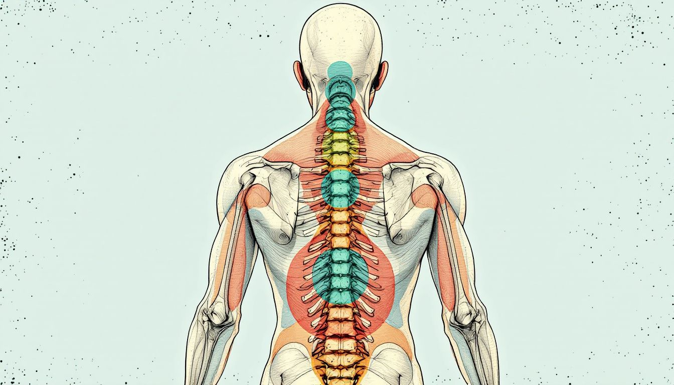
Anatomy of Lumbar Spinal Nerve Roots and Deramotomes
There are five pairs of lumbar spinal nerves (L1-L5). Each nerve is formed by the combination of roots from two levels: the transverse processes (sacral and coccygeal nerves) and the posterior elements (cauda equina).
The lumbar spinal nerve roots divide into anterior (ventral) and posterior (dorsal) divisions, which then recombine to form the final lumbar spinal nerve.
The anterior divisions of the lumbar spinal nerve roots thicken as they travel down the leg and eventually combine to form the lumbar plexus and the posterior femoral cutaneous nerve.
The posterior divisions of the lumbar spinal nerve roots combine to form the posterior femoral cutaneous nerve of the thigh and the nerve to the short heads of the biceps femoris (common peroneal nerve).
The lumbar plexus is formed by the L1, L2, L3, and a small part of L4. It gives rise to the ilioinguinal and genitofemoral nerves, as well as the obturator and femoral nerves.
The L5 nerve is primarily responsible for innervating the lower leg.
The lumbar plexus supplies many of the muscles in the lower back and legs, as well as the skin on the anterior and medial aspects of the legs.
Here’s a quick rundown of which lumbar nerve roots contribute to specific muscles and dermatomes:
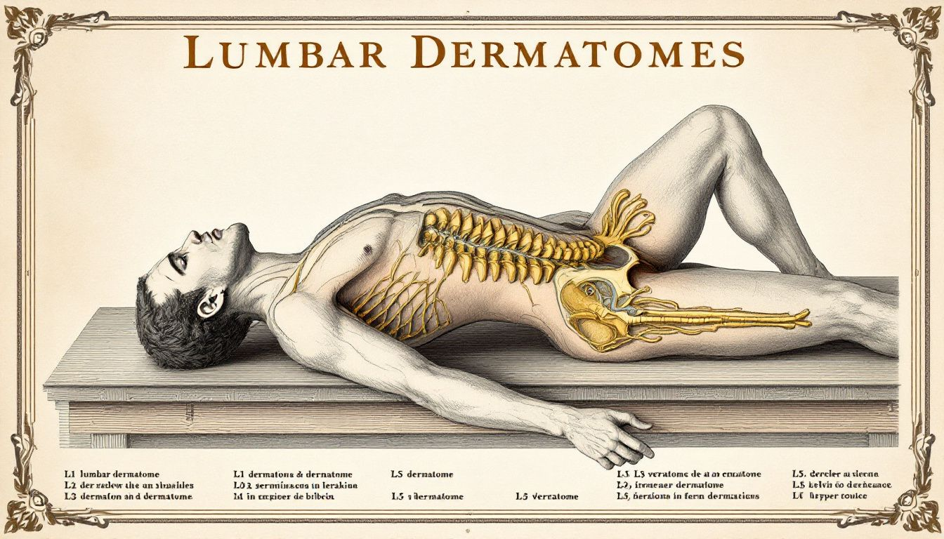
Lumbar Dermatome Map
The lumbar dermatome map shows the specific areas of skin supplied by each lumbar nerve. These mappings are important for understanding sensation in the hips, thighs, and lower legs.
Here’s a quick rundown of the lumbar dermatome map:
- L1 dermatome: upper pubic area and hips
- L2 dermatome: upper thigh muscles (adductors)
- L3 dermatome: front and outer aspect of the thigh
- L4 dermatome: front of the lower leg and medial (inside) aspect of the calf
- L5 dermatome: lateral (outside) and posterior aspects of the lower leg and foot
It’s important to remember that these are general guidelines, and there can be significant variation in how specific perceive sensation in their bodies. This is known as zone of referral.
Specific Lumbar Dermatome Patterns
Each lumbar nerve has a specific dermatome pattern. Here’s a quick look at the individual lumbar dermatomes:
- L1 dermatome: back and groin area
- L2 dermatome: anterior thigh to knee
- L3 dermatome: upper buttock and medial lower leg
- L4 dermatome: medial aspect of the leg and dorsum of the foot
- L5 dermatome: lateral aspect of the leg and first three toes
Sensory Changes and Lumbar Dermatomes
Sensory symptoms in lumbar dermatomes, like tingling sensations, often signal irritation or damage to the nerves.
Each lumbar dermatome is linked to particular regions in the hips and legs, helping in the understanding of pain and sensory loss. Tingling or numbness at the front of the thigh might indicate an issue with the L2 or L3 dermatome.
Loss of sensation in areas related to specific lumbar dermatomes can point to issues with the corresponding spinal nerves.
Healthcare professionals use this knowledge to locate the source of pain and sensory problems, improving diagnostic precision.
Compression of nerve roots often leads to radiating pain, tingling, and numbness along the affected dermatome, following specific dermatomal patterns.
Read More : Hand Dermatomes
Nerve Root Compression
You might feel tingling in your lower body due to pressure on lumbar spinal nerves affecting sensory pathways. Herniated discs often cause this by pressing on nerve roots, especially at L4/L5 and L5/S1 levels. This can lead to:
- Sharp or burning pain
- Numbness
- Muscle weakness in affected areas
Doctors usually prefer MRI to evaluate lumbar radiculopathy as it clearly shows nerve root compression.
When MRI isn’t possible, CT myelography offers an alternative for diagnosing lumbar spine issues.
These imaging methods are essential for pinpointing the source of nerve root compression and planning the right treatment.
Pain Dermatomes in the Lumbar Region
When your lumbar nerve roots are compressed, you might feel sharp or burning sensations running down your leg, often known as sciatica.
This pain starts in your lower back and travels down. The leg pain pattern depends on which lumbar nerve root is affected.
Here’s what happens:
- The severity and location of your pain change with the specific lumbar nerve involved.
- Recognizing these pain areas helps doctors identify the source of compression.
- This understanding is crucial for crafting effective treatment plans, boosting your recovery and comfort.
Treatment Options for Lumbar Dermatome-Related Conditions
Mapping lumbar dermatomes lets us pinpoint nerve root injuries, helping us tailor treatment strategies effectively.
Clinicians rely on these maps to assess nerve damage and plan rehabilitation.
This mapping predicts how patients might respond to treatments based on the affected nerve root, ensuring targeted interventions.
When you experience sensory loss in the lumbar region, it often shows up as reduced sensitivity to touch or temperature. Here are some treatment options:
- Conservative methods
- Physical therapy
- NSAIDs
- Invasive procedures
- Epidural steroid injections
- Surgery
By understanding and mapping lumbar dermatomes, doctors can craft personalized treatment plans that address each patient’s unique needs.
Final Verdicts
I hope this guide helps you better understand lumbar dermatomes, as they are key to diagnosing and managing lower back and leg conditions.
From the anatomy and mapping of lumbar spinal nerves to the clinical implications and treatment options, this knowledge will improve your diagnostic accuracy and patient care.