

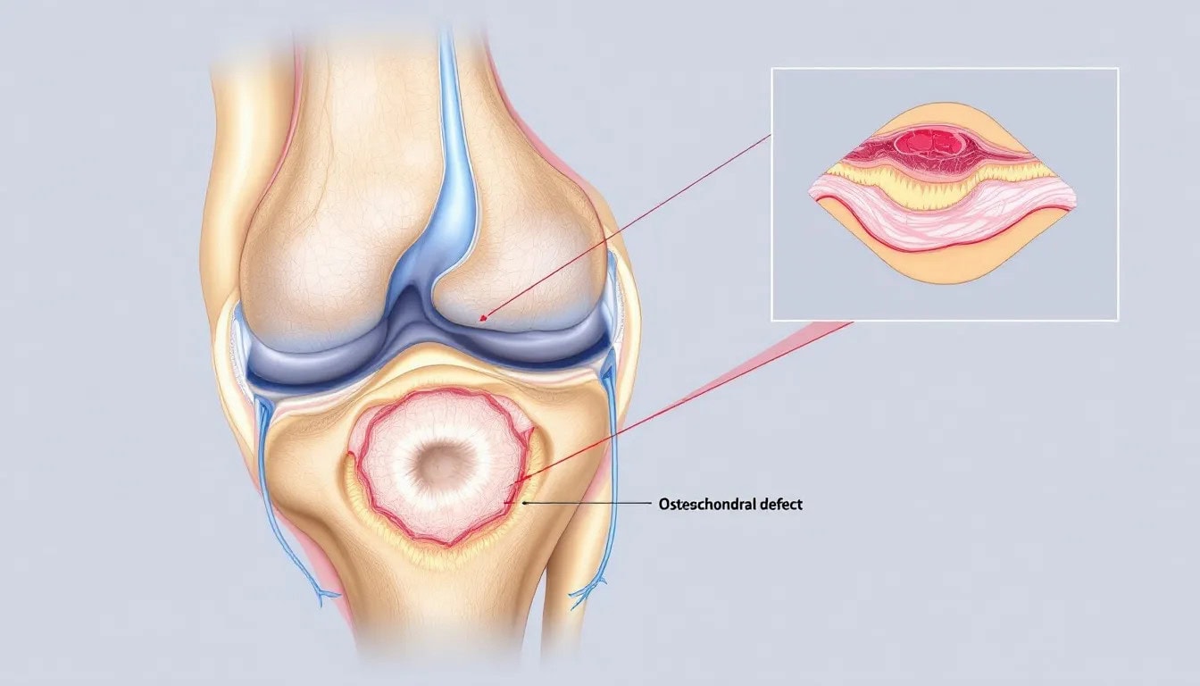
An osteochondral defect occurs when cartilage and the underlying bone in a joint are damaged, causing pain and swelling. It involves damage to both articular cartilage and the underlying bone, leading to significant pain and joint dysfunction.
This article will cover the causes, symptoms, and treatment options for this condition to help you understand how to manage it effectively.
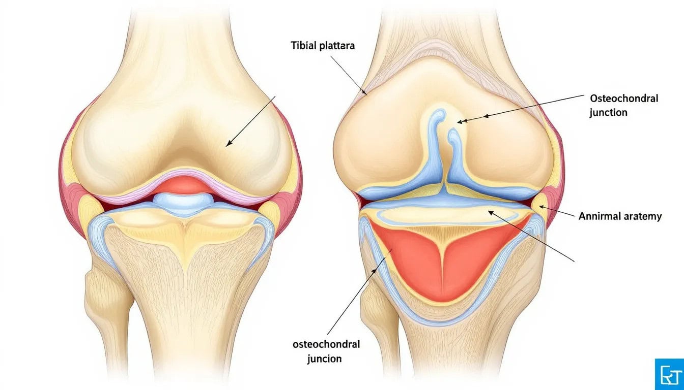
What is Osteochondral Defects
Osteochondral defects refer to damage that affects both the articular cartilage and the underlying bone. This condition is more severe than chondral lesions, which impact only the cartilage. The term ‘chondral’ is specific to cartilage, while ‘osteo’ refers to bone, underscoring the dual nature of osteochondral defects. These defects can lead to significant joint pain, swelling, and mechanical symptoms, complicating daily activities and athletic performance.
One specific type of osteochondral defect is Osteochondritis Dissecans (OCD), characterized by abnormalities in the bone beneath the cartilage. In OCD, the bone and the overlying cartilage can become detached, forming an osteochondral fragment that interferes with joint function.
Common Causes of Osteochondral Defects
The development of osteochondral defects and chondral defect can be attributed to multiple factors. These defects often result from a combination of:
- Trauma
- Repetitive stress
- Genetic predisposition
- Vascular insufficiency
- Inflammatory or degenerative conditions
Each of these causes can independently or collectively contribute to the deterioration of the articular cartilage and subchondral bone.
1. Trauma or Injury
Acute trauma is a significant cause of osteochondral defects. Events such as falls, accidents, and severe ankle sprains can lead to fractures or dislocations that damage both cartilage and bone. These injuries are particularly common in the talus of the ankle, where traumatic insult is considered the most critical factor.
Specific populations, such as athletes or individuals involved in high-risk activities, are at a higher risk for these traumatic injuries. For instance, knee injury in sports can result in damage to the femur and femoral head, leading to osteochondral lesions that may require surgical intervention.
2. Repetitive Stress or Overuse
Repetitive movements and overuse are also common culprits in the development of osteochondral defects. Activities that place continuous stress on the joints, such as running or jumping, can gradually wear down the cartilage and underlying bone. Over time, this repetitive stress leads to mechanical symptoms and significant cartilage damage.
Athletes engaged in high-impact sports are particularly susceptible to these injuries. Chronic overload due to malalignment or instability of the ankle joint can exacerbate the condition, leading to osteochondral lesions that require careful management.
3. Genetic Factors
Genetic predisposition plays a crucial role in the development of osteochondral defects. Certain inherited conditions, such as skeletal dysplasias, can lead to abnormal cartilage and bone development. These genetic factors contribute to the degeneration of cartilage cells, making individuals more susceptible to osteochondral lesions.
4. Vascular Insufficiency
Poor blood supply to the cartilage and subchondral bone, known as vascular insufficiency, can significantly impact the healing process and lead to osteochondral defects. Conditions such as avascular necrosis are associated with this insufficiency, where the reduced blood flow leads to bone tissue death and subsequent cartilage damage.
5. Inflammatory or Degenerative Conditions
Inflammatory conditions like rheumatoid arthritis can lead to chronic inflammation, which deteriorates cartilage integrity and contributes to osteochondral defects. Similarly, degenerative diseases such as osteoarthritis exacerbate the wear and tear of the joint, leading to significant articular cartilage damage and bone damage.
The continuous inflammation associated with these conditions can result in necrotic bone and further complicate the joint’s structural integrity. These inflammatory and degenerative processes highlight the importance of early intervention and appropriate management to prevent the progression of osteochondral lesions.
Symptoms of Osteochondral Defect
Osteochondral defects present with a range of symptoms that can significantly impact daily life. Common symptoms include:
- Pain
- Swelling
- Joint instability
- Mechanical symptoms like locking or clicking
1. Pain and Discomfort
Pain is the most commonly reported symptom associated with osteochondral defects. Patients often experience deep, disabling pain in the affected joint, which worsens with activity. This pain can be chronic or acute, varying in intensity based on the severity of the defect.
The persistent discomfort can significantly hinder mobility and quality of life.
2. Swelling and Joint Effusion
Swelling and joint effusion are also common symptoms. Fluid buildup within the joint occurs as a reaction to the cartilage and bone damage. This swelling can cause visible enlargement of the joint and lead to stiffness and reduced mobility.
3. Limited Range of Motion
Osteochondral defects can restrict the range of motion in the affected joint. Patients may find it difficult to fully extend or flex the joint, leading to stiffness and limited mobility. This restriction impacts daily activities, making simple tasks challenging.
4. Locking or Catching Sensation
A locking or catching sensation is indicative of significant osteochondral issues. Fragments of cartilage or bone can cause mechanical blockage, making it difficult to move the joint smoothly. These sensations are often triggered by specific activities or positions, leading to sudden immobility.
5. Instability or Weakness
Joint instability or weakness is another symptom of osteochondral defects. The damaged cartilage and bone can lead to loose flaps, causing a feeling of instability in the joint. This instability increases the risk of further injury and can lead to muscle weakness due to pain and inactivity.
Diagnosis Techniques
Accurate diagnosis is critical for planning effective treatment for osteochondral defects. Various diagnostic techniques are employed to evaluate the extent of cartilage damage and the condition of the underlying bone. Magnetic Resonance Imaging (MRI) and mr imaging are highly effective for detecting cartilage damage and assessing surrounding soft tissues. Conventional X-rays can initially identify osteochondral defects but are less effective than MRI in detecting cartilage issues.
Computed Tomography (CT) provides detailed images of bony lesions, making it the preferred method for visualizing the precise location and dimensions of the damage in bones. Skeletal radiol ultrasound can help identify surface irregularities and loose fragments, while Single Photon Emission Computed Tomography (SPECT) enhances understanding of subchondral activity.
Accurate diagnosis ensures that the chosen treatment options are tailored to the specific needs of the patient.
Non-Surgical Treatment Options
Non-surgical treatment aims to reduce pain and inflammation, allowing the joint to heal naturally through spontaneous healing. Common non-surgical treatments include pain reduction medications, hot and cold therapy, rest, activity modification, and physical therapy. Injections of platelet-rich plasma can also reduce pain and enhance functionality for several months.
Physical therapy plays a crucial role in strengthening muscles and retaining flexibility. Non-surgical treatment can be highly effective in healing osteochondral lesions, especially for younger patients like growing children or adolescents. Immobilization with a non-weight-bearing cast is often recommended for six weeks to manage acute lesions.
Longitudinal studies show that successful nonoperative treatment plans often result in minimal symptoms and a low rate of treatment failure.
Surgical Interventions for Osteochondral Defects
Surgical treatment is usually recommended for displaced talar osteochondral lesions, particularly those involving the medial talar dome. It is also considered when lesions do not improve with non-operative management. The choice of surgical intervention depends on factors such as the patient’s activity level, the size, location, and extent of the damage. Common procedures include arthroscopic, open, or combined approaches.
Microfracture is a minimally invasive technique that stimulates fibrocartilage formation by creating small holes in the subchondral plate of the subchondral bone. Mosaicplasty involves transferring osteochondral plugs from a healthy donor site to repair damaged cartilage, creating a mosaic-like pattern. Autologous Chondrocyte Implantation (ACI) is a two-step procedure where cartilage cells are harvested, expanded in a lab, and implanted back into the defect.
Surgery is often necessary for osteochondral defects when conservative treatment fails to improve symptoms. The goal of surgical intervention is to restore the joint’s function and alleviate pain, allowing patients to return to their daily activities with minimal discomfort.
Recovery and Rehabilitation
Post-operative rehabilitation is crucial in preventing complications and ensuring proper healing. The rehabilitation phase should be tailored to the specific surgical procedure and individual patient factors. Physical therapy is essential for maximizing the healing process and minimizing the chances of recurrence.
Pain management protocols are integral during the early stages of recovery to facilitate rehabilitation. Techniques such as soft tissue massage, electrotherapy, and joint mobilization are commonly used in physical therapy. Gradually returning to weight-bearing and athletic activities is essential after immobilization to ensure a successful recovery.
Long-Term Outcomes and Complications
Long-term outcomes for osteochondral defects vary based on several factors, including the patient’s age, activity level, and the size of the lesion. Clinical assessments over a 14-year follow-up indicate that many patients maintain good ankle function and have minimal pain. However, a small percentage of patients may experience a progression of ankle osteoarthritis even after initially successful treatment.
Surgical options like mosaicplasty and ACI may carry risks such as complications at the donor site and the need for future interventions. Additionally, maintaining healthy cartilage and a healthy body weight can help prevent the progression of articular cartilage lesions, which may vary depending on individual circumstances.
Preventative Measures
Preventing osteochondral defects involves a combination of proper training techniques, protective gear, and early intervention. Focusing on landing mechanics during training can reduce the risk of injuries that lead to osteochondral defects. Utilizing protective gear during sports activities can help mitigate the impact of injuries.
Wrap Up
Osteochondral defects and their treatment options is crucial for managing joint health and preventing long-term complications. Common causes include trauma, repetitive stress, genetic predisposition, vascular insufficiency, and inflammatory conditions, necessitating prompt diagnosis and tailored management.
Frequently Asked Questions
How long does it take for an osteochondral defect to heal?
An osteochondral defect typically requires about eight weeks of light rehabilitation for healing before more intensive exercises can commence. Engaging in guided physical therapy can facilitate a return to full athletic performance.


