

A pineal cyst, or pineal gland cyst, is a fluid-filled sac in the pineal gland of the brain. Often harmless and symptom-free, some cysts can cause problems. This guide explains what a pineal cyst is, its symptoms, diagnosis, and treatment options.
What is the Pineal Gland
The pineal gland, a small endocrine internal structure located deep within the brain, plays a crucial role in regulating our sleep-wake cycle and helps regulate the body’s hormone called melatonin. Nestled above the thalamus, its primary function is to produce a hormone called melatonin.
This hormone signals our bodies when it’s time to sleep and when to wake up, with melatonin production peaking during the night to promote sleep. The human pineal gland, with its inner layer, is essential for this process.
Melatonin production is highly responsive to light and darkness, with the pineal gland secreting more melatonin in the absence of light. This intricate mechanism helps regulate the body’s internal clock, ensuring that we maintain a consistent sleep-wake cycle by producing. Any disruption in this cycle can lead to significant sleep disorders and affect overall health.
What is a Pineal Cyst?
A pineal cyst is a small fluid filled sac filled with fluid. It forms in the pineal gland, situated deep within the center of the brain. These fluid filled sacs are classified as large pineal cysts when they exceed 2.0 mm in diameter and typically range from 2 to 14 mm. While they are more commonly found in adults, children and adolescents can also develop a pineal gland cyst.
Most pineal cysts are benign and asymptomatic, often discovered incidentally during brain imaging studies conducted for other reasons. It’s not uncommon for someone to go through life without ever knowing they have a pineal cyst. However, in rare cases a pineal cyst, larger cysts can cause symptoms that may require medical attention. Common are pineal cysts.
Causes of Pineal Cysts
Pineal cysts can arise from a variety of factors, often involving structural or functional changes in the pineal gland. These can include:
- Genetic predispositions
- Hormonal fluctuations
- Trauma
- Inflammation
- Blocked ducts
These causes explain why these cysts form and how they might impact individuals differently, including the potential for a mass effect.
The formation of pineal cysts is a complex process influenced by multiple variables. Each factor contributes in unique ways, and in some cases, the exact cause remains unidentified. Let’s delve deeper into these causes to better understand the origins of pineal cysts.
1. Congenital Causes
Some pineal cysts are present from birth, formed during early brain development. These congenital cysts can result from genetic factors and developmental anomalies. Inherited genetic predispositions may also play a role, suggesting that a family history of pineal cysts could increase the likelihood of their occurrence.
During fetal development, fluid-filled spaces naturally occur in the pineal gland. If these spaces do not resolve properly, they can lead to cyst formation. The congenital origins of pineal cysts underscore the importance of genetic and developmental factors in their occurrence.
2. Hormonal Changes
Hormonal changes, especially during puberty, can significantly influence the development of pineal cysts. Variations in hormone levels during this period can impact the pineal gland, leading to cyst formation. These hormonal shifts are a natural part of growth and development but can have unexpected effects on the pineal gland.
Significant hormonal transitions throughout life, such as those occurring during puberty, can thus be a key factor in the development of pineal cysts. Hormonal changes can explain why pineal cysts might develop during certain life stages.
3. Trauma or Injury
Head trauma and injuries are another potential cause of pineal cysts. Physical trauma can affect the pineal gland, creating conditions that foster cyst formation. Injuries to the head may lead to the development of cysts by causing structural changes or damage within the pineal region tumors.
The relationship between trauma and cyst formation is still an area of ongoing research, but it underscores the impact that physical injuries can have on brain structures. This connection can help in identifying and managing cysts that arise due to trauma.
4. Inflammation or Infection
Infections and inflammation can also contribute to the formation of pineal cysts. Infections affecting the brain can cause tissue damage, leading to cyst development within the pineal gland. Chronic inflammation in the pineal region may trigger cyst formation by disrupting normal tissue function.
Infections and inflammation in cyst development highlight the importance of managing brain and spinal cord health to prevent such occurrences. This understanding is crucial for both prevention and treatment strategies.
5. Cyst Formation Due to Blocked Ducts
Blocked ducts or fluid retention can lead to the formation of cysts in the pineal gland. When ducts within the gland become obstructed, fluid can accumulate, resulting in cyst formation. This mechanism is similar to how cysts form in other parts of the body due to fluid retention.
The process of cyst formation due to blocked ducts gives insight into the mechanical aspects of these cysts’ development. This knowledge can inform treatment strategies aimed at relieving duct obstructions to prevent or reduce cyst growth.
6. Unknown Causes
In many cases, the specific cause of pineal cysts remains unclear. Despite extensive research, some cysts develop without a clearly identifiable origin. Various theories have been proposed, but the exact mechanisms remain elusive.
This uncertainty highlights the need for ongoing research to uncover the unexplained reasons behind some pineal cyst occurrences. It also underscores the complexity of the human brain and the pineal gland’s role within it.
Symptoms of Pineal Cysts
The symptoms of pineal cysts can vary widely depending on their size and location. While most pineal cysts are asymptomatic, larger cysts can cause a range of neurological symptoms. Recognizing these symptoms is crucial for early detection and management.
Let’s explore the different symptoms of a pineal that can arise from pineal cysts, recognizing that each individual’s experience may vary. This knowledge can help in identifying when medical intervention is necessary.
1. Asymptomatic Cases
Most pineal cysts do not present noticeable symptoms. They are frequently found accidentally during brain imaging. Monitoring these asymptomatic cysts is important to ensure they remain stable over time.
2. Headaches and Migraines
Pineal cysts can be associated with frequent headaches or migraines, often due to pressure exerted by the cyst on surrounding brain structures. These chronic headaches may be persistent headaches and require medical evaluation.
3. Vision Problems
Larger pineal cysts can impact vision, leading to symptoms such as blurred vision or double vision. These visual disturbances occur due to the cyst pressing against visual pathways in the brain.
4. Hormonal Imbalances
Pineal cysts can affect melatonin production, leading to hormonal imbalances that disrupt sleep patterns and mood. This disruption can significantly impact daily functioning and overall well-being.
5. Nausea and Vomiting
Increased intracranial pressure from larger cysts can lead to nausea and vomiting. These symptoms occur due to the cyst pressing against nearby brain structures, affecting normal brain function.
6. Cognitive Impairment or Memory Issues
Pineal cysts can impact cognitive functions, causing memory issues and difficulty with concentration. Pressure on surrounding brain tissue can disrupt normal communication pathways, leading to these neurological symptoms.
7. Seizures or Epileptic Events
In rare cases a pineal, pineal cysts can lead to seizures or epileptic events. This occurs when the cyst creates abnormal pressure within the brain, triggering seizure activity.
Diagnosing Pineal Cysts
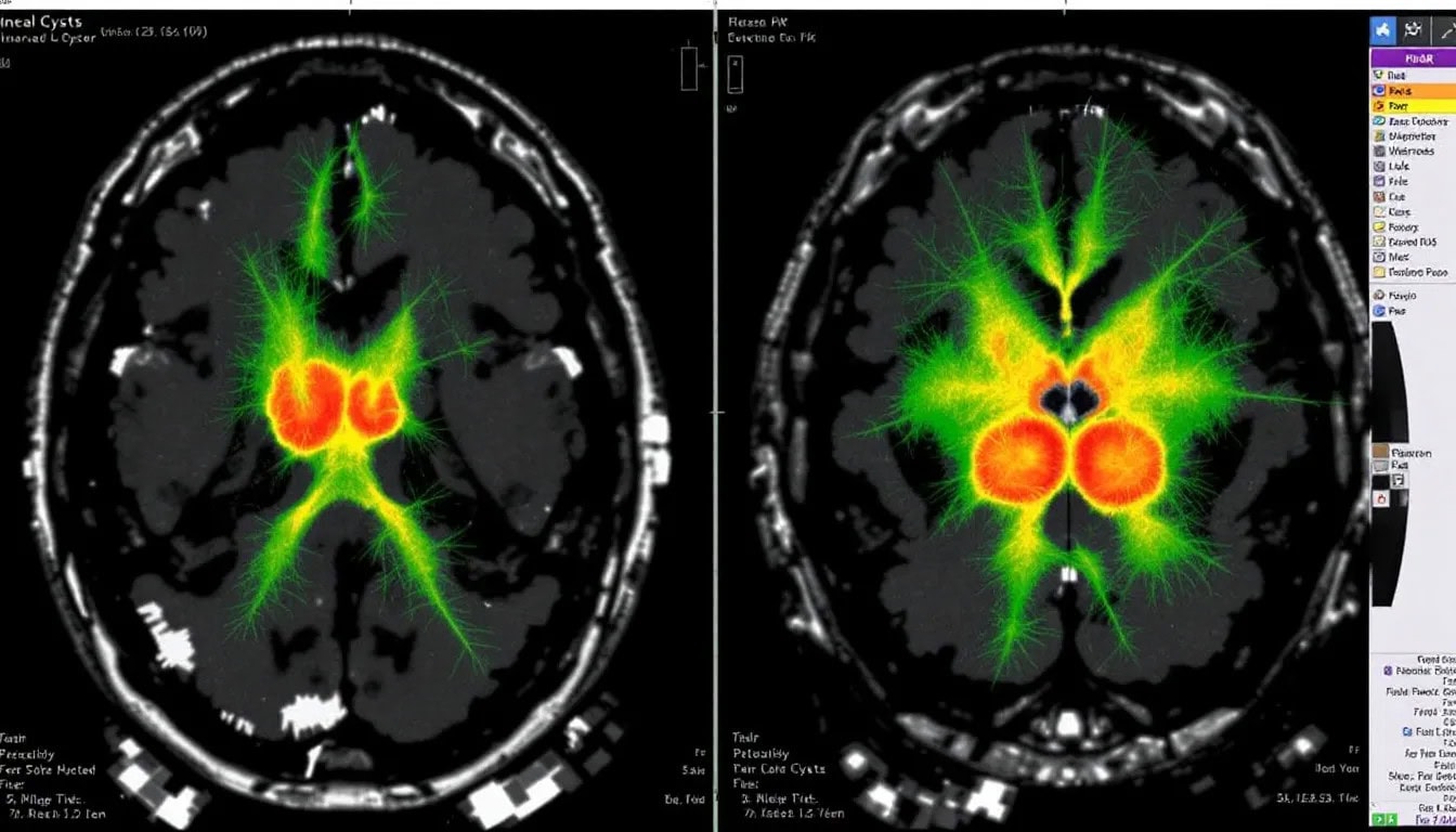
Diagnosing a pineal cyst typically involves imaging tests like CT scans and MRI. These scans reveal well-defined fluid density lesions on CT and homogeneous signals on MRI, with pineal cysts often appearing iso to hypointense on T1-weighted images. About 60% of pineal cysts demonstrate enhancement with contrast on T1-weighted imaging, providing additional diagnostic clarity. Nodular enhancement may indicate previous hemorrhage, which is an important detail for accurate diagnosis.
Pineal cysts are often discovered incidentally during imaging for unrelated issues. Regular imaging is crucial for monitoring the cyst’s stability and ensuring it does not lead to complications.
Treatment Options
Treatment for pineal cysts depends on their size and symptoms. Most pineal cysts are harmless and do not require treatment, especially if they are small and asymptomatic. However, larger cysts that cause symptoms may necessitate medical intervention. Additionally, a treatment for a pineal cyst may be necessary if the cysts are larger and symptomatic.
There are two primary approaches to managing pineal cysts: monitoring asymptomatic cysts through regular imaging and surgically intervening when necessary. Each approach aims to ensure the best possible outcome for the patient.
Monitoring Asymptomatic Cysts
For small, asymptomatic pineal cysts, the standard approach is regular monitoring through brain imaging. This involves periodic magnetic resonance imaging mri scans and brain scans to ensure the cyst remains stable and does not grow.
Regular imaging helps confirm the cyst’s condition over time, providing reassurance and preventing unnecessary interventions.
Surgical Interventions
Surgical intervention is considered for symptomatic pineal cysts that cause significant neurological symptoms or rapid enlargement. The primary surgical options include craniotomy and creating a drainage path to relieve pressure on surrounding structures. In some cases, endoscopic surgery offers a minimally invasive option with a faster recovery time.
Post-surgery, patients are closely monitored in the ICU for 24-48 hours to check for any complications. Patients usually remain in the hospital for several days. This duration is dependent on their recovery progress. Symptoms such as fatigue, mild headaches, or temporary vision changes may occur but generally improve over time.
Surgical intervention can significantly improve the quality of life for patients with symptomatic pineal cysts, providing relief from debilitating symptoms and preventing further complications.
Potential Complications
While most pineal cysts are benign and asymptomatic, there are potential complications that need to be considered. Pineal apoplexy, a rare but serious complication, involves sudden hemorrhage within the cyst, leading to acute symptoms such as severe headaches, neurological deficits, and even sudden death. Hydrocephalus, another severe complication, can occur if the cyst blocks the flow of cerebrospinal fluid in the brain, causing increased intracranial pressure.
Rapid enlargement of pineal cysts can compress surrounding brain structures, potentially leading to respiratory or cardiovascular failure due to midbrain compression. These complications highlight the importance of regular monitoring and timely intervention when necessary to prevent life-threatening outcomes.
Prognosis for Patients with Pineal Cysts
The prognosis for patients with pineal cysts is generally positive. This is especially true for small, asymptomatic, and stable cysts. Pineal cysts are typically benign and rarely lead to severe complications or sudden death. Most patients with stable pineal cysts do not experience significant health issues or symptoms.
Follow-up imaging is usually unwarranted for small and asymptomatic cysts, as around 75% remain the same size over time, with some even shrinking. For those undergoing surgery for symptomatic cysts, the outlook is very good, with significant relief from symptoms following removal.
Key Takeaways
Pineal cysts are mostly asymptomatic and benign; they are often discovered incidentally during imaging.
Common symptoms of larger pineal cysts include headaches, vision problems, and cognitive impairments, which require medical evaluation.


