

Need a clear small intestine diagram for better understanding? This article provides a detailed look at the small intestine’s anatomy with visuals, covering the duodenum, jejunum, and ileum. Learn how each part contributes to the digestive process.
Key Takeaways
The small intestine consists of three main parts: the duodenum, jejunum, and ileum, each playing specific roles in digestion and nutrient absorption.
Nutrient absorption is facilitated by specialized structures like villi and microvilli in the mucosal layer, while the digestive process is supported by pancreatic enzymes and blood supply from the superior mesenteric artery.
Common disorders affecting the small intestine, such as duodenal ulcers, IBS, and Crohn’s disease, can significantly impact digestive health and require proper management.
Anatomy of the Small Intestine
The small intestine is a marvel of biological engineering, consisting of three main parts: the duodenum, jejunum, and ileum. Each segment plays a distinct role in the digestive process, working in harmony to ensure that nutrients are efficiently absorbed and waste is moved along.
This long, coiled structure, nestled within the abdominal cavity, is a hub of digestive activity in the digestive tract that operates with precision and efficiency.
Diagram of Small Intestine

Duodenum
The duodenum is the first section of the small intestine and is approximately 25 cm in length. It wraps around the pancreas, creating a vital connection between the stomach and the rest of the small intestines. This section consists of four distinct parts: the superior, descending, inferior, and ascending sections, each contributing to its critical functions.
One of the primary roles of the duodenum is to mix the partially digested food from the stomach with bile and pancreatic enzymes, which are essential for breaking down fats, proteins, and carbohydrates. The major duodenal papilla, located in the descending part, is a key player in regulating the release of these digestive juices. Brunner’s glands, extending from the muscularis mucosa to the submucosal layer, secrete mucus and enzymes to protect the intestinal lining and aid in digestion.
The blood supply to the duodenum is rich and complex, drawing from the celiac trunk, superior mesenteric artery, and pancreaticoduodenal arteries. This ensures that the duodenum receives the nutrients and oxygen it needs to function effectively. Venous drainage occurs via the hepatic portal vein, while lymphatic drainage goes to the pancreatoduodenal and superior mesenteric nodes.
Jejunum
Beginning at the duodenojejunal flexure, the jejunum extends for approximately 8 feet, making up the proximal two-fifths of the small intestine. Located in the lower abdominal cavity, the jejunum is suspended by the mesentery proper, which attaches it to the posterior abdominal wall.
This section of the small intestine is characterized by its thick small intestine wall and wide lumen, which are essential for its role in nutrient absorption. The jejunum’s dark red color is due to its rich blood supply, which facilitates the efficient uptake of nutrients into the bloodstream.
Ileum
Measuring approximately 10 to 12 feet in length, the ileum is the longest segment of the small intestine. Food spends the most time in the ileum during digestion, underscoring its crucial role in nutrient absorption.
The ileum ends at the ileocecal junction, where it connects to the large intestine. This section is vital for the absorption of vitamins and minerals, including vitamin B12 and bile salts, which are essential for maintaining overall health.
Crohn’s disease, a common disorder, often affects this part of the gastrointestinal tract, causing inflammation and other symptoms.
Histological Features of the Small Intestine
The small intestine’s histological structure is a testament to its efficiency in nutrient absorption. It comprises four layers: the mucosa, submucosa, muscularis externa, and serosa. These layers work in unison to facilitate digestion and protect the intestinal wall.
Remember these layers with the mnemonic M.S.M.S..
GI Tract on Small Intestine Lining
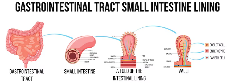
Mucosa and Submucosa
The mucosal layer is the innermost layer, featuring villi and microvilli that significantly increase the surface area for absorption. The enterocytes in the mucosa have microvilli, which enhance nutrient absorption by providing a larger surface area. Goblet cells, present in the intestinal glands, secrete mucus to lubricate and protect the intestinal lining.
Beneath the mucosa lies the submucosa, a dense layer of connective tissue containing blood vessels and nerves that support the mucosa. This layer delivers nutrients and oxygen to the mucosal cells, ensuring their proper function.
Muscularis Externa and Serosa
The muscularis externa consists of two layers of smooth muscle: an inner circular layer and an outer longitudinal layer. These muscle layers facilitate peristalsis, the wave-like contractions moving food through the small intestine.
The outermost layer, the serosa, serves as a protective barrier and reduces friction between the small intestine and surrounding structures. This layer allows the small intestine to move freely within the abdominal cavity without causing damage to itself or other organs.
Blood Supply and Innervation
A robust blood supply and intricate innervation are critical for the small intestine’s function. Blood and nerve supply are crucial for maintaining the health and efficiency of this vital organ.
Blood Vessels
The arterial supply for the jejunum and ileum comes from 15-18 branches of the superior mesenteric artery, known as jejunal and ileal arteries. These arteries form a network known as arterial arcades, sending straight arteries to supply the jejunum and ileum. Venous drainage occurs through the superior mesenteric vein, leading to the hepatic portal vein and eventually to the liver.
The duodenum receives blood from both the gastroduodenal artery, branching from the celiac trunk, and the superior mesenteric artery. Venous drainage for the duodenum occurs through the hepatic portal vein.
Nervous System
The small intestine’s innervation is handled by the vagus nerve and thoracic splanchnic nerves, which are essential for regulating intestinal movements and digestive processes. Parasympathetic fibers from the vagus nerve enhance peristalsis and secretions, while the sympathetic nervous system reduces these activities.
Afferent fibers from the small intestine transmit pain signals through sympathetic nerves to the central nervous system. The jejunum and ileum are also innervated by lesser splanchnic nerves from thoracic spinal segments T9 to T10.
Small intestine diagram with key areas
The small intestine is a long, coiled structure measuring about 22 feet, with a narrow diameter of about 2 centimeters. It consists of three main parts: the duodenum, jejunum, and ileum, each playing distinct roles in digestion.
The duodenum, about 10 inches long, is where digestive juices and enzymes from the pancreas and bile mix with food. This initial mixing breaks down fats, proteins, and carbohydrates, setting the stage for further digestion and absorption.
The jejunum, roughly 8 feet long, has a rich blood supply, allowing for efficient nutrient absorption. The ileum, the longest part, absorbs vitamins and minerals before moving waste to the large intestine.
Villi, tiny finger-like projections on the inner lining, significantly increase the surface area for absorb nutrients, enhancing the small intestine’s efficiency at extracting nutrients from food.
Digestive Processes in the Small Intestine
The small intestine is the powerhouse of the digestive system, breaking down food and absorbs nutrients into the bloodstream. The enzymatic activity and nutrient absorption processes within this vital organ are explored below.
Enzymatic Activity
Pancreatic digestive enzymes break down fats, carbohydrates, and proteins in the small intestine. The major duodenal papilla in the descending part of the duodenum regulates the release of these enzymes for proper mixing with food.
Brush border enzymes on the microvilli of the small intestine are essential for the final stages of chemical digestion. These enzymes break down nutrients into their simplest forms, making absorption easier for the body.
Nutrient Absorption
Carbohydrates are primarily absorbed in the duodenum, while proteins are absorbed in the jejunum. Fatty acids and monoglycerides are absorbed through the intestinal mucosa into the lymphatic system, highlighting the small intestine’s efficiency in nutrient absorption.
All three segments of the small intestine absorb water and electrolytes, keeping the body hydrated and maintaining electrolyte balance.
Common Disorders Affecting the Small Intestine
Despite its efficiency, the small intestine is susceptible to various disorders that can significantly impact its function. Understanding these disorders is crucial for maintaining the health and proper function of the small intestine.
Duodenal Ulcers
Helicobacter pylori infection commonly causes duodenal ulcers. Chronic NSAID therapy can also lead to their development. These ulcers cause burning pain in the epigastric region, nausea, and vomiting, and are more prevalent in the younger population.
Untreated duodenal ulcers can lead to complications such as bleeding and small intestine obstruction, emphasizing the importance of early diagnosis and treatment.
Irritable Bowel Syndrome (IBS)
Irritable Bowel Syndrome (IBS) is a common gastrointestinal disorder characterized by abdominal discomfort and altered bowel habits. Symptoms include cramping, bloating, gas, diarrhea, and constipation. IBS symptoms can be triggered by stress, certain foods, and hormonal changes.
Management strategies include dietary changes, stress management techniques, and medication to alleviate symptoms.
Crohn’s Disease
Crohn’s disease can cause inflammation in any part of the gastrointestinal tract, but it is most commonly found in the ileum. Symptoms often include:
severe abdominal pain
diarrhea
fatigue
weight loss
This chronic condition can lead to complications such as small bowel obstruction and requires ongoing management to control symptoms and prevent flare-ups.


