The stomach is a J-shaped, muscular organ located in the upper abdomen that plays a crucial role in the digestive process by mixing food with digestive juices and regulating its passage into the small intestine.
The stomach diagram is a valuable tool for understanding the anatomy and functions of the stomach. By looking at a detailed stomach diagram, you can pinpoint where you’re experiencing discomfort and potentially identify the underlying cause of your symptoms. Whether it’s pain, bloating, or indigestion, knowing the specific location and type of discomfort can help distinguish between issues like gastritis, acid reflux, ulcers, or even digestive disorders.
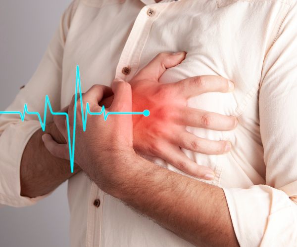
Common stomach-related conditions include indigestion, heartburn, acid reflux, and peptic ulcers, all of which affect millions of people worldwide.
This article will break down these components to give you a clearer picture of how your stomach operates.
Anatomy of the Stomach
The stomach, an integral part of the digestive system, is located in the upper abdomen between the esophagus and the duodenum. It is shaped like a J and functions as a flexible pouch, accommodating varying amounts of food and liquid.
The stomach’s primary role is to mix food with digestive juices, facilitating the breakdown and absorption of nutrients in the small intestine. It can hold approximately 1.5 liters of food and liquid, changing size based on its content volume.
Location of the Stomach in the Human Body
Located on the left side of the upper abdomen, the stomach connects to the esophagus through the esophageal sphincter at its top and to the small intestine at its bottom. This placement permits efficient food processing, making it integral to the digestive tract.
Its J-shaped structure aids in accommodating its functions during digestion.
Size and Shape of the Stomach
The size and shape of the stomach are variable, influenced by factors like fullness, eating habits, and body size. Generally J-shaped, the stomach expands when filled with food and contracts when empty. It includes a long, convex greater curvature and a shorter, concave lesser curvature.
These size and shape variations are essential for storing and processing different food amounts.
Layers of the Stomach Wall
The stomach wall comprises four primary layers: mucosa, submucosa, muscularis externa, and serosa. Each layer has a unique structure and function, aiding in the stomach’s role in digestion.
The mucosa, the innermost layer, contains gastric glands that secrete digestive juices. The submucosa provides support and contains blood vessels and nerves. The muscularis externa, with its three layers of muscle, facilitates the mixing and movement of food. The serosa, the outermost layer, protects the stomach and allows it to move smoothly against other organs.
Mucosa
The innermost lining of the stomach, the mucosa, features folds called rugae that flatten when the stomach distends. It has three sub-layers: epithelium, lamina propria, and muscularis mucosae.
The epithelial layer secretes mucus for lubrication and protection. The lamina propria houses blood vessels and immune cells, maintaining tissue integrity and responding to pathogens. The muscularis mucosae, a smooth muscle layer, enables movement and flexibility, aiding digestion.
The mucosa serves as a barrier against the stomach’s acidic environment, protecting underlying tissues.
Submucosa
The submucosa, a supportive layer, contains dense connective tissue, blood vessels, lymphatic vessels, and nerve cells. It nourishes the mucosa and maintains the stomach’s structural integrity, supporting functions like the secretion of digestive juices and food movement.
The submucosa also contains Meissner’s plexus, a network of nerves that regulate digestive activity.
Muscularis Externa
The muscularis externa has three layers of smooth muscle: inner oblique, middle circular, and outer longitudinal. These muscles facilitate stomach contractions, mixing and breaking down food. Coordinated contractions, known as peristalsis, propel food through the digestive tract.
The muscularis externa’s thickness varies, being especially thick in the stomach to manage denser food contents. It also houses the myenteric plexus, which regulates peristalsis and overall gastric motility.
Serosa
The serosa, the stomach’s outermost layer, consists of squamous epithelial and connective tissue. This smooth membrane encases the stomach, allowing it to move within the abdominal cavity without friction. It produces a lubricating fluid that minimizes friction with adjacent organs, facilitating smooth movement during digestion.
This layer directly contacts surrounding abdominal organs and tissues, providing structural support and protection.
Components of the Stomach
The stomach comprises five distinct areas: cardia, fundus, body, antrum, and pylorus. Each region plays a crucial role in the digestive process, contributing to the stomach’s overall function.
Understanding these components clarifies how the stomach processes and moves food through the digestive system.
Cardia
The cardia is the initial stomach region connecting to the esophagus. It contains the cardiac sphincter, a muscle that prevents stomach contents from flowing back into the esophagus. The cardia controls food entry from the esophagus into the stomach and maintains stomach pressure.
Located just below the diaphragm, the cardia’s anatomical position influences its function and interaction with nearby organs.
Fundus
The fundus is the upper, rounded portion of the stomach, situated to the left of the cardia. It primarily serves as a storage chamber for undigested food and gases released during digestion. Typically, the fundus stores gas unless the stomach is fully occupied.
The fundus aids in the stomach’s overall function by storing and processing food before it moves to the stomach’s body.
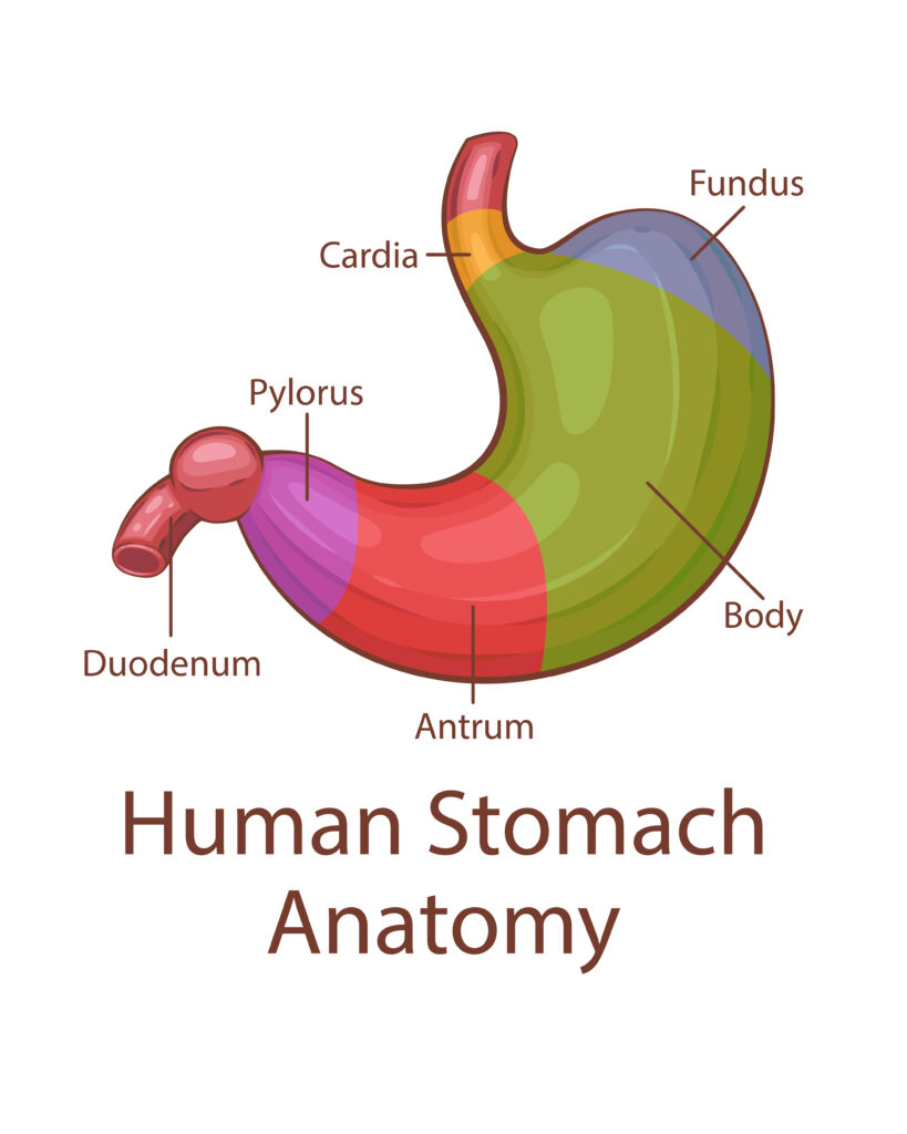
Body of the Stomach
The body, or corpus, is the largest stomach section where food is mixed with digestive juices. It temporarily stores food after ingestion, allowing for initial digestion before moving to the next part of the gastrointestinal tract.
The body contains specialized cells that produce digestive enzymes and acids, crucial for the chemical breakdown of food. Muscular contractions in the body also play a significant role in mixing and breaking down food.
Pyloric Region
The pyloric region connects the stomach to the small intestine and includes the pyloric sphincter, which regulates the passage of digested food into the duodenum. This region consists of the pyloric antrum and the pyloric canal, leading to the duodenum.
The pylorus acts as a gateway between the stomach and small intestine, regulating the flow of chyme for further digestion and nutrient absorption.
Pyloric Sphincter and its Role
The pyloric sphincter acts as a vital valve controlling the passage of partially digested food, or chyme, from the stomach to the duodenum. It ensures proper food processing and prevents intestinal contents from backflowing into the stomach.
The sphincter’s contraction and relaxation are influenced by hormones like gastrin released during digestion. Proper functioning of the pyloric sphincter is crucial for optimal digestion and nutrient absorption in the small intestine.
The Digestive Process in the Stomach
The stomach plays a vital role in digestion by acting as a reservoir for food and initiating its breakdown. Through mechanical and chemical processes, the stomach transforms food into a semi-liquid substance called chyme.
This involves mixing food with gastric juices, including hydrochloric acid and enzymes, and the rhythmic contractions of stomach muscles.
How the Stomach Breaks Down Food
Food enters the stomach through the lower esophageal sphincter, which relaxes to allow passage. Inside, stomach muscles mix the food with digestive juices, forming a semi-liquid mixture called chyme.
Peristalsis, a series of muscle contractions, helps break down food and mix it with gastric juices. This mechanical process, combined with enzymes and acids, breaks food into smaller, more manageable particles.
Role of Gastric Juices
Gastric juices, produced by stomach lining glands, contain hydrochloric acid, pepsin, and lipase. These components break down food and kill harmful bacteria. Hydrochloric acid decomposes food and creates an acidic environment for enzyme activation. Pepsin, an enzyme, starts protein digestion into smaller peptides.
Mucus produced by specific glands protects the stomach lining from gastric acid’s corrosive effects, while the alkaline bicarbonate in mucus neutralizes the acid before it reaches the stomach wall.
Mechanical Digestion (Churning)
Mechanical digestion in the stomach is facilitated by its muscular walls, which rhythmically contract to mix food with gastric juices. This churning process turns food into a semi-liquid substance called chyme, increasing its surface area for better exposure to digestive enzymes.
Peristaltic contractions move food from the fundus towards the pylorus, grinding food particles and aiding their breakdown. Retropulsion pushes larger food particles back towards the stomach’s body for further digestion.
Enzymatic Breakdown in the Stomach
Enzymatic breakdown in the stomach involves enzymes like pepsin, which begin digesting proteins into smaller peptides. The acidic environment created by gastric juices significantly enhances these enzymes’ action. Lipase, another enzyme secreted in the stomach, aids fat digestion, though its activity is limited compared to pancreatic lipase.
Enzymatic activity in the stomach is crucial for digestion, preparing food for further breakdown in the small intestine.
Pepsin
Pepsin is an enzyme secreted in its inactive form, pepsinogen, which activates in the stomach’s acidic environment. This enzyme breaks down dietary proteins into smaller peptides and amino acids. Pepsin, secreted by chief cells, is activated by hydrochloric acid.
The optimal pH for pepsin activity is between 1.5 and 2, enabling it to cleave from pepsinogen and function effectively. Despite its importance, pepsin is not essential for life, as other enzymes in the small intestine can continue protein digestion.
Hydrochloric Acid
Hydrochloric acid plays a crucial role in the digestive process by providing an acidic environment necessary for pepsin activation and optimal digestion. It aids in breaking down food, activating digestive enzymes, and destroying harmful bacteria. Glands in the stomach lining produce hydrochloric acid, contributing to the overall gastric juice.
The production of hydrochloric acid can reach up to three to four liters daily, ensuring that the stomach maintains the necessary acidic environment for effective digestion.
Mucus
Mucus in the stomach serves as a protective barrier against the corrosive effects of stomach acid and enzymes. Secreted by the surface mucous cells in the gastric mucosa, mucus provides lubrication and facilitates the movement of food. It also helps to neutralize the acidity of the stomach contents, reducing damage to the stomach lining.
The bicarbonate component of mucus increases the pH near the gastric epithelium, enhancing mucosal defense and maintaining a stable internal environment for digestion.
Blood Supply and Nerve Supply to the Stomach
The stomach’s blood supply is primarily derived from the celiac trunk and its branches, organized into two main anastomotic systems along its curvatures. This rich blood supply is essential for delivering nutrients and oxygen to the stomach tissues, supporting its functions.
The stomach also receives nerve supply from both the parasympathetic and sympathetic branches of the autonomic nervous system, which regulate its functions.
Arterial Supply
The arterial supply to the stomach is provided by the right and left gastric arteries, which run along the lesser curvature, and the right and left gastro-omental arteries, which run along the greater curvature. The celiac trunk branches into the left gastric, splenic, and common hepatic arteries, supplying the stomach with blood.
The left gastric artery supplies the lesser curvature and gives rise to esophageal branches, while the splenic artery provides blood to the greater curvature through the left gastroepiploic artery.
Gastric Arteries
The right gastric artery originates from the common hepatic artery, while the left gastric artery comes from the celiac trunk. These arteries run along the lesser curvature of the stomach, providing essential blood supply.
The splenic artery, running posterior to the stomach, gives off branches such as the left gastroepiploic and short gastric arteries, enhancing the blood supply to the greater curvature and fundus. The right gastroepiploic artery, originating from the gastroduodenal artery, also supplies the greater curvature of the stomach.
Venous Drainage
Venous drainage from the stomach closely follows arterial paths and primarily channels into the hepatic portal vein. The left and right gastric veins drain into the portal vein, while the short gastric veins drain the fundus of the stomach into the splenic vein. The right gastroomental vein drains into the superior mesenteric vein.
This venous drainage system ensures that blood from the stomach is directed to the liver for processing before returning to the heart.
Portal Vein System
The portal vein system collects all blood from the gastrointestinal tract, including the stomach, and directs it to the liver. The superior mesenteric vein collects drainage from the short gastric and gastro-omental veins, forming the portal vein at the junction with the splenic vein. After filtering in the liver, the blood returns to the heart via the inferior vena cava.
This system is essential for the detoxification and processing of nutrients absorbed from the digestive tract.
Nervous Control
The stomach receives its nerve supply from both the parasympathetic and sympathetic branches of the autonomic nervous system. The enteric nervous system, along with the autonomic nervous system, regulates the stomach’s functions, such as motility and secretion. Vagal fibers play a crucial role in stomach relaxation, allowing it to accommodate food.
The stomach generates a basal electrical rhythm through pacemaker cells, promoting contractions at a rate of 3 to 8 per minute. Mechanoreceptors in the gastric wall trigger responses that stimulate the pyloric pump and regulate food movement through the pylorus.
Role of the Autonomic Nervous System
The autonomic nervous system regulates stomach functions such as secretion, motility, and blood flow. Parasympathetic innervation from the vagus nerve promotes digestive processes, while sympathetic innervation can inhibit digestive functions, particularly during stress. The interplay between these systems allows for a finely tuned regulation of gastric functions, adapting to the physiological demands of digestion.
The enteric system specifically manages digestive processes, further enhancing the stomach’s ability to process and move food.
Vagus Nerve and its Function
The vagus nerve plays a crucial role in stimulating gastric secretions and enhancing gastric motility. It is involved in the parasympathetic nervous system, which is responsible for ‘rest and digest’ functions. The vagus nerve communicates information between the brain and the digestive system, influencing gastric secretions and gut motility.
Damage to the vagus nerve can lead to conditions such as gastroparesis, where food moves slowly from the stomach to the intestines.
Common Disorders of the Stomach
Many gastric disorders can be categorized as either acute, occurring suddenly, or chronic, persisting for months or years. Common disorders include gastritis, ulcers, and gastroesophageal reflux disease (GERD), each with distinct causes, symptoms, and treatments.
Understanding these disorders is crucial for maintaining stomach health and seeking appropriate medical care when necessary.
Gastritis
Gastritis involves the inflammation of the stomach lining, often leading to symptoms such as nausea and loss of appetite. It can result from bacterial infections, especially by Helicobacter pylori, or from excessive alcohol consumption and the regular use of certain pain medications.
Acute gastritis occurs suddenly, while chronic gastritis develops gradually and can lead to complications such as stomach ulcers and an increased risk of gastric cancer. The risk of gastritis increases with age, as older adults are more likely to experience a thinning of the stomach lining.
Types and Symptoms
Symptoms of gastritis can vary based on the underlying cause, including irritation from food, chemicals, or infections. Common symptoms include a burning sensation in the stomach, nausea, and feelings of fullness after eating.
Gastroenteritis, characterized by inflammation that extends to the intestines, causes pain and diarrhea. Acid reflux, or GERD, occurs when stomach acid flows back into the esophagus, causing a burning sensation.
Other conditions like irritable bowel syndrome (IBS) and celiac disease also manifest through digestive issues, including stomach pain and bloating.
Ulcers
Stomach ulcers, also known as gastric ulcers, occur when the stomach lining is damaged, often due to H. pylori infection or long-term use of NSAIDs.
Symptoms can include:
a burning sensation in the stomach
indigestion
bloating
in severe cases, bleeding.
Treatment typically involves medications that reduce stomach acid and antibiotics if an H. pylori infection is present. Complications from untreated ulcers can lead to severe conditions such as internal bleeding or perforation of the stomach wall. Lifestyle factors like smoking and excessive alcohol consumption can exacerbate ulcer symptoms.
Peptic Ulcers
Peptic ulcers can affect the stomach (gastric) or the upper part of the small intestine (duodenal), causing similar pain and discomfort. These ulcers result from a breakdown in the protective mucous layer due to excessive stomach acid or bacterial infections like H. pylori. Symptoms often include dull or burning stomach pain, particularly between meals or at night.
Complications from untreated peptic ulcers may include severe bleeding, perforation of the stomach wall, blockage in the digestive tract, and an increased risk of stomach cancer. While stress and spicy foods do not cause peptic ulcers, they can exacerbate existing symptoms.
Causes and Risk Factors
Risk factors for stomach disorders include:
Excessive alcohol consumption
Smoking
Prolonged usage of pain relievers like aspirin
Helicobacter pylori infection
Regular use of NSAIDs, which are significant contributors to ulcers
Certain lifestyle choices, such as poor diet and lack of exercise, which can lead to functional gastrointestinal disorders.
Stress and anxiety can exacerbate symptoms, and structural gastrointestinal diseases may arise from chronic inflammation or blockages. Genetic predispositions and infections also play a role in the development of stomach disorders.
Gastroesophageal Reflux Disease (GERD)
Gastroesophageal reflux disease (GERD) occurs when stomach acid frequently flows back into the esophagus, leading to symptoms such as heartburn and acid regurgitation. This condition often results from a weakened lower esophageal sphincter, which typically prevents stomach contents from backing up into the esophagus.
Risk factors for GERD include obesity, pregnancy, and certain lifestyle choices like smoking and eating large meals. If left untreated, GERD can lead to complications such as esophagitis, esophageal strictures, and an increased risk of Barrett’s esophagus, which may lead to cancer.
Management strategies for GERD involve lifestyle modifications, medications, and in severe cases, surgical interventions.
When to See a Doctor
It is essential to consult a healthcare professional if you experience persistent stomach issues such as chronic abdominal pain, frequent indigestion, or persistent diarrhea. Frequent bloating may indicate food sensitivities or gastrointestinal disorders, which should be assessed by a healthcare provider.
Recognizing persistent symptoms can help in deciding when to seek medical advice, ensuring timely diagnosis and treatment of potential digestive disorders.
Bottom Line
The structure of the stomach includes distinct regions (cardia, fundus, body, antrum, pylorus) and multiple layers (mucosa, submucosa, muscularis externa, serosa), each serving specific functions essential for digestion. Also Common stomach disorders, such as gastritis, ulcers, and GERD, can arise due to various factors, including bacterial infections and lifestyle choices, emphasizing the importance of maintaining stomach health through diet and regular medical check-ups.