Envision you’re in the operating room, and the surgeon points out the falciform ligament. This vital structure doesn’t just hold your liver in place; it also tells a story about your health. Whether it’s guiding surgical techniques or revealing signs of diseases, comprehending this ligament is essential. So, why does it matter to you? Let’s examine its anatomy, clinical significance, and what you should know about its role in surgeries.
Anatomy and Structure of the Falciform Ligament
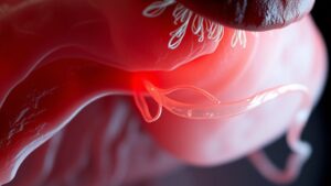
The falciform ligament could seem like a complex term, but it’s actually just a slender, sickle-shaped fold of tissue in your abdomen that serves a vital function in maintaining your liver in position. This ligament extends from your anterior abdominal wall to your liver, dividing it into left and right lobes.
It’s made up of a bilayer of parietal peritoneum, with the right layer covering the anterior abdominal wall and the left layer becoming visceral peritoneum over your left liver lobe. Within the falciform ligament, you’ll find the round ligament, or ligamentum teres hepatis, which is a remnant of the fetal umbilical vein.
Importantly, its bare area lies between the coronary ligament’s layers, lacking any peritoneal covering.
Embryology and Development of the Falciform Ligament
During your development in the womb, the falciform ligament begins to take shape from the ventral mesentery, which is attached to your foregut’s ventral border. This ligament forms as the ventral mesogastrium splits into the falciform ligament and the lesser omentum during embryogenesis.
Through the seventh week, while the right umbilical vein degenerates, the left umbilical vein remains, eventually turning into the ligamentum teres hepatis within the falciform ligament. As liver development occurs and the foregut rotates, the ventral mesogastrium stretches, defining the ligament’s final position.
Sometimes, congenital variants like an obliterated or absent liver fissure might arise, but these incidental findings typically don’t have clinical significance. It’s fascinating how our bodies develop, isn’t it?
Vascular and Lymphatic Supply
At the time you consider how your body is wired, it’s intriguing to realize that the falciform ligament benefits from a sturdy vascular and lymphatic supply. Here’s what you should know:
- The falciform artery, branching from the internal thoracic artery and inferior phrenic artery, supplies this ligament and connects to other vessels.
- Venous drainage primarily relies on the internal thoracic and inferior phrenic veins, while the umbilical vein remnant can recanalize during portal hypertension.
- Superficial lymphatics manage liver lymph drainage but could permit infections to spread, particularly should paraumbilical veins dilate in cases like caput medusae.
Understanding this vascular network can improve your awareness of potential complications, like those encountered during chemoembolization.
Clinical Significance and Common Pathologies
Comprehending the clinical significance of the falciform ligament is key for anyone involved in health care.
In conditions like portal hypertension, you could observe changes such as caput medusae, a visible sign of foundational liver issues.
Plus, being aware of potential complications like hernias or infections can aid in making timely decisions during surgical procedures.
Portal Hypertension Indicators
At the time the pressure in your portal vein rises, it can lead to some telltale signs that hint at portal hypertension-a condition that can impact your liver’s health. You could observe:
- Engorged periumbilical veins, known as caput medusae, which signal vascular redistribution.
- A dilated paraumbilical vein in the falciform ligament can result from increased pressure, observed during an ultrasound.
- Cullen’s sign, or periumbilical ecchymosis, could appear with serious issues like a variceal rupture.
If you’re experiencing any of these indicators, it’s essential to talk with your healthcare provider.
Managing portal hypertension at the initial stage can help you control its effects and safeguard your liver from further complications related to cirrhosis and other conditions.
Herniation and Obstruction Risks
Herniation and obstruction risks associated with the falciform ligament are more significant than you could believe, particularly since internal herniation can lead to small bowel obstruction in rare cases.
Internal hernias involving the falciform ligament can occasionally be linked to congenital defects, where improper peritoneal fusion occurs. Should the ligament experience nonfixation due to developmental issues, you could find yourself at higher risk for these hernias.
Diagnosis can be tricky; many times, preoperative imaging like CT scans could miss the issue, especially should ascites be present. Surgical intervention might become necessary once symptoms like pain or bowel compromise arise.
It’s essential to stay informed about these risks and discuss any concerns with your healthcare provider.
Role in Surgical Procedures
Surgery often hinges on precise anatomical landmarks, and the falciform ligament stands out as a significant guide in various procedures.
- In laparoscopic cholecystectomy, it helps identify the gallbladder’s position relative to the liver.
- During liver resection, surgeons often divide the falciform ligament to mobilize the left lobe, while being careful not to injure the falciform artery, found in 67% of cases.
- In portal hypertension, paraumbilical veins can form collaterals, leading to visible caput medusae.
Additionally, the falciform ligament might serve as a vascularized flap for duodenal perforation repairs, often reducing complications compared to omental patches.
Plus, non-target embolization during hepatic chemoembolization can lead to skin necrosis in some cases. Grasping these aspects can markedly improve surgical results.
Surgical Applications and Techniques
In any surgical setting, having precise anatomical landmarks can make all the difference in achieving successful results, and that’s where the falciform ligament comes into play. Serving as a vital surgical landmark, it helps you distinguish between liver segments during laparoscopic cholecystectomy.
While performing liver resection, you could divide the ligament to improve access to hepatic veins and the inferior vena cava. Its unique structures can also act as a vascularized flap in reconstructive procedures, minimizing donor-site complications.
However, be cautious during chemoembolization-complications can arise should the falciform artery be unintentionally embolized. Additionally, in cases of portal hypertension, you’ll want to recall that the paraumbilical veins could recanalize, necessitating careful dissection to prevent hemorrhage.
Imaging Considerations for the Falciform Ligament
When it comes to imaging the falciform ligament, you’ve got several techniques at your disposal.
Each method, whether it’s a CT scan, MRI, or ultrasound, allows you to identify various abnormalities and understand the ligament’s role in your body better.
Knowing what these images can reveal will help you feel more informed and prepared for any medical discussions.
Imaging Techniques Overview
Grasping how imaging techniques assess the falciform ligament can enable you in tackling potential health concerns. Here’s a quick overview of useful modalities:
- Ultrasound: Detects abnormalities like cysts with 85-90% sensitivity for identifying paraumbilical vein dilation in cases of portal hypertension.
- CT Imaging: The gold standard due to its contrast improvement and high spatial resolution, allowing precise visualization of ligament pathologies.
- MRI: Offers superior soft-tissue contrast, particularly utilizing T1- and T2-weighted sequences, making it ideal for distinguishing between benign and malignant lesions.
Doppler ultrasound is crucial too, measuring blood flow in recanalized paraumbilical veins, which signals significant shunting. Understanding these techniques helps strengthen your health decisions and promotes better patient results.
Pathological Findings Identification
Identifying pathological findings related to the falciform ligament can markedly aid in diagnosing fundamental health issues. For instance, CT scans are effective at detecting abscesses, which often appear as hypodense lesions with surrounding fat stranding.
Should you be considering a lipoma, MRI is your go-to; it reveals high signal intensity on T1-weighted images. Ultrasound can help identify cysts, showing anechoic structures with posterior enhancement.
In cases of portal hypertension, you’ll see tubular structures inside the falciform ligament, indicating paraumbilical vein recanalization on a contrast-enhanced CT.
Variants and Anomalies
The falciform ligament, while often overlooked, can exhibit fascinating variants and anomalies that provide insight into our anatomy’s complexity.
Here are some intriguing characteristics:
- Congenital defects could lead to internal herniation of the small bowel, although it’s rare.
- The falciform artery, found in 67% of cadavers, can arise variably from the left or middle hepatic artery.
- Variants of the paraumbilical vein can recanalize in portal hypertension, contributing to conditions like caput medusae.
Nonfixation of the falciform ligament due to failed peritoneal fusion can also predispose neonates to rare hernias.
These subtleties in the falciform ligament highlight how details in our anatomy can impact health in unexpected ways, reminding us of the body’s extraordinary complexity.
Complications Related to Falciform Ligament
Although often regarded as a mere anatomical feature, complications related to the falciform ligament can lead to significant health issues that could catch you off guard. For instance, portal hypertension could cause dilation of the paraumbilical vein, resulting in caput medusae, a visible network of veins around your belly button.
You should also be aware that internal herniation, while rare, can lead to small bowel obstruction. In case you undergo chemoembolization targeting the falciform artery, watch for complications like skin rashes or necrosis, which can trigger acute abdomen symptoms.
Whenever necrosis occurs, immediate surgical intervention becomes essential to avoid further complications. Identifying these potential complications is critical for timely medical assistance and maintaining your health.
Future Perspectives in Falciform Ligament Research
As researchers explore further into the complexities of the falciform ligament, it becomes clear that there’s so much more to this anatomical structure than meets the eye. Future studies could pave the way for revolutionary advancements in surgical practices. Here are some exciting prospects:
- Biomechanical research could deepen our grasp of the ligament’s tensile strength, aiding safer surgical techniques.
- 3D reconstruction might refine preoperative mapping, helping to pinpoint vascular anomalies and reduce complications.
- Genetic studies could reveal connections to congenital defects, enabling timely identification of internal herniation risks.
Additionally, ongoing clinical trials examining ligament grafts in reconstructive surgeries could standardize techniques, while investigations into lymphatic pathways might uncover fresh perspectives on metastatic spread.
Each step forward holds the promise of improved patient results.