You may question whether gallstones show up on imaging, and the answer isn’t straightforward—it depends on what they’re made of. While cholesterol stones often remain obscure on X-rays, pigmented or calcified ones can appear clearly. Ultrasound usually steals the spotlight for identification, but CT and MRI step in when complications arise. Understanding how these imaging tools work gives you insight into diagnosing and managing gallstones before they cause trouble. Curious which stones play hide-and-seek? Let’s break it down.
Types of Gallstones and Their Radiographic Properties
While you’re managing gallstones, comprehending what type you could have can make a significant difference in how they appear on imaging. The three main types of gallstones—cholesterol, pigment, and mixed—vary in their imaging properties.
Cholesterol stones, making up about 10% of cases, are usually radiopaque on X-rays due to low calcium content, so they mightn’t show up clearly.
Pigment stones, another 10%, can be black or brown; black ones often contain calcium bilirubinate, making them more visible.
Mixed stones, the most common at 80%, may show both solid and faint spots on scans, depending on their makeup.
Half of gallstones can be seen on standard X-rays when they’re calcified, while others need advanced imaging.
Recognizing these details helps your doctor pick the right tests for clearer results.
Common Imaging Modalities for Gallstone Detection
Should you be managing gallstones, recognizing which imaging tests can identify them makes a significant difference in obtaining the correct diagnosis.
Cholesterol gallstones, the most common type, often don’t show up on standard X-rays because they’re radiolucent. That’s why doctors rely on other imaging modalities.
Computed tomography (CT) scans can spot some gallstones, especially calcified ones, but they may miss softer cholesterol stones.
MRI, particularly MRCP, is another option—it’s great for seeing bile ducts and concealed stones without radiation.
Dual-energy CT (DECT) improves identification by telling apart calcified and non-calcified stones.
Each test has strengths, so your doctor will choose based on your symptoms and stone type.
Recognizing these options helps you feel more informed and confident in your care.
Ultrasound: The Gold Standard for Gallstone Diagnosis
As other imaging tests could overlook gallstones, ultrasound steps in as the most reliable way to spot them. It’s quick, painless, and doesn’t use radiation, making it a go-to for doctors.
With over 95% accuracy, ultrasound finds gallstones as bright spots with dark shadows behind them—like tiny echoes in your gallbladder. Should you press on the area and it hurts during the scan (sonographic Murphy’s sign), it could mean inflammation from a stuck stone.
While most gallstones show up clearly, some cholesterol ones can hide, so careful imaging matters. Ultrasound also checks for complications like thickened gallbladder walls or fluid buildup, warning signs of trouble.
You’ll get answers fast, without invasive steps, which is why it’s the gold standard.
CT Scans and Gallstone Visibility
CT scans aren’t always the best at spotting gallstones, but they can still play a key role whenever complications are suspected.
While ultrasound is the go-to for diagnosis, CT visibility of gallstones depends on their type. Calcified stones show up clearly, but cholesterol stones often blend in, making them harder to notice.
Here’s what you should know:
- Size matters: Tiny gallstones (under 3mm) may not appear on CT scans, especially in cases where they’re not calcified.
- Dual-energy CT helps: This advanced technique improves identification by analyzing stone composition better than standard CT.
- Not perfect for diagnosis: CT excels at spotting complications like infections or blockages but misses some gallstones ultrasound would catch.
In the event your doctor orders a CT, it’s likely to check for bigger issues, not just the stones themselves.
MRI and MRCP in Evaluating Gallstones
You’ll find MRI and MRCP incredibly useful for detecting gallstones without radiation, giving you high accuracy in spotting even small stones.
These scans also help pinpoint the stone type, so your doctor can tailor treatment without invasive tests.
Plus, they’re great for checking whether complications, like blockages or inflammation, are causing your symptoms.
High Sensitivity Detection
Whenever gallstones don’t show up clearly on other scans, MRI and MRCP step in as reliable tools that spot them with impressive accuracy—without radiation.
Should you have tricky-to-detect stones slipping past X-rays or CT scans, these imaging methods offer high sensitivity, catching even non-calcified ones lurking in your gallbladder or bile ducts.
With rates like 90-94% accuracy, you can trust MRI to uncover what other tests miss—no harmful rays needed.
- Detailed imaging: MRI and MRCP give clear visuals of your biliary system, making concealed gallstones obvious.
- No radiation worry: Unlike CT scans, these scans use magnets and radio waves, keeping you safe.
- Better for complications: MRCP excels at spotting blockages or stones in ducts, helping doctors plan treatment smarter.
Should gallstones play hide-and-seek, MRI won’t let them win.
Non-Invasive Imaging Benefits
Even though other scans miss them, MRI and MRCP make spotting gallstones safer and easier—no needles, no radiation, just clear, detailed depictions of what’s happening in your biliary system.
Unlike CT scans, these non-invasive imaging techniques don’t expose you to radiation, making them ideal should you need repeated checks. MRI highlights gallstones as dark spots on T2-weighted images, while MRCP zooms in on bile ducts, showing blockages without invasive procedures.
You won’t feel discomfort, and there’s no recovery time. Plus, they catch complications like obstructions promptly.
For those wary of risks, these methods offer peace of mind with high accuracy. No dyes, no incisions—just a precise look at trouble spots so you and your doctor can plan the next steps confidently.
Accurate Stone Characterization
Because gallstones aren’t all the same, MRI and MRCP step in to give doctors a clearer image of what’s really going on.
These imaging modalities help distinguish between cholesterol and pigmented stones, even though they’re concealed in bile sludge.
MRI’s high sensitivity means it won’t miss much, and MRCP maps out your bile ducts without radiation.
- Spot the difference: MRI shows gallstones as dark spots on T2-weighted images, making them stand out against bright bile.
- No guesswork: MRCP pinpoints stone size and location, helping plan treatment without invasive tests.
- Safe and detailed: Unlike X-rays, MRI doesn’t use radiation, so it’s safer for repeated scans if needed.
With these tools, doctors get a precise look at your gallstones, ensuring the right care for you.
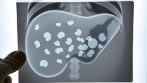
Plain Radiography: When Gallstones Appear Radiopaque
Should you’ve ever had an abdominal X-ray, you could question why some gallstones show up clearly while others don’t.
On plain radiography, only about 15-20% of gallstones appear radiopaque—meaning they’re visible because they contain enough calcium to block X-rays. Cholesterol stones, the most common type, usually don’t show up because they lack calcium.
But black pigment stones, which have more calcium bilirubinate, often do.
In case your X-ray reveals small, dense spots in the right upper quadrant, those could be radiopaque gallstones.
While plain radiography isn’t the best test for gallstones (most slip under the radar), it’s sometimes the initial clue.
Should your doctor suspect gallstones but your X-ray’s clear, don’t worry—other imaging tests can catch what plain films miss.
Factors Affecting Gallstone Detection on Imaging
Should you have ever pondered why some imaging tests miss gallstones while others catch them easily, it’s because several factors play a role in how they show up.
The type of imaging modality used, the size and composition of the gallstones, and even the presence of sludge in your gallbladder can all affect identification.
- Imaging Modalities: Ultrasound excels at spotting gallstones, even radiolucent ones, while plain X-rays often miss them unless they’re calcified.
- Stone Composition: Cholesterol stones blend in on X-rays but stand out on ultrasound, whereas calcified stones might appear on both.
- Size Matters: Tiny gallstones (<2mm) can hide in sludge or mimic other structures, making them trickier to identify.
Choosing the right test matters—your doctor will pick the best one based on these factors.
Differential Diagnoses: Mimics of Gallstones
Gallstones aren’t always the only thing showing up on your imaging tests—sometimes other conditions can look just like them. Should you have gallbladder polyps or tumors, they could mimic stones on ultrasound, especially in cases where they’re small or concealed in folds.
Sludge, a thick bile mixture, can also appear similar, making it tricky to spot the real issue. Porcelain gallbladder, where the wall hardens and calcifies, could even resemble large stones on X-rays.
Adenomyomatosis, a benign condition causing wall thickening, can confuse imaging features too. MRI helps clarify things by showing signal voids that point to stones.
Whenever your doctor checks your scans, they’ll compare these differential diagnoses to make certain they’re not missing something else. It’s all about getting the right depiction of what’s really going on in your gallbladder.
Complications of Gallstones Visible on Imaging
- Acute cholecystitis: Inflammation shows up as a thickened gallbladder wall and fluid around it.
- Mirizzi Syndrome: Impacted stones squeeze the bile duct, visible as dilation on scans.
- Gallstone ileus: A rare blockage in the intestines, seen as air in the bile ducts on CT.
Early discovery helps you avoid worse problems, so don’t ignore persistent belly pain.
Interventional Radiology Approaches for Gallstone Management
Should you be facing gallstones and surgery isn’t an option, interventional radiology offers solutions like percutaneous cholecystostomy to drain your gallbladder safely.
Biliary stenting procedures can also relieve blockages caused by stones, helping you feel better without major surgery.
These techniques give doctors a way to manage complications while keeping risks low.
Percutaneous Cholecystostomy Techniques
- How it works: A radiologist guides the catheter into your gallbladder, draining infected bile to relieve pain and prevent complications.
- Who needs it: Critically ill patients or those with high surgery risks.
- What to expect: Local anesthesia, minimal downtime, and follow-up imaging to check progress.
You’ll feel relief fast, and it’s often the bridge to safer, long-term solutions.
Biliary Stenting Procedures
Whenever percutaneous cholecystostomy isn’t enough to manage gallstone complications, biliary stenting offers another way to ease blockages in your bile ducts.
This procedure uses a small tube (stent) to keep the narrowed or blocked biliary pathways open, letting bile flow normally again. Imaging tools like fluoroscopy or ultrasound guide the stent placement, ensuring precision and safety.
It’s often a good option should you not be ready for surgery or need quick relief from gallstone-related pain. Temporary or permanent stents can be used, depending on your condition.
While complications like infection or stent movement are possible, they’re rare with careful monitoring.
Biliary stenting can improve your quality of life by reducing symptoms like jaundice or discomfort, making it a key tool in gallstone management.