A bone diagram shows the structure and function of the human skeleton. The adult human skeleton consists of 206 bones, categorized into the axial and appendicular skeleton, serving essential functions such as support, protection, movement, and blood cell production.
Bone tissue is classified into compact and spongy bone, with distinct types of bones including long, short, flat, irregular, and sesamoid bones, each fulfilling unique roles in the skeletal system.
This article will show detailed diagrams of the bone axial and appendicular skeletons and explain the role of each bone.
Overview of the Human Skeleton
The adult human skeleton is a marvel of biological engineering, comprising 206 bones that form the bony framework of the human body. These bones are divided into two main categories: the axial skeleton and the appendicular skeleton.
The skeletal system serves multiple purposes, including support, protection, movement, blood cell production, and mineral storage, playing a crucial role in maintaining posture and overall bodily functions.
Appreciating the skeletal system anatomy reveals its complexity and functionality.
Axial Skeleton
The axial skeleton forms the central core of the human body, consisting of 80 bones that include the skull, vertebral column, ribs, and sternum. Its primary function is to provide support and protection for the brain, spinal cord, and thoracic organs.
The five main parts of the axial skeleton are:
- The skull
- Ossicles of the middle ear
- Hyoid bone
- Vertebrae
- Thoracic cage
This central structure supports posture and shields internal organs.
The skull, composed of several bones fused together, houses and protects the brain, while the vertebral column, made up of individual vertebrae, encases the spinal cord. The ribs and sternum form the thoracic cage, safeguarding the heart and lungs. Together, these components illustrate the axial skeleton’s vital role in the body’s structural integrity and protection of critical systems.
Appendicular Skeleton
The appendicular skeleton, consisting of 126 bones, includes the bones of the pectoral girdle, pelvic girdle, and limbs. This part of the skeleton is designed to facilitate a wide range of movements, making it essential for tasks such as walking, lifting, and manipulating objects.
The appendicular skeleton’s primary function is to provide mobility and dexterity, enabling the human body to interact dynamically with its environment.
Detailed Bone Structures
Bone tissue is categorized into two primary types: compact bone and spongy bone, each differing in density and structural organization.
This section will explore the various types of bones, including long bones, short bones, flat bones, irregular bones, and sesamoid bones, highlighting their unique structures and functions within the skeletal system.
Long Bones
Long bones, characterized by their elongated shape, are longer than they are wide and primarily consist of compact bone. Bones like the femur and humerus support body weight and facilitate movement.
Long bones feature a diaphysis (shaft) and epiphyses (ends) that contain bone marrow and serve as sites for growth and development.
Short, Flat, and Irregular Bones
Short bones, such as carpals and tarsals, are roughly equal in length and width, providing stability and some mobility. These bones support body weight and enable fine motor movements.
Flat bones, like the parietal bones of the skull and the sternum, provide protection for internal organs and serve as attachment points for muscles. Irregular bones, such as the vertebrae, have complex shapes that allow them to fulfill protective roles and bear weight, contributing to the overall stability and functionality of the skeletal system.
Sesamoid Bones
Sesamoid bones, such as the patella, are small, round bones embedded within tendons. These bones serve to reduce friction and enhance mechanical leverage, improving the efficiency of muscle movements and reducing the wear on tendons.
Microscopic Structure of Bone Tissue
Bone tissue, primarily made up of osseous tissue, includes a collagen matrix mineralized with calcium and phosphorus, giving bones their strength and resilience.
Delving into the microscopic anatomy of bone tissue, including compact bone, spongy bone, and the bone matrix and cells, provides a deeper understanding of bone structure and function.
Compact Bone
Compact bone is characterized by its dense structure, composed of cylindrical units called osteons, which contain a central canal for blood supply. These tightly packed osteons provide strength and rigidity, enabling bones to withstand significant stress.
Each osteon contains concentric layers of bone matrix surrounding a central canal, which houses blood vessels and nerves. This arrangement enables compact bone to support and protect the body’s weight and vital structures efficiently.
Spongy Bone
Spongy bone, also known as cancellous bone, is composed of a network of trabeculae, which are aligned according to stress lines to enhance strength while remaining lightweight. Spongy bone supports red bone marrow and adapts to mechanical stress, offering structural integrity and flexibility.
Bone Matrix and Cells
The bone matrix is primarily composed of collagen fibers and minerals like calcium phosphate, ensuring its strength and resilience. This matrix provides the framework for bone tissue, allowing it to withstand compressive forces and maintain structural integrity.
Key bone cells include osteoblasts, which build bone; osteoclasts, which break down bone; and osteocytes, which maintain the bone matrix. These cells work together to ensure continuous bone growth, remodeling, and repair, maintaining the overall health and functionality of the skeletal system.
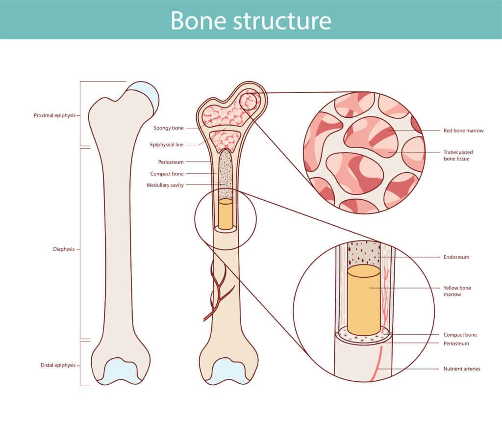
Bone Diagram with Key Locations
A diagram can clarify the anatomy of bones. For example, a long bone has two main parts: the diaphysis (tubular shaft) and the epiphysis (wider end section filled with spongy bone). The diaphysis contains the medullary cavity, which in adults, is filled with yellow bone marrow.
The epiphyseal plate, present in growing bones, is located at the junction of the diaphysis and epiphysis, replaced by an epiphyseal line in adulthood. Inside the bone, the endosteum lines the medullary cavity and contains bone cells that play a role in growth and repair, while the periosteum envelops the outer surface of bones, facilitating attachment for tendons and ligaments.
Articular cartilage covers the surfaces of the epiphyses where they meet other bones, reducing friction and acting as a shock absorber during movement.
Bone Marrow
Bone marrow is classified into two main types: red bone marrow and yellow bone marrow. Red bone marrow, located in spongy bone, is responsible for hematopoiesis, producing red blood cells, white blood cells, and platelets.
Yellow bone marrow, found in the medullary cavity of long bones, primarily stores lipids, serving as an energy reserve.
Red Bone Marrow
Red bone marrow is crucial for hematopoiesis, generating red blood cells, white blood cells, and platelets essential for immune response and oxygen transport. This marrow is rich in blood stem cells and is primarily located in spongy bone, playing a vital role in maintaining the body’s blood cell supply.
Yellow Bone Marrow
Yellow bone marrow exists mainly in the medullary cavity of long bones, primarily serving the purpose of lipid storage and energy reserve. Composed mainly of adipose tissue, yellow bone marrow functions as an energy store that can be converted into fuel for the body, particularly during periods of low energy intake.
Joints and Articulations
Joints are the connections between bones, allowing them to work together in movement and stability. They play a crucial role in facilitating movement and responding to various conditions that may affect mobility.
Exploring different types of joints-synovial, fibrous, and cartilaginous-highlights their unique structures and functions.
Synovial Joints
Synovial joints are characterized by a fluid-filled cavity, allowing for extensive movement and greatly enhancing their range of motion. Examples of synovial joints include the knee and elbow, which are crucial for mobility.
These joints are lined with articular cartilage, which reduces friction and acts as a shock absorber during movement.
Fibrous Joints
Fibrous joints are primarily immovable and are held together by dense irregular connective tissue. These joints, such as the sutures in the skull, provide stability and protection but allow very little movement. The dense irregular connective tissue prevents significant movement between the connected bones, ensuring structural integrity.
Cartilaginous Joints
Cartilaginous joints involve bones connected by cartilage, allowing for limited movement while providing stability. Intervertebral discs are a prime example, allowing slight movement and providing cushioning between the vertebrae.
These joints are essential for maintaining flexibility and support in the skeletal system.
Bone Development and Growth
Bone development and growth are complex processes that involve various mechanisms, such as endochondral ossification and intramembranous ossification. These processes ensure that bones grow and develop properly, adapting to mechanical stress and repairing damage.
Bone remodeling is also crucial, allowing the skeleton to maintain its strength and integrity over time.
Endochondral Ossification
Endochondral ossification is a key process in bone development, where mesenchymal tissue transforms into a cartilage model that is later converted to bone. This process begins with the formation of cartilage models, which are then infiltrated by blood vessels and osteoblasts.
The primary ossification center forms in the diaphysis, with growth continuing at the growth plate epiphyseal plates.
Intramembranous Ossification
Intramembranous ossification transforms connective tissue membranes into bony tissue, forming specific flat bones. This process involves the direct conversion of mesenchymal stem cells into osteoblasts, which then form bone tissue.
This process primarily forms flat bones, such as those in the skull.
Bone Remodeling
Bone remodeling is a continuous process involving the breakdown of bone by osteoclasts and the formation of new bone by osteoblasts. This process helps the skeleton adapt to stress, repair damage, and maintain strength and resilience.
Osteoclasts resorb old or damaged bone, while osteoblasts build new bone, ensuring the skeletal system’s dynamic adaptability.
Functions of the Skeletal System
The skeletal system serves multiple essential functions, including providing support and protection, enabling movement, and storing and releasing minerals. It plays a crucial role in maintaining the body’s shape, protecting vital organs, and facilitating blood cell production and mineral storage.
This section will explore these functions in detail.
Support and Protection
The skeleton serves as a sturdy framework that maintains body posture and safeguards internal organs from injury. Bones shield vital organs, like the skull protecting the brain and the ribcage shielding the heart and lungs. This protective function minimizes potential injuries during impacts, ensuring the body’s critical systems remain intact.
Movement
Bones play a crucial role in facilitating movement by acting as attachment points for muscles. Skeletal muscles attach to bones, providing anchor points and enabling movement through leverage.
This interaction between bones and muscles allows for a variety of movements across joints, supporting physical activities and overall mobility.
Storage and Release
Bones serve as reservoirs for essential nutrients like calcium and phosphorus, which can be released into the bloodstream to maintain mineral balance in the body. This storage ensures a steady supply of vital minerals for various physiological processes.
Common Bone Diseases and Conditions
Bone diseases and conditions can significantly impact the skeletal system’s functionality and overall health. Common issues include:
- Osteoporosis, which weakens bones and makes them more susceptible to fractures
- Arthritis, which causes inflammation and pain in the joints
- Bone cancer, which can affect the integrity and function of bone tissue
Each of these conditions affects bone tissue, structure, and function in different ways.
Osteoporosis
Osteoporosis leads to a reduction in bone density, resulting in fragile bones that are more susceptible to fractures. Often referred to as a ‘silent disease,’ it can progress without symptoms until a fracture occurs. Weakened bones in individuals with osteoporosis lead to fractures from minimal trauma, making it a significant health concern.
Arthritis
Arthritis is characterized by inflammation in the joints, causing chronic pain and limiting mobility. With over 100 types, the most common are osteoarthritis and rheumatoid arthritis.
Chronic inflammation and pain from arthritis can significantly impair joint mobility and quality of life, potentially leading to permanent joint changes and reduced mobility.
Bone Cancer
Primary bone cancer is rare and can originate in the bone, while metastatic bone cancer arises when cancer cells spread to the bone from other parts of the body. Malignant tumors in bone cancer significantly affect bone health and structure.
Both types of bone cancer can compromize bone integrity, leading to pain, fractures, and other serious health issues.
Wrap Up
Finally we can say, The human skeletal system is an intricate and essential part of the body, providing support, protection, movement, and storage of vital minerals.