The knee is stabilized by four main ligaments: the ACL, PCL, MCL, and LCL, each serving specific functions to control movement and prevent injuries. Also Knee ligament injuries are common in sports and can range from mild sprains to complete tears, with ACL and MCL injuries being the most frequent.
Need to see how knee ligaments look and what they do? A knee ligaments diagram can help. This article explains each ligament’s role and common injuries.
What are Knee Ligaments?
Knee ligaments are tough bands of tissue that connect the femur (thigh bone) to the tibia and fibula (lower leg bones), providing stability to the knee joint.
The knee joint consists of three main bones: the femur, tibia, and fibula, all held together by these crucial ligaments. Among the primary ligaments, the Anterior Cruciate Ligament (ACL), Posterior Cruciate Ligament (PCL), Medial Collateral Ligament (MCL), and Lateral Collateral Ligament (LCL) are essential for maintaining the knee’s stability and function.
The collateral ligaments, including the MCL and LCL, prevent side-to-side motion of the knee. The MCL connects the femur to the tibia, stabilizing the inside of the knee, while the LCL connects the femur to the fibula, providing stability against lateral forces.
The cruciate ligaments, namely the ACL and PCL, cross each other in the center of the knee to control back-and-forth motion and ensure the knee bends properly.
Anterior Cruciate Ligament (ACL)
The anterior cruciate ligament (ACL) is located in the center of the knee, running diagonally from the front of the tibia to the back of the femur.
This positioning allows the ACL to play a vital role in controlling the knee’s back-and-forth motion, providing essential stability during activities that involve sudden stops, changes in direction, or jumping. The ACL works in conjunction with the posterior cruciate ligament (PCL) to form an X-shape within the knee, which is crucial for maintaining overall knee stability.
Common activities that lead to ACL injuries include sports such as football, basketball, and soccer, where sudden movements are frequent. Individuals often report hearing a popping sound or sensation at the time of injury, followed by knee pain and swelling.
Posterior Cruciate Ligament (PCL)
The posterior cruciate ligament (PCL) is another critical stabilizing ligament of the knee, running diagonally behind the ACL and forming an X-shape as they cross each other.
The PCL’s primary function is to prevent the tibia from moving too far backward in relation to the femur, which is essential for maintaining proper knee mechanics during movement. This ligament is composed of two functional bundles: the larger anterolateral bundle (ALB) and the smaller posteromedial bundle (PMB).
PCL injuries are less common than ACL or MCL injuries and often occur due to significant trauma, such as a dashboard injury in a car accident where a large force is applied to the shins while the knee is flexed. Symptoms of a PCL injury include swelling, pain, and instability, which can be tested using the posterior drawer test.
Medial Collateral Ligament (MCL)
The medial collateral ligament (MCL) is a broad, flat ligament located on the inner part of the knee, primarily responsible for stabilizing the knee against excessive valgus stress, which occurs when the knee is pushed inward.
This ligament is composed of two layers: a superficial layer and a deep layer, both of which work together to maintain knee stability. The MCL connects the femur to the tibia, playing a crucial role in preventing side-to-side motion.
MCL injuries commonly occur due to direct impact to the outer side of the knee, such as during contact sports like football or rugby. Rehabilitation for MCL injuries often emphasizes strengthening the surrounding muscles to support the knee and restore function.
Lateral Collateral Ligament (LCL)
The lateral collateral ligament (LCL) is a thinner, more rope-like structure compared to the MCL, located on the outer side of the knee. The LCL connects the femur to the fibula and plays a crucial role in stabilizing the knee against excessive lateral movement, preventing the knee from bending outward.
Injuries to the LCL typically occur due to a direct impact on the inside of the knee or from excessive outward forces, such as during a fall or collision. Despite its important role, LCL injuries are relatively rare, occurring in isolation in less than 2% of all knee injuries.
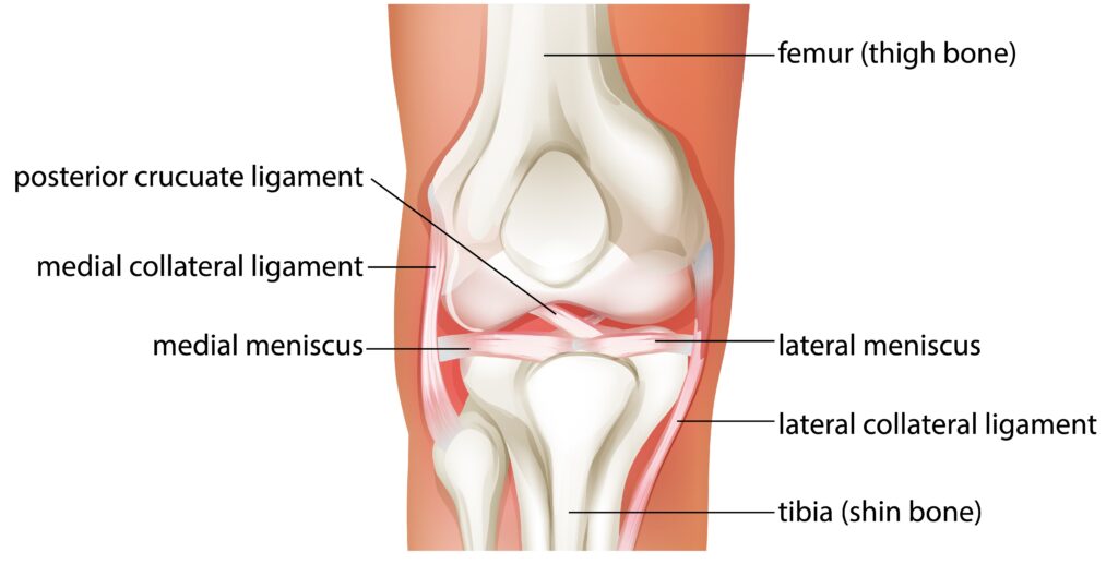
Knee Ligaments Diagram
Menisci and Their Relationship with Ligaments
The menisci are two C-shaped pieces of cartilage located between the femur and tibia, known as the medial and lateral meniscus. These structures act as shock absorbers, helping to transmit loads and absorb impact within the tibiofemoral joint. The outer parts of the menisci are thicker, which enhances joint stability and congruence, allowing for better distribution of weight across the knee.
The medial meniscus is more commonly injured than the lateral meniscus, partly due to its reduced mobility.
Meniscal injuries frequently occur alongside ligament injuries, such as the ‘Unhappy Triad,’ which involves tears to the ACL, MCL, and medial meniscus.
Joint Capsule and Supporting Structures
The knee joint capsule is a strong, fibrous covering that surrounds the knee joint, composed primarily of tendons. This capsule has an outer fibrous layer that merges with surrounding tendons and a synovial membrane on the inside that facilitates lubrication, allowing for smooth movement within the joint. Additionally, fluid-filled sacs known as bursae are formed within the knee joint capsule to help reduce friction during movement.
The patellar tendon, located on the anterior aspect of the knee joint, plays a crucial role in knee stability and function.
Common Knee Ligament Injuries
Knee ligament injuries can range from mild sprains, characterized by stretched ligaments, to complete tears, indicating a more severe knee injury.
The most commonly injured ligaments are the ACL and MCL, often occurring in contact sports where sudden movements and impacts are frequent. Symptoms of knee ligament injuries include sudden swelling, pain, and sometimes a popping sound at the time of injury.
PCL injuries, although less common, often occur alongside other knee injuries, particularly after significant trauma such as falls or vehicle accidents.
Symptoms include swelling, pain, and instability, which can affect the knee’s range of motion. LCL injuries, while rare, can result in lateral knee pain and instability, complicating knee function.
Athletes involved in high-impact sports, such as football and basketball, are at a heightened risk for ACL injuries due to the dynamic nature of these activities. Treatment plans for knee ligament injuries vary significantly depending on individual factors, including age, health status, and the severity of the injury.
Knee Ligaments Diagram
The knee joint consists of two main parts: the tibiofemoral joint between the femur and tibia, and the patellofemoral joint between the femur and patella. The anterior cruciate ligament (ACL) and posterior cruciate ligament (PCL) cross each other, forming an ‘X’ inside the knee joint, providing crucial stability.
The medial collateral ligament (MCL) links the femur to the tibia, while the lateral collateral ligament (LCL) connects the femur to the fibula, both stabilizing the knee by controlling medial and lateral rotation as well as sideways movements.
Menisci are two cartilages in the knee that act as shock absorbers between the femur and tibia.
Diagnosis and Treatment of Knee Ligament Injuries
Accurate diagnosis of knee ligament injuries often requires imaging tests such as X-rays and MRIs. These tests help in identifying the extent of the injury and planning the appropriate treatment. Initial treatment may involve rest, bracing, and pain management using medications.
Physical therapy is often a primary treatment method for minor MCL and PCL injuries, focusing on strengthening the muscles around the knee to support recovery. For more severe injuries, surgical intervention may be necessary to restore knee stability and function.
Prevention Tips for Knee Injuries
Preventing knee injuries is essential for maintaining knee health and avoiding long-term complications. Proper training and conditioning can significantly reduce the risk of suffering an ACL injury. Here are some detailed tips to help you keep your knees healthy and strong:
- Focus on Proper Alignment: When you’re doing exercises like wall squats, make sure to maintain proper knee alignment. This helps strengthen specific muscle groups crucial for knee health and stability.
- Incorporate Lunges with Care: Performing lunges with the correct form can enhance muscle endurance and flexibility in your knee area. This reduces the risk of injury by ensuring your knee joint is well-supported.
- Mind Your Knee Extension: Avoid extending your knees beyond your toes during exercises. This minimizes undue stress on the knee joint, protecting it from potential damage.
- Stretch Your Hamstrings Regularly: Regular hamstring stretches, while maintaining muscle contraction, can improve flexibility and support knee stability. This is key to preventing injuries.
Wrap Up
In this comprehensive guide, we’ve explored the intricate anatomy of knee ligaments, their crucial functions, and common injury mechanisms.