

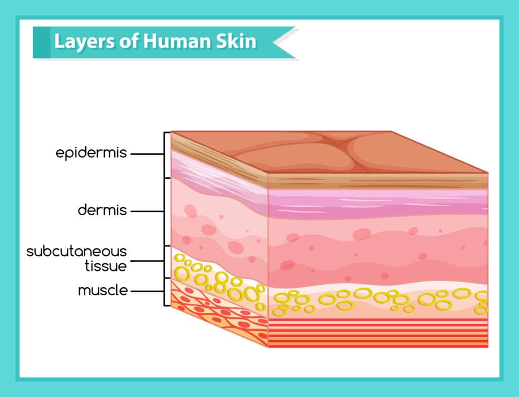
Our skin is composed of three primary layers: the epidermis, dermis, and hypodermis. This skin layers diagram clearly illustrates each layer’s structure and function. The skin consists of three primary layers: epidermis, dermis, and hypodermis, each with specific functions vital for protection and health.
Check out how these layers protect and maintain your body in this detailed overview.
What are Skin Layers ?
The skin is a complex organ composed of three primary layers: the epidermis, dermis, and hypodermis. These layers work in unison to protect internal organs from environmental hazards, maintain hydration, and regulate body temperature. Each layer has unique structural components and functions that contribute to overall skin health and protection.
The epidermis, the outermost layer, serves as a protective shield against pathogens and external elements.
Beneath it lies the dermis, which provides structural support through collagen and elastin fibers. The deepest layer, the hypodermis, primarily consists of fatty tissue that insulates the body and cushions underlying structures.
Skin Layers Diagram
Here is the detailed skin layers diagram for you.
Epidermis: The Outer Protective Barrier
The epidermis serves as the body’s first line of defense, shielding against environmental threats and pathogens. It continuously renews itself by shedding dead epidermal cells and replacing them with new ones to maintain integrity.
The epidermis comprises five distinct sublayers, each contributing to the skin’s protective and regenerative functions.
These sublayers include the stratum basale, stratum spinosum, stratum granulosum, stratum lucidum, and stratum corneum.
Each plays a unique role in skin health, from cell production to barrier formation. Collectively, these sublayers ensure the epidermis effectively protects the body while maintaining its appearance and functionality.
Stratum Basale
As the deepest layer of the epidermis, the stratum basale is where basal cells divide to produce new keratinocytes.
These keratinocytes migrate upwards, replenishing the skin’s outer layers and maintaining its protective function. This layer also houses melanocytes, specialized cells that produce melanin, the pigment responsible for skin color.
Melanin protects the skin from UV radiation, reducing the risk of damage and cancer. The stratum basale’s activity is vital for skin regeneration and pigmentation, making it a cornerstone of the epidermis’ protective abilities.
Stratum Spinosum
Above the stratum basale is the stratum spinosum, known as the prickle cell layer due to its spiny appearance from protruding cell processes.
This layer provides structural support and contributes to the skin’s strength and flexibility. It is populated by keratinocytes and Langerhans cells, which play a role in the immune response by acting as macrophages.
Langerhans cells in this layer help defend against infections and injuries, emphasizing the stratum spinosum’s role in protection and immune function.
Stratum Granulosum
Keratinocytes undergo significant changes in the stratum granulosum. They begin to flatten and accumulate keratohyalin granules, which are essential for keratin formation. This transformation is vital for developing the stratum corneum, the outermost protective layer.
The keratinocytes in this layer also produce lipids that contribute to the skin’s barrier function and hydration, preventing water loss and maintaining healthy skin. The stratum granulosum’s role in forming these crucial components underscores its importance in skin protection and hydration.
Stratum Lucidum
The stratum lucidum is a clear, thin skin layer found only in thick skin areas such as the palms of the hands and soles of the feet. It provides an additional layer of protection and is located between the stratum granulosum and the stratum corneum.
This layer enhances the skin’s ability to withstand friction and shear forces in these high-contact areas.
Stratum Corneum
The stratum corneum is the outermost layer of the epidermis, consisting of dead keratinized cells that form a durable, protective barrier. This layer is composed of 15 to 30 layers of dead keratinocytes, which are regularly shed and replaced approximately every four weeks.
The keratin and lipids present in the stratum corneum are essential for preventing water loss and protecting against environmental damage. This layer’s continuous renewal ensures the skin remains resilient and effective in its protective role.
Dermis: The Supportive Middle Layer
Beneath the epidermis lies the dermis, which accounts for about 90% of the skin’s thickness. This layer is rich in collagen and elastin fibers, providing strength, elasticity, and structural support. The dermis houses vital structures such as blood vessels, nerves, hair follicles, and glands, all essential for skin function and health.
The dermis is divided into two sublayers: the papillary layer and the reticular layer. These layers work together to support the epidermis and maintain skin integrity, playing a crucial role in the skin’s ability to protect and heal itself.
Papillary Dermis
The papillary layer is the uppermost part of the dermis, composed of loose connective tissue that provides flexibility and support. This layer contains capillary loops that supply nutrients to the epidermis, ensuring its survival and function.
Ruffini corpuscles, found in the papillary dermis, are mechanoreceptors that respond to skin stretch and pressure, contributing to the skin’s sensory capabilities.
Reticular Dermis
The reticular dermis is the deeper layer of the dermis, composed of dense irregular connective tissue rich in collagen and elastin fibers. These fibers provide the skin with strength and elasticity, allowing it to withstand physical stress and recover from injuries.
The dense network of fibers in this layer forms a supportive meshwork of dense connective tissue, crucial for maintaining skin’s structural integrity and resilience.
Hypodermis: The Insulating Layer
The hypodermis, or subcutaneous layer, is the skin’s deepest part, primarily made up of fat cells. This layer provides cushioning for muscles and bones, protecting them from external impacts and mechanical stress. Besides providing protection, the hypodermis insulates the body, regulating temperature and storing energy.
Blood vessels and connective tissue in the hypodermis connect the skin to underlying tissues, providing stability and support.
Blood Supply and Nerve Endings
The skin’s blood supply is crucial for its function and health. It is provided by two main plexuses: one located between the papillary and reticular layers, and another between the dermis and the subcutaneous tissue. These plexuses play a vital role in supplying nutrients, regulating temperature, facilitating wound healing, and ensuring proper blood flow.
Nerve endings in the skin detect sensations such as touch, pain, and temperature. These sensory functions protect the body from harm and maintain homeostasis. The presence of both somatic and autonomic nerves ensures the skin can respond effectively to environmental changes.
Skin Appendages: Hair Follicles and Glands
Embedded in the dermis, hair follicles produce hair. These follicles are associated with sebaceous glands, which produce sebum to moisturize hair and skin, and arrector pili muscles that can cause hair to stand on end.
Sweat glands, including eccrine and apocrine types, regulate body temperature and excrete waste products. Eccrine glands are the most common, secreting a watery fluid to cool the body, while apocrine glands release a thicker fluid that can produce a strong odor.
Clinical Significance of Skin Layers
Skin cancer, for example, primarily arises in areas frequently exposed to UV radiation, emphasizing the importance of protective measures. Non-melanoma skin cancers are more common but generally have a better prognosis compared to melanoma.
Variations in genetic factors like the MC1R gene can increase UV sensitivity and the risk of skin cancer. Overexposure to UV light can cause skin conditions like pigmentation changes and malignancies, emphasizing the need for effective sun protection strategies.


