

The cecum is a critical component of the digestive system, marking the beginning of the large intestine. Located in the lower right abdomen, it connects the small intestine to the colon, playing a vital role in waste processing and nutrient absorption. This article explores the cecum’s anatomy, functions, common conditions, and treatments.
Key Takeaways
The cecum connects the small intestine to the large intestine, facilitating waste processing and nutrient absorption.
It plays vital roles in fluid absorption, fermentation of fibers, and supporting immune health through beneficial gut bacteria.
Cecal conditions such as volvulus or colorectal cancer underscore the importance of early detection and regular screening for maintaining digestive health.
What is Cecum
The cecum is the first section of the large intestine, positioned between the ileum and the ascending colon and the descending colon. The cecum serves as a junction between the small and large intestines, managing the flow of waste from the small to the large intestine. This transition is essential for starting the absorption of fluids and nutrients from waste material.
The cecum plays an indispensable role in the digestive system, ensuring proper processing of waste and facilitating the absorption of remaining fluids and nutrients.
Cecum Diagram
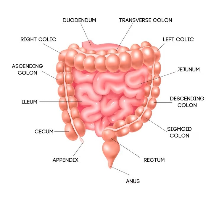
We’ll delve deeper into the cecum’s location, structure, and role.
Location of the Cecum
Located in the lower right abdomen, the cecum resides in the right iliac fossa, nestled adjacent to the appendix. This strategic position allows it to serve as the entry point where the small intestine, specifically the ileum, delivers digested material into the large intestine.
Its proximity to other abdominal organs and its lower abdominal location are key for its digestive role.
Structure of the Cecum
The cecum resembles a pouch and connects directly to the ileum, the final segment of the small intestine. This blind-end structure, which is the origin of its name derived from the Latin word meaning ‘blind,’ plays a key role in the digestive system.
This unique shape facilitates the initial processing of waste as it enters the large intestine.
Role in Digestion
The cecum plays a critical role in the digestive system, acting as an entry point for material from the small intestine and participating in various digestive processes. It is involved in the initial processing of waste, receiving material from the small intestine and preparing it for further digestion in the large intestine.
Its role in fermentation and nutrient absorption further highlights its importance in digestion.
Heading
The cecum functions as a pouch that serves as the initial segment of the large intestine, linking the small intestine and the colon. Positioned on the right side of the body, the cecum is integral to the digestive system, ensuring that waste material is efficiently processed as it transitions from the small intestine to the colon.
This crucial link sets the stage for understanding the cecum’s multifaceted roles in digestion and overall health.
Functions of the Cecum
The cecum’s functions are essential for maintaining a healthy digestive system. It plays a crucial role in absorbing remaining fluids and salts after the digestive process in the intestines. This absorption is vital for preventing dehydration and maintaining the body’s electrolyte balance. Additionally, the cecum is involved in the fermentation of undigested food particles, particularly fibers, contributing to nutrient breakdown and the production of short-chain fatty acids.
The cecum’s functions are essential for maintaining a healthy digestive system. It plays a crucial role in:
Absorbing remaining fluids and salts after the digestive process in the intestines
Preventing dehydration and maintaining the body’s electrolyte balance
Fermenting undigested food particles, particularly fibers
Contributing to nutrient breakdown and the production of short-chain fatty acids
Moreover, the cecum hosts beneficial bacteria that aid in digestion and support immune health. These bacteria help maintain a balanced gut microbiome, which is critical for overall health and wellbeing.
We will explore the specific functions of the cecum, including fluid absorption, food fermentation, and immune support.
Absorption of Fluids
Fluid absorption in the cecum occurs primarily through osmosis, driven by the absorption of electrolytes such as sodium. The cecum is essential for reclaiming water and electrolytes from undigested food, contributing to the body’s hydration and electrolyte balance. This process helps regulate fluid balance in the body, preventing dehydration and maintaining overall health.
The absorption of excess water and electrolytes from the chyme highlights the cecum’s role in digestion and fluid management.
Fermentation of Food
The cecum hosts a diverse array of bacteria that ferment undigested carbohydrates, aiding in the breakdown of fibrous food. In many mammals, the cecum serves as a critical site for fermenting dietary fibers, aiding in nutrient breakdown and the production of short-chain fatty acids. These beneficial bacteria assist in the breakdown of fibrous food components, facilitating further nutrient absorption and contributing to the overall digestive process.
Water absorption and fermentation underscore the cecum’s importance in digestion.
Immune Function
The cecum contributes to immune health by harboring beneficial bacteria that help maintain a balanced gut microbiome. These bacteria interact with immune cells, playing a significant role in maintaining immune tolerance and protecting against pathogens.
A diverse bacterial population in the cecum supports the immune system and protects against harmful microorganisms.
Gross Anatomy
The cecum is situated in the right lower quadrant of the abdomen and is the initial segment of the large intestine. As part of the digestive system, it plays a pivotal role in processing waste material. Its location and anatomical relations are crucial for its function in digestion and interaction with other abdominal organs.
We’ll examine the shape, size, and anatomical relations of the cecum.
Shape and Size
The cecum typically measures around 6 cm in length, with a potential maximum diameter of 9 cm. Its shape and size can vary among individuals, but it generally resembles a pouch.
Grasping the cecum’s dimensions helps understand its digestive role and interaction with other abdominal structures.
Anatomical Relations
Anteriorly, the cecum is positioned in relation to the abdominal wall and adjacent loops of the small intestine. Posteriorly, it is in contact with the iliacus and psoas major muscles. Medially, it is related to the ileocecal valve and the terminal part of the ileum. Laterally, the cecum is located near the right paracolic gutter and the anterior superior iliac spine.
These relations are key to understanding the cecum’s function and its interactions with surrounding structures.
Internal Structure
The cecum’s internal structure includes distinct wall layers that play specific functional roles. It consists of four main layers:
Mucosa
Submucosa
Muscularis
Serosa
Each layer contributes to the cecum’s overall functionality, from absorption and secretion to muscle contraction and protection.
We’ll explore the details of these layers and their specific roles.
Cecal Walls
The cecal wall is composed of four main layers: mucosa, submucosa, muscularis, and serosa. The mucosal layer is responsible for absorption and secretion, playing a vital role in the cecum’s function.
The multi-layered structure allows the cecum to process and move waste effectively through the digestive system.
Ileocecal Valve
The ileocecal valve, located between the cecum and the terminal ileum, plays a critical role in regulating material passage. This valve prevents the backflow of contents, ensuring a one-way flow from the cecum to the ileum.
Composed of two muscular layers, the ileocecal valve controls the flow of intestinal contents and prevents their reverse movement.
Blood Supply
The blood supply to the cecum is crucial for its function, ensuring that it receives the necessary nutrients and oxygen. The cecum receives its blood supply primarily from the cecal arteries, which branch from the superior mesenteric artery.
We will examine the arterial supply, venous drainage, and lymphatic drainage of the cecum.
Arterial Supply
The arterial supply to the cecum is primarily provided by the ileocolic artery, a branch of the superior mesenteric artery. The anterior and posterior cecal arteries, which arise from the ileocolic artery, are responsible for supplying blood to the cecum.
The ileocolic artery also gives rise to the appendicular artery, supplying the appendix.
Venous Drainage
The veins draining the cecum correspond to the cecal arteries and drain into the superior mesenteric vein. This venous blood then flows into the portal vein, which transports it to the liver for detoxification and nutrient processing.
Efficient venous drainage promptly removes the cecum’s metabolic waste.
Lymphatic Drainage
Lymphatic drainage from the cecum involves lymph nodes associated with its arterial supply. These lymphatic vessels drain into the lymph nodes that follow the path of the ileocolic artery. This system is vital for immune function, managing liquid waste and protecting against infections.
Nerve Supply
The nerve supply to the cecum is primarily through the autonomic nervous system, specifically via sympathetic and parasympathetic fibers.
We’ll explore how sympathetic, parasympathetic, and sensory innervation contribute to the cecum’s function.
Sympathetic Innervation
The lumbar splanchnic nerves primarily provide sympathetic innervation to the cecum. These nerves originate from the superior mesenteric plexus and play a crucial role in regulating blood flow and muscle contraction within the cecum.
Parasympathetic Innervation
The parasympathetic innervation of the cecum is largely mediated by the vagus nerve. The vagus nerve promotes digestion by stimulating muscle contractions and secretions within the cecum.
Sensory Innervation
Sensory fibers responsible for pain and reflex sensations in the cecum travel through the pelvic splanchnic nerves. These fibers transmit pain and other sensations, allowing the body to respond to stimuli affecting the cecum.
Histology
The cecum’s histological structure consists of four primary layers, each contributing to its functions. These layers include the mucosa, submucosa, muscularis, and serosa, each playing a specific role in the cecum’s overall functionality.
We’ll examine each layer’s details and their contributions to the cecum’s functions.
Mucosal Layer
The mucosal layer features a smooth mucous membrane rich in goblet cells that produce mucus. These goblet cells secrete mucus for lubrication, facilitating the movement of waste material through the cecum.
Additionally, the mucosal layer features lymphoid tissue, which plays a role in immune function.
Submucosal Layer
The submucosal layer, containing essential blood vessels and nerve structures, supports the cecum’s functions. This layer is composed of connective tissue, blood vessels, and nerves, providing structural support and facilitating nutrient exchange within the cecum.
Muscularis Layer
The muscularis layer features both longitudinal and circular smooth muscle bands that facilitate chyme movement in the cecum. Arranged in specific patterns, these smooth muscle layers promote peristalsis, ensuring efficient waste movement through the cecum.
Serosal Layer
The serosal layer, known as the serosa, is the outermost layer of the cecum and consists of simple squamous epithelium. This layer secretes fluid to reduce friction with surrounding tissues, protecting the cecum and facilitating its movement.
Embryology
The cecum originates from the midgut during embryonic development. Understanding its development provides insights into its final anatomical position and potential congenital anomalies.
We will explore the development of the cecum and common congenital anomalies affecting it.
Development of the Cecum
The cecum develops as a part of the midgut, which evolves from the embryonic gut tube. The midgut undergoes a 270° rotation during embryonic development, positioning the cecum in the right iliac fossa.
This rotation and fixation process properly position the cecum within the abdomen.
Congenital Anomalies
Common congenital anomalies affecting the cecum include cecal malrotation, mobile cecum, and cecal atresia. These anomalies can lead to various complications, such as bowel obstruction or increased risk of volvulus due to abnormal mobility.
Understanding these anomalies helps diagnose and manage potential cecal issues in newborns.
Clinical Significance
Conditions involving the cecum can lead to serious complications, including bowel obstruction and ischemia. We will delve into clinical conditions like cecal volvulus, appendicitis, cecal carcinoma, and diagnostic imaging.
Cecal Volvulus
Cecal volvulus involves the cecum twisting around its mesenteric axis, potentially causing obstruction and compromised blood flow. This rare condition is often misdiagnosed due to symptoms that resemble other gastrointestinal disorders. Symptoms include severe abdominal pain, vomiting, and abdominal distension, often mistaken for conditions like IBS. Colon muscle weakness, pregnancy, and previous abdominal surgeries can contribute to cecal volvulus.
Untreated cecal volvulus can cause severe complications, potentially leading to a mortality rate of up to 40 percent.
Appendicitis
Appendicitis can sometimes present with symptoms similar to those of cecal volvulus, making differential diagnosis essential. The relationship between the cecum and the appendix is crucial, as inflammation of the appendix can affect the cecum and lead to severe abdominal pain and other symptoms.
Accurate diagnosis is crucial for effective management.
Cecal Carcinoma
Cecal carcinoma is often diagnosed at advanced stages due to its subtle early symptoms. Risk factors for cecal carcinoma include age, family history of colorectal cancer, and certain lifestyle factors.
Routine screenings are essential for early detection and effective treatment, improving patient outcomes.
Diagnostic Imaging
Diagnostic imaging techniques, such as CT scans, MRI, and ultrasound, are highly effective in visualizing the cecum and diagnosing conditions like cecal volvulus. CT scans are particularly effective in diagnosing cecal conditions, making them valuable in clinical practice.
Regular screening tests can help detect issues early, leading to better treatment outcomes.
Surgical Considerations
Surgical interventions are often necessary for treating cecal conditions. We will explore surgical procedures like cecostomy, right hemicolectomy, and laparoscopic approaches, highlighting their indications and benefits.
Cecostomy
Cecostomy is performed to provide an alternative pathway for fecal elimination when the normal route is obstructed. This procedure can be indicated in patients with severe constipation or fecal incontinence.
Cecostomy creates an opening in the cecum for direct solid waste elimination, bypassing the rectum and anal canal.
Right Hemicolectomy
Right hemicolectomy generally involves excising the cecum along with portions of the ascending colon. This surgery treats conditions like colon cancer, severe cecal volvulus, or inflammatory diseases of the right colon.
Right hemicolectomy restores normal bowel function and prevents further complications by removing affected areas.
Laparoscopic Approaches
Laparoscopic techniques can reduce recovery time and hospital stays. These minimally invasive methods use small incisions and a camera to guide the surgery. Laparoscopic surgery is designed to minimize recovery time and reduce postoperative pain, making it a preferred option for many patients.
Comparative Anatomy
The cecum’s structure and function can vary significantly among different mammalian species. We will compare the cecum in humans and other mammals, exploring the evolutionary significance of these differences.
Cecum in Other Mammals
The cecum’s size and function can vary significantly among different mammalian species. In herbivores, the cecum is often larger and plays a critical role in fermenting plant material, aiding in the breakdown of fibrous food. In contrast, carnivores have a smaller cecum, reflecting their different dietary needs.
These differences offer insights into the cecum’s adaptability and its role in diverse digestive processes.
Evolutionary Perspective
The cecum has evolved to adapt to various dietary needs in different species. Its evolutionary significance lies in its ability to support diverse diets, from plant-based to meat-based.
Examining its evolution helps us understand the cecum’s role in human anatomy and its digestive importance.
Common Conditions Affecting the Cecum
The cecum can be affected by various health issues, including volvulus, inflammation, and cancer. Early identification of these conditions is crucial to prevent complications such as gangrene or perforation from obstructions.
We will discuss common conditions affecting the cecum, their symptoms, and treatment options.
Cecal Volvulus
Cecal volvulus involves the cecum twisting, leading to obstruction and necessitating prompt medical treatment.
Symptoms include:
severe abdominal pain
vomiting
constipation
an inflated abdomen
Severe cases may require surgical options like cecopexy or intestinal resection to correct cecal volvulus. In certain cases, the entire cecum may be removed to prevent further complications.
Inflammation and Infection
Cecal diverticulitis involves inflammation of the diverticula in the cecum, which can lead to abdominal pain and fever. Appendicitis can present similar symptoms to cecal conditions, making accurate diagnosis crucial for proper treatment.
Infections affecting the cecum may be treated with antibiotics.
Colorectal Cancer
The risk of colorectal cancer is notably high in the cecum, with early detection being crucial for effective treatment. Cecal cancer, a type of colorectal cancer, is often detected through routine screenings and can be asymptomatic in early stages.
Regular screenings significantly enhance early detection and successful treatment outcomes for colorectal cancer.
Surgical Interventions
Surgical intervention is the primary treatment for cecal volvulus, with procedures such as cecopexy and intestinal resection being common. Treatment for cecal volvulus may involve surgery to untwist the cecum and ensure proper blood flow.
Surgical options vary based on the cecum’s condition, focusing on restoring normal anatomy and function.
Preventative Measures and Healthy Practices
Maintaining a healthy diet and active lifestyle is essential to lower the risk of cecal disorders. We will discuss ways to promote cecal health through diet, regular screenings, and lifestyle changes.
Diet and Nutrition
A high-fiber diet is recommended; aiming for at least 30 grams of fiber daily can promote cecal function. Including high-fiber foods like fruits, vegetables, and whole grains is essential for optimal cecal function.
Modifying your diet can alleviate symptoms and improve cecal conditions.
Regular Screening
Regular screening tests are crucial for early detection of colorectal cancer as they can identify cancer before symptoms appear. Recommended screenings include colonoscopy, which allows direct observation and biopsy of the cecum and surrounding areas.
Consult with healthcare providers to personalize your screening schedule based on risk factors like family history.
Lifestyle Changes
Regular exercise improves digestive health and reduces the risk of conditions affecting the cecum. Avoiding smoking is vital, as it can lead to various gastrointestinal issues, including those related to the cecum.
A balanced diet rich in fiber, probiotics, and proper hydration supports overall gut health and cecal function.
Bottom Line
Throughout this guide, we’ve delved into the intricate world of the cecum, an essential yet often overlooked part of the large intestine. From its anatomy and location in the lower right abdomen to its vital roles in fluid absorption, fermentation of food, and immune function, the cecum is integral to our digestive health. Understanding the cecum’s various functions helps us appreciate its importance in maintaining overall well-being.
We’ve also explored the clinical significance of the cecum, highlighting conditions such as cecal volvulus, appendicitis, and cecal carcinoma. Early detection and appropriate treatment of these conditions are crucial for preventing severe complications. Diagnostic imaging techniques and surgical interventions play a significant role in managing cecal disorders, emphasizing the need for regular medical check-ups and screenings.


