Forearm anatomy involves two main bones, key muscles, and important joints. Understanding these components is vital for comprehending forearm functions and movements. This article breaks down each element, explaining their roles in everyday tasks.
Key Takeaways
The forearm is made up of two main bones, the radius and ulna, allowing for flexible movement through various joints.
Muscles in the forearm are categorized into flexors and extensors, enabling actions like wrist flexion, extension, pronation, and supination.
Key nerves, including the median, ulnar, and radial nerves, provide motor and sensory functions, while maintaining proper forearm health is crucial to prevent injuries like carpal tunnel syndrome.
Bones of the Forearm
The forearm’s structural integrity depends on two primary bones: the radius and the ulna. These bones are like the pillars of a bridge, providing support and facilitating movement. The radius and ulna are held together by the interosseous membrane, a fibrous sheet that adds stability and allows for the attachment of muscles and ligaments.
These bones articulate at the elbow joint and two crucial radioulnar joints – the proximal and distal radioulnar joints. This articulation enables a wide range of forearm movements, essential for tasks ranging from lifting objects to rotating the wrist.
Forearm Bone Diagram
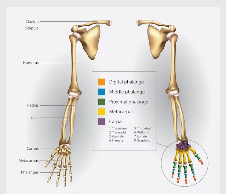
Radius
The radius is a remarkable bone located on the lateral side of the forearm, extending from the elbow to the wrist. It plays a pivotal role in forearm movement, particularly in pronation and supination, where the palm turns up or down.
The radial artery, a critical blood vessel, runs alongside the radius, ensuring that the forearm muscles receive adequate blood supply.
Ulna
The ulna, positioned on the medial side of the forearm, is key to forearm stability. It extends from the elbow to the wrist, where it forms a joint with the radius. This bone maintains the forearm’s structural integrity and provides attachment points for muscles like the extensor carpi radialis longus and flexor carpi ulnaris.
Joints of the Forearm
Joints are the hinges and pivots that allow our forearm to perform its intricate dance of movements. The forearm boasts several critical joints, each playing a unique role in mobility and function. The proximal and distal radioulnar joints enable the rotation of the forearm, while the elbow joint connects the upper arm to the forearm, facilitating flexion and extension movements.
Proximal Radioulnar Joint
The proximal radioulnar joint enables the forearm to perform pronation and supination-the rotational movements turning the palm up or down. It is stabilized by multiple ligaments, including the annular ligament, which encircles the radial head for stability during rotation.
The range of motion provided by this joint is crucial for everyday tasks like turning a doorknob or twisting a jar open, relying on the smooth functioning of the proximal radioulnar joint.
Distal Radioulnar Joint
The distal radioulnar joint, located near the wrist, complements its proximal counterpart by allowing the radius and ulna to pivot around each other. This joint’s design, supported by the triangular fibrocartilage complex, ensures stability and cushioning during forearm rotation.
Proper alignment and movement at this joint ensure smooth execution of tasks requiring forearm rotation, such as using a screwdriver or playing a musical instrument.
Elbow Joint and its Relation to the Forearm
The elbow joint connects the upper arm to the forearm, primarily allowing flexion and extension. Its mechanics depend on the interaction between the humerus, radius, and ulna, significantly influencing forearm movement.
The elbow joint functions as both a hinge and a pivot joint, accommodating a wide range of movements. Muscles like the biceps brachii and triceps brachii control these movements, allowing us to bend and straighten our arms with ease.
Classified as a synovial joint, the elbow joint has a cavity filled with synovial fluid that facilitates smooth movement. It is supported by several ligaments, including the medial and lateral collateral ligaments, which stabilize it during movement.
Muscles of the Forearm
The forearm is home to a plethora of muscles, each contributing to its versatility and strength. These muscles are categorized into flexors, extensors, and specific muscles responsible for pronation and supination. They enable a vast array of movements, from flexing the wrist to extending the fingers.
Knowing these muscle groups and their functions helps optimize forearm performance, whether for athletic pursuits, daily tasks, or rehabilitation.
Flexor Muscles of the Forearm
Located on the anterior side of the forearm, the flexor muscles primarily flex the wrist and fingers. The flexor carpi radialis and flexor carpi ulnaris are key in wrist flexion, while the flexor digitorum superficialis and flexor digitorum profundus flex the fingers.
The palmaris longus, although absent in about 15% of individuals, is involved in wrist flexion. Deep flexors like the flexor pollicis longus play a crucial role in thumb flexion, showcasing the intricate design of the forearm’s muscle system.
Extensor Muscles of the Forearm
On the posterior side of the forearm, the extensor muscles extend the wrist and fingers. The key muscles include:
Extensor carpi radialis longus, which handles wrist extension and abduction
Extensor carpi radialis brevis, which also assists in wrist extension and abduction
Extensor digitorum, which extends the fingers and is crucial for hand function
Deep extensors such as the extensor pollicis longus muscle, extensor pollicis brevis, and abductor pollicis longus are essential for thumb movements, highlighting the complexity of the forearm’s muscular anatomy.
Pronators and Supinators
Pronation and supination, the rotational movements of the forearm, are facilitated by specific muscles. The pronator teres and pronator quadratus muscles primarily turn the palm downward, a motion known as pronation.
Conversely, the supinator muscle and biceps brachii work together to turn the palm upward, a motion called supination. Their coordinated actions enable a wide range of forearm movements essential for daily tasks.
Nerves of the Forearm
The forearm receives nerve supply from three main nerves. These are the median, ulnar, and radial nerves. These nerves, originating from the brachial plexus, are responsible for both motor and sensory functions, ensuring that the forearm muscles receive the necessary signals to perform their tasks.
Knowing the pathways and functions of these nerves is crucial, especially for conditions like carpal tunnel syndrome or nerve injuries, which significantly impact forearm functionality.
Median Nerve
The median nerve arises from the spinal nerves C5-T1 and travels down the arm, passing through the carpal tunnel at the wrist. It is primarily responsible for controlling most of the flexor muscles in the forearm, except for the flexor carpi ulnaris and part of the flexor digitorum profundus.
Compression of the median nerve can lead to carpal tunnel syndrome, characterized by pain, tingling, and numbness, especially in the thumb and adjacent fingers. This condition often requires medical intervention, including wrist splints or surgical release.
Ulnar Nerve
The ulnar nerve originates from the C8-T1 nerve roots and runs along the inner side of the forearm. It is responsible for innervating several muscles in the forearm and hand, particularly those involved in finger movements.
Ulnar nerve compression, often occurring at the elbow, can lead to symptoms such as tingling, numbness, and weakness in the ring and little fingers, a condition known as cubital tunnel syndrome. Addressing this condition typically involves avoiding prolonged elbow flexion and sometimes surgical intervention.
Radial Nerve
The radial nerve, originating from the C5-T1 roots, travels down the outer side of the forearm. It is essential for extending the wrist and fingers, innervating the extensor muscles of the forearm.
Injuries to the radial nerve can result in wrist drop, a condition where the individual cannot extend the wrist and fingers. Rehabilitation for radial nerve injury often involves physical therapy to restore strength and mobility.
Blood Supply to the Forearm
The forearm receives its blood supply primarily from the radial and ulnar arteries, both of which branch from the brachial artery at the cubital fossa. These arteries are essential for delivering oxygen-rich blood to the forearm muscles and other tissues, ensuring they function optimally.
Proper blood circulation is crucial for muscle performance and recovery, making the health of these arteries vital for overall forearm health.
Blood Supply to the Forearm Diagram
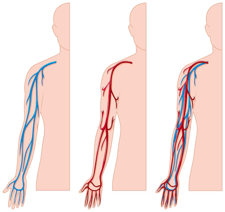
Brachial Artery
The brachial artery, originating from the axillary artery, is the main vessel that supplies blood to the upper extremity. At the cubital fossa, it divides into the radial and ulnar arteries, ensuring that the forearm and hand receive adequate blood flow.
Radial Artery
Running along the lateral side of the forearm, the radial artery is easily palpated near the wrist, making it a common site for checking pulse. It supplies the lateral forearm muscles and plays a crucial role in hand circulation.
Ulnar Artery
The ulnar artery travels along the medial side of the forearm, supplying blood to the medial aspect and the hand. It branches into several smaller arteries that ensure the forearm and hand receive sufficient blood supply.
Movements of the Forearm
The forearm is capable of a variety of movements essential for hand positioning and function. These include pronation and supination – rotational movements – as well as flexion and extension of the wrist.
Pronation and Supination
Pronation involves the radius rotating over the ulna, turning the palm downward. This movement is primarily facilitated by the pronator teres and pronator quadratus muscles. Supination, which turns the palm upward, is achieved by the supinator muscle and the biceps brachii.
These rotational movements are crucial for tasks such as turning a key or using a screwdriver, showcasing the forearm’s versatility.
Flexion and Extension of the Wrist
Wrist flexion and extension are driven by the coordinated actions of multiple muscles. The flexor carpi radialis and flexor carpi ulnaris are key players in wrist flexion, while the extensor carpi radialis longus and extensor carpi radialis brevis facilitate wrist extension.
These movements are essential for actions ranging from typing to lifting objects, highlighting the forearm’s role in everyday activities.
Muscles of the Forearm
The forearm muscles consist of two primary compartments. These are the anterior (flexor) compartment and the posterior (extensor) compartment of the forearm. These muscles enable a range of movements, from flexing and extending the wrist to intricate finger motions.
Anterior Compartment
The anterior compartment contains important flexor muscles. These include the flexor carpi ulnaris, palmaris longus, flexor carpi radialis, and pronator teres. These muscles are primarily involved in wrist flexion, finger flexion, and forearm pronation.
Posterior Compartment
The posterior compartment contains the extensor muscles, such as the extensor carpi radialis longus, extensor digitorum, and extensor carpi ulnaris. These muscles are crucial for wrist and finger extension and forearm supination.
Deep Muscles
Deep muscles in the forearm, including the flexor pollicis longus and flexor digitorum profundus, are essential for fine motor tasks and stabilization. These muscles play a significant role in executing precise movements, such as writing or threading a needle.
Major Nerves of the Forearm
The forearm has several major nerves. These include the median, ulnar, and radial nerves. These nerves are responsible for the motor and sensory functions of the forearm muscles, enabling a wide range of movements and sensations.
Blood Supply to the Forearm
The radial and ulnar arteries are vital for supplying blood to the forearm and hand. These arteries originate from the brachial artery and provide essential circulation to the forearm muscles.
Common Conditions Affecting the Forearm
Common conditions affecting the forearm often involve muscle strains or nerve injuries. These conditions can significantly impact forearm function and require appropriate treatment to restore normalcy.
Muscle Strains
Muscle strains are commonly caused by overuse or lifting heavy objects. Symptoms include swelling, tenderness, and pain, which can range from minor discomfort to severe muscle tears. Chronic muscle strains develop gradually due to repetitive use or overtraining, highlighting the importance of proper technique and adequate rest.
Carpal Tunnel Syndrome
Carpal tunnel syndrome occurs when the median nerve is compressed at the wrist, leading to symptoms like pain, numbness, and tingling. These symptoms often worsen with repetitive wrist movements.
Treatment options may involve wrist splints, corticosteroid injections, or surgical release, depending on the severity.
Radial Nerve Injury
Radial nerve injury can severely impair wrist and finger movements, resulting in a condition known as wrist drop. This injury often requires physical therapy to restore strength and mobility.
Rehabilitation strategies focus on exercises to strengthen the forearm muscles and improve function, showcasing the importance of addressing nerve injuries promptly.
Tips for Maintaining Forearm Health
Forearm health is vital for overall upper limb functionality. Regular exercise strengthens the forearm muscles, enhancing performance and reducing injury risk. Warm up before exercising to prepare the muscles and avoid strains. Gentle wrist rotations improve blood circulation, and taking breaks during prolonged computer use helps prevent discomfort.
Avoid bouncing during stretches to maintain proper technique and prevent injury. Incorporating these practices into your routine enhances forearm health and functionality.