

The femur is the longest and strongest bone in your body. Understanding femur anatomy helps explain how it supports movement and handles stress. This article covers the femur’s key parts, common injuries, and related conditions.
Key Takeaways
- The femur is crucial for mobility, with its proximal, shaft, and distal regions each having unique anatomical features that support hip and knee functions.
- Common injuries include neck of femur fractures in the elderly, which require prompt diagnosis and treatment to prevent complications like avascular necrosis.
- Maintaining femur health through proper nutrition, weight-bearing exercises, and regular health check-ups is essential for preventing fractures and ensuring overall bone density.
Proximal Femur Anatomy
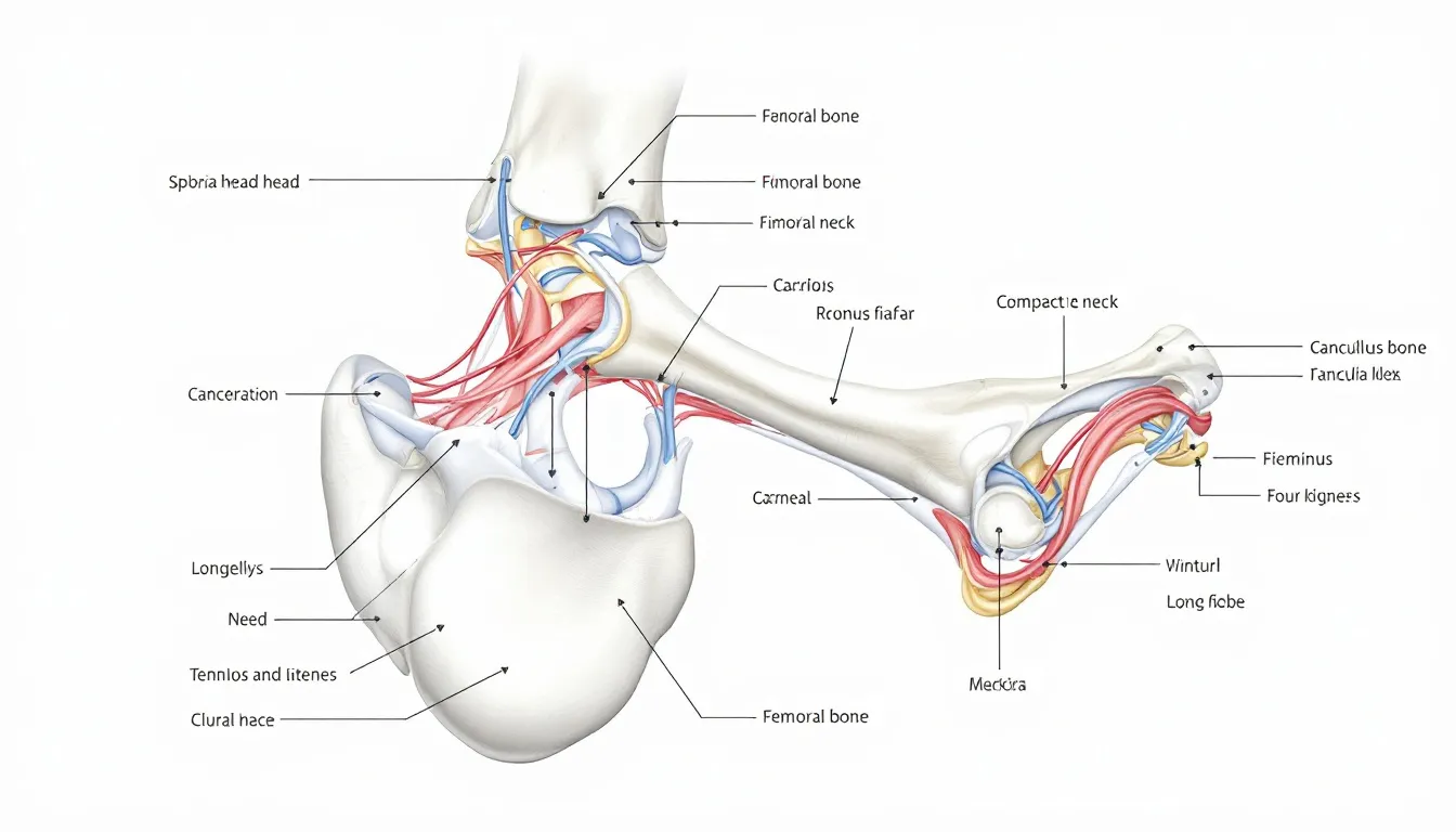
The proximal femur has several key components. These include the head, neck, greater trochanter, and lesser trochanter. The head of the femur fits into the acetabulum of the pelvis to form the hip joint, a ball-and-socket joint that allows for a wide range of motion. The femoral neck, which connects the head to the shaft, is pyramid-shaped and forms an angle of about 128 degrees with the shaft in an adult. This angle is crucial for the proper alignment and functioning of the lower limb.
The greater and lesser trochanters are prominent bony protrusions on the proximal femur. These structures serve as attachment points for various muscles, playing a vital role in the movement and stability of the hip joint.
The greater trochanter, located on the lateral aspect of the femur, serves as an attachment for muscles such as the gluteus medius and minimus. Meanwhile, the lesser trochanter, found on the medial side, provides attachment for the iliopsoas muscle. Together, these components enable the femur to withstand the stresses and forces exerted during activities like walking, running, and jumping.
Common Proximal Femur Injuries
The femoral neck, a crucial connector between the femoral head and shaft, is particularly prone to fractures. One of the most common types of femur fractures in the proximal region is the neck of femur fracture (NOF), which often occurs in elderly individuals following low-energy falls. Women are more susceptible to these fractures due to factors like osteoporosis, making prevention and early diagnosis essential.
Neck of femur fractures can lead to serious complications, such as avascular necrosis, where the blood supply to the femoral head is compromised. This condition can significantly impact recovery and mobility.
Clinically, these fractures often present with a shortened and externally rotated lower limb. Understanding the types and implications of proximal femur injuries is crucial for implementing effective treatment strategies, ensuring better outcomes for patients.
Femoral Shaft Structure
The femoral shaft, or diaphysis, is a cylindrical structure that slightly widens at the top and curves forward. This unique shape allows the femur to bear the body’s weight and absorb the impact during physical activities. The shaft’s direction descends slightly medially, aligning with the body’s natural biomechanics. In cross-section, the femoral shaft is circular in the middle but flattens posteriorly, which provides additional strength and stability.
The medial supracondylar line, a ridge on the posterior surface of the shaft, ends at the adductor tubercle. This ridge, along with the linea aspera, serves as an attachment point for several thigh muscles, contributing to the femur’s function in movement and support.
Understanding the femoral shaft’s structure is key to appreciating its role in the overall function of the lower limb.
Femoral Shaft Fractures
Femoral shaft fractures can result from high-energy injuries, such as car accidents, or low-energy falls, particularly in the elderly. These fractures are often severe due to the significant forces involved, leading to substantial blood loss, estimated between 1000 to 1500 ml. Such hemorrhage can pose a serious risk to the patient’s health and requires prompt medical attention.
Complications from femoral shaft fractures can be extensive, including damage to nearby soft tissues and potential leg shortening, especially in spiral fractures. Effective management of these fractures is crucial to minimize complications and ensure proper healing. Surgical intervention is often necessary to stabilize the bone and restore its function.
Femur Diagram
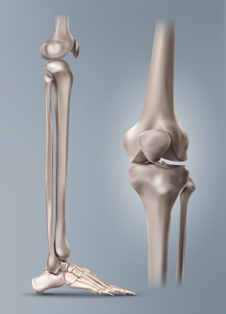
Distal Femur Anatomy
The distal femur includes critical structures that play a vital role in the knee joint’s function:
- The medial and lateral condyles, which are large, rounded protrusions that articulate with the tibia, forming the knee joint.
- The intercondylar fossa, located between the condyles, serves as an attachment point for the cruciate ligaments, which are essential for knee stability.
- The patellar surface, which is also included in the structure of the distal femur.
The patellar surface, where the patella (kneecap) rests, facilitates the smooth movement of the knee joint during flexion and extension. The distal femur’s anatomy is crucial for the knee joints’ overall function and stability, making it a key area of focus for understanding knee-related conditions and injuries.
Conditions Affecting the Distal Femur
One common condition affecting the distal femur is patellofemoral pain syndrome (PFPS), also known as runner’s or jumper’s knee.
This syndrome is characterized by:
- Pain around and under the kneecap
- Often resulting from overuse
- Improper alignment
- Changes in footwear
PFPS can significantly impact an individual’s ability to perform daily activities and engage in physical exercise.
It’s essential to consult a healthcare provider if new knee pain arises, to accurately diagnose and manage the condition. Proper treatment and preventative measures can help alleviate pain and improve knee function, ensuring a higher quality of life.
Blood Supply to the Femur
The primary blood supply to the femur comes from the femoral artery. This artery is the main supplier of blood to the lower extremity. The medial and lateral circumflex arteries, which branch from the femoral artery, provide blood to the femur’s head and neck. These arteries, including the medial femoral circumflex artery, are crucial for maintaining the health and function of the proximal femur.
The perforating branches of the deep femoral artery supply blood to the shaft of the femur. They also provide blood to the distal portion of the femur. Additionally, the femoral head is supplied via anastomotic connections from the obturator artery.
The periosteum, a membrane surrounding the femur, also plays a vital role in nourishing the bone through its adjacent blood supply. Understanding the femur’s blood supply is essential for diagnosing and treating conditions that affect this vital bone.
Muscle Attachments on the Femur
The femur serves as an attachment point for various muscle groups, which are divided into four main compartments:
- Anterior
- Medial
- Posterior
- Gluteal
These muscle groups play critical roles in movement and stability. For instance, the anterior compartment includes muscles like the pectineus and iliopsoas, which originate from pelvic structures and insert on the femur.
The vastus medialis and vastus lateralis muscles, part of the anterior and gluteal compartments respectively, attach to the femur’s linea aspera. These muscles are essential for extending the knee and stabilizing the hip joint. The intricate network of muscle attachments on the femur highlights the bone’s importance in supporting and facilitating movement.
Nerve Innervation of the Femur
The femoral nerve, the largest branch of the lumbar plexus, originates from the anterior rami of L2, L3, and L4. This nerve provides motor innervation to the hip flexors and knee extensors, including the quadriceps femoris. The femoral nerve also has sensory branches that supply the anteromedial thigh and medial leg and foot.
The femoral nerve traverses the psoas major muscle before passing under the inguinal ligament to reach the femoral triangle, where it divides into anterior and posterior divisions. Understanding the nerve innervation of the femur is crucial for diagnosing and treating nerve-related issues that affect lower limb function.
Embryological Development of the Femur
The development of the femur starts with the lateral plate mesoderm cells. These cells play a crucial role in initiating limb bud development. The cartilaginous form of the femur appears between Carnegie stages 16 and 17 through mesenchymal condensation. Chondrification, the process of cartilage formation, occurs from Carnegie stage 17 to stage 18.
Ossification, the process of bone formation, begins around Carnegie stages 22 to 23, progressing from the center of the femoral diaphysis towards both ends. During fetal development, there is a strong correlation between femur length and the length of the ossified shaft.
Understanding the femur’s embryological development provides insights into congenital conditions and growth abnormalities.
Clinical Significance of Femur Anatomy
The clinical significance of femur anatomy is highlighted by conditions like slipped capital femoral epiphysis (SCFE), a hip disorder causing anterior and superior displacement of the femoral head. Delayed diagnosis of SCFE can lead to severe complications, including femoral head osteonecrosis. Effective management often involves surgical fixation to prevent further displacement.
High-energy trauma, such as from falls or accidents, frequently results in neck of femur fractures among younger individuals. Complications from femur fracture surgery can include acute compartment syndrome and fat embolism syndrome. Recovery from a broken femur can take several months, requiring comprehensive post-operative care and physical therapy.
Surgical Considerations for Femur Injuries
Surgical procedures for femur fractures often involve Open Reduction and Internal Fixation (ORIF) or external fixation. The primary goal of surgical management is to allow patients earlier ambulation and improve their quality of life. The choice between arthroplasty and internal fixation depends on the fracture pattern, with intracapsular fractures disrupting blood supply more than extracapsular fractures.
There are potential complications that could require surgical revision. These include avascular necrosis, aseptic nonunion, and periprosthetic fracture. Understanding these surgical considerations is essential for optimizing patient outcomes and ensuring successful recovery.
Bone Health and the Femur
Maintaining bone health is crucial for preventing femur fractures and ensuring overall well-being. A common test to assess femur health is a bone density scan (DEXA or DXA), which helps detect osteoporosis early. Adequate calcium intake is essential for maintaining bone density, with adults aged 19 to 50 needing 1,000 mg daily. Vitamin D is also vital for calcium absorption, with an RDA of 600 IUs for adults.
Weight-bearing exercises, such as walking and jogging, enhance bone strength and help prevent osteoporosis. Avoiding smoking and excessive alcohol consumption is also important for bone health. Regular check-ups with healthcare professionals can help monitor bone health and develop customized osteoporosis treatment plans.


