

Hand muscles allow us to perform everyday tasks like gripping, typing, and lifting. These muscles are divided into intrinsic and extrinsic groups. Intrinsic muscles are within the hand, while extrinsic muscles start in the forearm. This article explains the anatomy and function of these muscles.
Key Takeaways
The human hand has 34 muscles divided into intrinsic (fine motor skills) and extrinsic (gross movements) groups, each crucial for different tasks.
Key muscle groups like the thenar and hypothenar muscles are essential for thumb and little finger movements, enabling grips and dexterity.
Proper nerve health is vital for hand functionality, as conditions like carpal tunnel syndrome highlight the significant impact of nerve damage on muscle activity.
Overview of Hand Muscles
The human hand is a marvel of biological engineering, containing 34 different muscles that work in harmony to provide both power and precision. The muscles are divided into two primary categories. These are known as intrinsic and extrinsic muscle groups.
Intrinsic muscles, located entirely within the hand, enable fine motor skills and dexterity, such as holding a pen or buttoning a shirt. Extrinsic muscles, which originate in the forearm, control broader movements and strength. Together, these muscles facilitate various types of grips, from the powerful grasp needed to lift a heavy object to the delicate precision required for threading a needle.
Hand Muscle Diagram
Thenar Muscle Group
Located at the base of the thumb, the thenar muscle group enables the thumb’s diverse range of movements. This group includes the abductor pollicis brevis, flexor pollicis brevis, and opponents pollicis. The abductor pollicis brevis is responsible for moving the thumb away from the hand, a motion crucial for grasping objects. The flexor pollicis brevis aids in flexing the thumb, making it easier to hold and manipulate items.
The opponens pollicis plays a pivotal role by allowing the thumb to touch the tips of other fingers, a movement known as opposition. This movement is vital for gripping and handling small objects. All these muscles are innervated by the median nerve, which, if compromised due to conditions like carpal tunnel syndrome, can severely impact thumb functionality.
Knowledge of the thenar muscle group helps appreciate the thumb’s versatility and the intricate tasks it can perform. Whether you’re texting, writing, or cooking, these muscles are hard at work, ensuring your thumb moves with precision and strength.
Hypothenar Muscle Group
The hypothenar muscle group, located on the opposite side of the palm, controls the intricate movements of the little finger. The hypothenar eminence consists of three muscles. These are the abductor digiti minimi, flexor digiti minimi brevis, and opponens digiti minimi. The abductor digiti minimi helps spread the fingers apart, a movement necessary for tasks requiring finger separation.
The flexor digiti minimi brevis bends the little finger at the metacarpophalangeal joint, aiding in grip formation and stability. The opponens digiti minimi allows the little finger to touch the thumb, enhancing the hand’s ability to grasp and manipulate objects.
Innervated by the ulnar nerve, these muscles maintain the little finger’s dexterity and strength.
Lumbrical Muscles
The lumbrical muscles are unique in their structure and function. They link to the tendons of the flexor digitorum profundus and the extensor expansions instead of attaching directly to bones. This connection allows them to flex the metacarpophalangeal joints while extending the proximal and distal interphalangeal joints.
These four muscles, numbered from lateral to medial, are essential for precise finger movements, such as typing or playing a musical instrument. The lumbricals’ ability to coordinate finger flexion and extension is vital for many fine motor tasks, making them indispensable for daily activities.
Interossei Muscles
The interossei muscles, located between the metacarpal bones, are divided into two groups: dorsal and palmar. The four dorsal interossei muscles are responsible for abducting the fingers, moving them away from the hand’s midline. This action is crucial for tasks that require finger spreading, such as playing the piano.
On the other hand, the three palmar interossei muscles perform the opposite function, adducting the fingers and pulling them towards the midline. This movement is essential for gripping and holding objects securely. Both groups of interossei muscles are innervated by the deep branch of the ulnar nerve, highlighting their importance in finger movements and hand functionality.
Adductor Pollicis Muscle
Located in the hand’s adductor compartment, the adductor pollicis muscle facilitates thumb adduction. It has two heads: the oblique head, originating from the capitate bone and the bases of the second and third metacarpals, and the transverse head, originating from the third metacarpal. These two heads converge and insert at the medial base of the first proximal phalanx.
This muscle’s primary function is to provide the power needed for pinching and gripping tasks. It is more robust than other intrinsic hand muscles, contributing significantly to thumb opposition and overall hand strength.
The adductor pollicis is innervated by the ulnar nerve, ensuring its proper function in hand movements.
Extrinsic Muscles of the Hand
The extrinsic muscles of the hand, originating in the forearm, are responsible for broader hand and wrist movements. These muscles, both flexors and extensors, collaborate to enable complex hand actions. The flexor digitorum profundus, for instance, enables deep finger flexion, while the extensor digitorum communis allows for finger extension.
These muscles’ tendons extend into the hand, connecting to bones and enabling precise control over hand movements. The radial nerve significantly influences motor functions, especially for extensor muscles, emphasizing the importance of nerve health for proper hand functionality.
Together, the extrinsic and intrinsic muscles provide the strength and dexterity needed for a wide range of tasks.
Tendons and Their Role
Tendons are the vital links between muscles and bones, allowing for the precise movements of the hand. Composed of collagen, these strong, flexible structures transmit the force from muscle contractions to bones, enabling movement. Flexor tendons, housed within the carpal tunnel, are essential for bending the fingers and enabling gripping actions.
The flexor retinaculum, a fibrous band, holds these tendons in place as they pass through the wrist, ensuring smooth and efficient movement. Extensor tendons, located along the back of the hand, allow for finger straightening and play a critical role in hand functionality. Together, these tendons enable the intricate and powerful movements that our hands perform daily.
Nerve Supply to Hand Muscles
Hand functionality depends significantly on its nerve supply, mainly from the median, ulnar, and radial nerves. The median nerve, originating from the lateral and medial cords of the brachial plexus, innervates the thenar muscles and the lateral three and a half fingers. This nerve is crucial for thumb movements and fine motor skills.
The ulnar nerve, arising from the C8 and T1 nerve roots, supplies the hypothenar muscles and the interossei muscles, facilitating finger movements such as abduction and adduction. The ulnar nerve innervates the interossei muscles, and injuries to the ulnar nerve can lead to significant functional deficits, including weakened finger movements and muscle atrophy.
The radial nerve, serving the extensor muscles, is vital for wrist and finger extension, highlighting the importance of nerve health for overall hand function.
Blood Supply and Lymphatics
A strong blood supply is vital for the health and functionality of hand muscles. The primary arteries supplying blood to the hand are the radial and ulnar arteries, forming the deep palmar and superficial palmar arches. These arches ensure a continuous blood flow, even if one of the main arteries is compromised, thanks to collateral circulation.
Lymphatic drainage in the hand is equally important, with both superficial and deep lymphatic systems following the venous structures. This drainage pathway extends from the fingertips to the lateral axilla lymph nodes, playing a crucial role in maintaining hand health and preventing infections.
Anatomy of Hand Muscles
The anatomy of hand muscles is categorized into intrinsic and extrinsic groups, each playing distinct roles in hand movements. Extrinsic muscles originate in the forearm and are responsible for gross movements, while intrinsic muscles are located entirely within the hand and are crucial for fine motor skills.
This classification aids in understanding the complexity and functionality of hand muscles.
Extrinsic Hand Muscles
Extrinsic hand muscles, including both flexors and extensors, are primarily responsible for broad movements of the hand and wrist. These muscles originate in the forearm and extend their tendons into the hand, allowing for powerful and controlled movements.
Flexor carpi radialis, for instance, aids in wrist flexion and abduction, while flexor carpi ulnaris assists with wrist flexion and adduction.
Intrinsic Hand Muscles
Intrinsic hand muscles, located entirely within the hand, are vital for fine motor control and dexterity. These muscles include the thenar and hypothenar muscles, lumbricals, and interossei muscles. They play critical roles in precise hand movements, such as gripping and pinching, which are crucial for everyday tasks.
The lumbrical muscles, for example, are responsible for flexing the metacarpophalangeal joints while extending the interphalangeal joints, allowing for coordinated finger movements. Similarly, the interossei muscles facilitate finger abduction and adduction, enhancing the hand’s ability to perform complex tasks.
Flexor Muscles of the Hand
The flexor muscles of the hand are primarily responsible for bending the fingers and thumb, enabling activities such as gripping and holding objects. These muscles originate in the forearm and extend into the hand, providing the strength and control needed for these movements.
The main flexor muscles include the flexor digitorum superficialis and flexor digitorum profundus, which play crucial roles in finger flexion. Additionally, the flexor pollicis longus and flexor carpi radialis contribute to thumb and wrist movements, respectively, highlighting the complexity and importance of these muscles in hand functionality.
Flexor Digitorum Superficialis
The flexor digitorum superficialis is a key muscle for bending the fingers at the middle phalanx joints. It originates from two heads: one from the medial epicondyle of the humerus and the other from the upper half of the radial shaft. This muscle is located between the superficial and deep flexor muscle layers of the forearm.
The muscle consists of four tendons that insert on the middle phalanges of the fingers, allowing for flexion of the proximal interphalangeal joints. The flexor digitorum superficialis is innervated by the median nerve, which is crucial for its movement capabilities.
Variations in this muscle can lead to complications such as carpal tunnel syndrome, emphasizing the need for understanding its anatomy and function.
Flexor Digitorum Profundus
The flexor digitorum profundus (FDP) is essential for flexing the distal interphalangeal joints of the fingers, allowing for deeper finger bending. It originates from the upper three-fourths of the ulna and interosseous membrane, positioning it deep in the forearm.
The FDP works in conjunction with the flexor digitorum superficialis to enhance grip strength and finger flexion. Blood supply to the FDP comes from the anterior interosseous artery, while its innervation involves both the median and ulnar nerves.
Knowledge of the FDP’s function aids in diagnosing and treating conditions like ‘jersey finger,’ characterized by the inability to flex the affected finger.
Flexor Pollicis Longus
The flexor pollicis longus is a long muscle in the forearm that primarily flexes the thumb at the interphalangeal joint, enabling grip and pinching movements. It originates from the anterior surface of the radius and the interosseous membrane, and inserts on the distal phalanx of the thumb.
This muscle is classified as an extrinsic muscle of the hand because it lies in the forearm but functions in the hand. Innervation of the flexor pollicis longus occurs through the anterior interosseous branch of the median nerve, highlighting its importance in thumb movements.
This muscle receives blood from both the anterior interosseous artery and the radial artery.
Flexor Carpi Radialis and Flexor Carpi Ulnaris
The flexor carpi radialis and flexor carpi ulnaris are crucial for wrist flexion and side-to-side movements of the hand. Originating from the medial epicondyle of the humerus, the flexor carpi radialis muscle primarily flexes and abducts the wrist. It also helps stabilize the scaphoid bone in the wrist, preventing excessive rotation during movement.
The flexor carpi ulnaris aids in wrist adduction as well as flexion, working alongside other wrist flexors. This muscle is the only one in the anterior compartment of the forearm that is entirely innervated by the ulnar nerve. Both flexor muscles receive blood supply from the anterior and posterior ulnar recurrent arteries, ensuring their functionality.
Extensor Muscles of the Hand
The extensor muscles primarily extend the wrist and fingers, enabling movements like opening the hand. These muscles originate in the forearm and have long tendons that connect to bones in the hand, facilitating precise control over hand movements.
The main extensor muscles include the extensor digitorum, extensor pollicis longus, and extensor carpi radialis longus and brevis. Each of these muscles plays a crucial role in hand and wrist extension, highlighting the importance of understanding their anatomy and function for maintaining hand health and functionality.
Extensor Digitorum
The extensor digitorum muscle is responsible for extending the four medial fingers at the metacarpophalangeal joints and assisting in extending the wrist. It originates from the lateral epicondyle of the humerus and inserts into the extensor expansions of the four medial digits.
This muscle is innervated by the posterior interosseous nerve, a branch of the radial nerve, ensuring its proper function in finger and wrist extension. The extensor digitorum also aids in separating the fingers due to its anatomical structure, highlighting its importance in hand movements.
Extensor Pollicis Longus
The extensor pollicis longus (EPL) is crucial for extending and adducting the thumb at both the metacarpophalangeal and interphalangeal joints. It originates from the posterior surface of the ulnar diaphysis and traverses over Lister’s tubercle before inserting at the distal phalanx of the thumb.
The EPL tendon runs through a synovial sheath that protects it as it crosses the wrist. This muscle is innervated by the posterior interosseous nerve, a branch of the radial nerve, and plays a vital role in thumb extension, particularly at the interphalangeal joint.
Extensor Pollicis Brevis
The extensor pollicis brevis primarily extends the thumb at the metacarpophalangeal joint, contributing to thumb movements. This slender muscle is found in the forearm’s posterior compartment, originating from the distal radius. It is innervated by the posterior interosseous nerve, which derives from the radial nerve, ensuring its proper function in thumb extension.
This muscle’s tendon forms part of the anatomical snuffbox, a key landmark for locating the radial pulse.
Extensor Indicis
The extensor indicis is a slender muscle located in the deep layer of the forearm’s posterior compartment, primarily facilitating the extension of the index finger. It originates from the posterior surface of the ulna and the interosseous membrane, contributing to its unique positioning and function.
This muscle inserts into the base of the second proximal phalanx and integrates into the tendon of the extensor digitorum. The extensor indicis is innervated by the posterior interosseous nerve, which is a branch of the radial nerve, ensuring its proper function in extending the index finger.
Extensor Carpi Radialis Longus and Brevis
The extensor carpi radialis longus and brevis are involved in extending and abducting the wrist, allowing for movements like lifting and carrying. These muscles originate from the lateral epicondyle of the humerus and insert on the dorsal aspect of the second and third metacarpals, respectively.
The extensor carpi radialis brevis primarily provides stability to the wrist during gripping actions, highlighting its importance in hand movements. Both muscles are innervated by the radial nerve, ensuring their proper function in wrist extension.
Intrinsic Muscles of the Hand
The intrinsic hand muscles are vital for fine motor skills and maintaining grip strength. These muscles, located entirely within the hand, enable intricate hand functions, such as:
writing
buttoning clothes
playing musical instruments
manipulating small objects
By providing precise control and dexterity, they play a crucial role in everyday tasks.
The intrinsic muscle groups include:
The thenar muscles
The hypothenar muscles
The lumbricals
The interossei muscles
Each of these groups plays a specific role in hand movements, highlighting the complexity and importance of intrinsic muscles in daily activities.
Thenar Muscles
The thenar muscles are found at the base of the thumb. They play a crucial role in enabling thumb movements. This group includes the abductor pollicis brevis, flexor pollicis brevis, and opponens pollicis. The abductor pollicis brevis moves the thumb away from the hand, while the flexor pollicis brevis aids in thumb flexion.
The opponens pollicis plays a pivotal role in thumb opposition, allowing the thumb to touch the tips of other fingers. These movements are crucial for gripping and handling small objects, highlighting the importance of the thenar muscles in daily activities.
Hypothenar Muscles
The hypothenar muscles, located at the base of the little finger, are responsible for its movements. The group consists of the abductor digiti minimi and the flexor digiti minimi brevis. It also includes the opponens digiti minimi. The abductor digiti minimi spreads the fingers apart, while the flexor digiti minimi brevis bends the little finger at the metacarpophalangeal joint.
The opponens digiti minimi allows the little finger to touch the thumb, enhancing the hand’s ability to grasp and manipulate objects. Innervated by the ulnar nerve, these muscles maintain the little finger’s dexterity and strength.
Lumbrical Muscles
The lumbrical muscles are unique in their structure and function. They link to the tendons of the flexor digitorum profundus and the extensor expansions instead of attaching directly to bones. This connection allows them to flex the metacarpophalangeal joints while extending the proximal and distal interphalangeal joints.
These four muscles are essential for precise finger movements, such as typing or playing a musical instrument. The lumbricals’ ability to coordinate finger flexion and extension is vital for many fine motor tasks, making them indispensable for daily activities.
Interossei Muscles
The interossei muscles, located between the metacarpal bones, play a critical role in finger abduction and adduction. The dorsal interossei muscles are responsible for spreading the fingers apart, while the palmar interossei muscles bring them together.
These movements are essential for tasks that require finger separation and gripping. Both groups of interossei muscles are innervated by the deep branch of the ulnar nerve, highlighting their importance in finger movements and hand functionality.
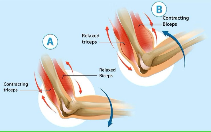
Muscular Coordination and Movements of the Hand
The coordination of hand movements relies on both intrinsic and extrinsic muscles, with intrinsic muscles crucial for precise actions. This coordination is essential for performing tasks ranging from simple to complex movements.
Understanding how these muscles work together helps in appreciating the complexity and functionality of the hand.
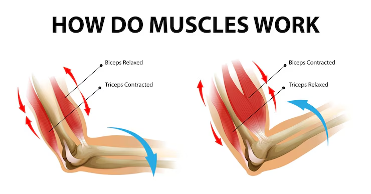
Diagram of Hand Muscle Work
Fine Motor Skills
Fine motor skills primarily rely on intrinsic hand muscles, enabling delicate movements like writing and buttoning. These skills are developed through coordinated movements of small muscles, particularly in the hands, and are essential for daily activities.
Practicing tasks like drawing or using scissors can enhance dexterity and control associated with fine motor skills.
Gripping and Power Movements
Extrinsic muscles largely facilitate gripping and power movements, providing the strength needed for tasks like lifting and holding objects. The power grip engages all four fingers through strong flexion and relies on the thumb for stability, enhancing grip strength through wrist dorsiflexion.
The hypothenar eminence plays a vital role in grip formation by stabilizing objects against the palm. Wrist extension is crucial for achieving a powerful grip, as it stabilizes the hand and optimizes the tension in the finger tendons during gripping activities.
Disorders Affecting Hand Muscles
Hand disorders can include various conditions such as fractures, deformities, and tendon injuries, leading to symptoms like pain, weakness, and reduced range of motion. Knowledge of these disorders is essential for proper diagnosis and treatment, ensuring the maintenance of hand functionality.
Carpal Tunnel Syndrome
Carpal tunnel syndrome arises when pressure is applied to the median nerve, leading to symptoms like tingling and pain. The condition occurs when the median nerve is compressed in a narrow passageway of the wrist, leading to numbness and tingling in the fingers. Women are generally more susceptible to carpal tunnel syndrome, likely due to a smaller carpal tunnel size and hormonal influences.
Symptoms typically start gradually, and as the condition progresses, the numbness can become constant and may lead to weakness, making it difficult to grasp objects. If untreated, carpal tunnel syndrome can result in permanent nerve and muscle damage, making timely medical intervention crucial.
Tendonitis
Tendonitis in the hand occurs due to inflammation of the tendons, often from repetitive use or strain. Hand tendonitis symptoms include pain, swelling, and a grinding sensation during movement. Repetitive motions and overuse are common causes of tendonitis in the hand.
Preventive measures against tendonitis include proper technique in repetitive activities, adequate rest, and stretching exercises. Without appropriate treatment, tendonitis can lead to more severe conditions, including tendon tears that may require surgical intervention.
Muscle Strain and Injury
Muscle strains in the hand can result from overexertion, causing pain and limiting movement. Symptoms of muscle injury in the hand can include:
localized pain
swelling
weakness
a limited range of motion
Treating hand muscle strains typically involves rest, ice application, compression, and elevation to alleviate symptoms. In more severe cases, surgical intervention may be necessary to properly repair the damaged tissue.
Common Hand Muscle Disorders
Common hand muscle disorders include:
Carpal tunnel syndrome, characterized by pressure on the median nerve, leading to symptoms such as tingling, numbness, and weakness in the hand.
Tendonitis, which involves inflammation of the tendons in the hand.
Muscle strains, which occur when muscles are overstretched or torn.
Carpal tunnel syndrome can arise from various factors, including anatomical differences, repetitive wrist movements, and health conditions like rheumatoid arthritis.
Fluid retention related to pregnancy or menopause and workplace factors like repetitive wrist movements may further exacerbate median nerve issues. Untreated intrinsic muscle injuries can lead to significant functional limitations, emphasizing the need for proper evaluation and prompt medical intervention.
Surgical Considerations for Hand Muscles
Surgical interventions for hand muscle disorders often involve tendon transfers to address functional deficits due to nerve damage. For example, tendon transfer surgery for ulnar nerve palsy aims to restore pinch and grip strength, correcting clawing. Typically, tendon transfer is considered three months after a muscle injury, allowing time for evaluation and planning.
The tenodesis effect can enhance finger tendon excursion in tendon transfer surgeries, improving functional outcomes. Complications in tendon transfer surgeries often arise from inadequate tensioning or failure at the repair site, emphasizing the importance of careful planning and execution.
Intrinsic muscles can also be used for surgical procedures, like muscle flaps to treat various conditions.
Clinical Significance of Hand Muscles
Knowledge of the clinical significance of hand muscles is vital for diagnosing and treating various conditions. Damage to the median and ulnar nerves can lead to conditions such as ulnar clawing and Klumpke palsy. Loss of nerve innervation to the lumbricals leads to a loss of equilibrium between intrinsic and extrinsic muscles, resulting in hand distortion.
A thorough physical exam, combined with knowledge of intrinsic hand muscles, can help physicians recognize anatomical distortions in hand function. Here are some key points to note: Median nerve palsy leads to the inability to abduct and oppose the thumb. It also causes sensory loss in the first three digits and results in weak forearm pronation.
Ulnar clawing is a result of a distal ulnar nerve lesion, causing paralysis of the interossei.
Intersection syndrome is a condition that can occur due to inflammation of the extensor carpi radialis brevis, highlighting the importance of hand muscle health.
Knowledge of these clinical aspects helps provide effective treatment and improve patient outcomes.


