

Understanding tongue anatomy is essential for grasping how this versatile organ aids in speech, taste, and swallowing. The tongue comprises distinct regions and surfaces, each with unique functions. This article delves into the tongue’s muscles, sensory functions, and blood supply, providing a comprehensive overview of its anatomy.
Key Takeaways
The tongue has two main parts, with the anterior part being highly mobile for speech and eating, and the posterior part fixed for swallowing.
The tongue’s functioning relies on intrinsic muscles for shape changes and extrinsic muscles for positioning, enabling diverse movements essential for talking and eating.
Taste buds on the tongue, located within different papillae, allow us to experience flavors and are connected to various nerves that transmit taste sensations to the brain.
Structural Overview of the Tongue
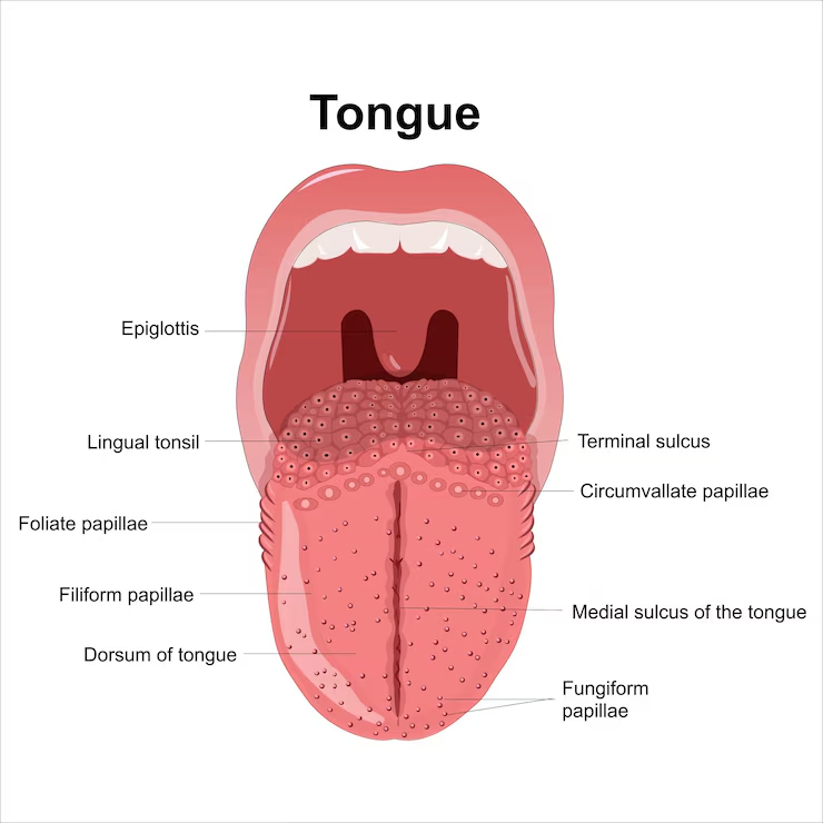
The tongue is divided into two main regions: the mobile anterior two-thirds, known as the oral part, and the fixed posterior one-third, called the pharyngeal part. The terminal sulcus is a V-shaped groove that plays a role in distinguishing these sections. It separates the body from the base of the tongue. The tongue has three main surfaces: the tip, body, and base, each contributing to its functions. The body aids in speech and eating, while the base is essential for swallowing and anchoring the tongue.
The dorsal surface of the tongue features specialized structures known as lingual papillae, which include filiform, fungiform, foliate, and vallate types. Filiform papillae are the most numerous and serve a mechanical function, as they do not contain taste buds.
Vallate papillae, typically numbering 8 to 12, are located just anterior to the terminal sulcus and are surrounded by troughs that help in taste sensation. Fungiform papillae are mushroom-shaped and are concentrated on the tip and sides of the tongue, containing taste buds.
Tongue Diagram
Muscles of the Tongue
The tongue is a fascinating organ made up entirely of muscle fibers, divided into intrinsic and extrinsic groups. These muscles work together to enable a wide range of movements and functions, from shaping the tongue for speech to positioning it for swallowing.
The coordination between these muscle groups gives the tongue its remarkable versatility and precision.
Intrinsic Muscles
The intrinsic muscles of the tongue are responsible for changing its shape, enabling movements crucial for speech and swallowing. The muscles that are included are:
Superior longitudinal muscle, which helps in shortening and elevating the tip of the tongue
Inferior longitudinal muscle, which retracts and lowers the apex of the tongue
Transverse muscle, which narrows and elongates the tongue
Vertical muscle, which flattens and broadens the tongue
Each muscle has a specific role in facilitating the complex movements of the tongue.
The transverse muscle narrows and elongates the tongue during movements, whereas the vertical muscle works to broaden and elongate the tongue as it contracts. Together, these intrinsic muscles enable complex movements like curling, flattening, and twisting, crucial for clear articulation and effective food manipulation.
Extrinsic Muscles
Intrinsic muscles modify the tongue’s shape, while extrinsic muscles position it within the oral cavity.
The four primary extrinsic muscles are:
Genioglossus, which is crucial for depressing and protruding the tongue, helping to prevent airway obstruction during sleep
Hyoglossus
Styloglossus
Palatoglossus
The hyoglossus muscle retracts and depresses the tongue, significantly aiding in its movement. The styloglossus muscle retracts the tongue and elevates its sides, making it vital for swallowing.
Lastly, the palatoglossus muscle, which is part of the soft palate, elevates the root of the tongue and constricts the isthmus of the fauces. These extrinsic muscles work together to ensure the tongue can perform its diverse functions effectively.
Structure of the Tongue
The tongue is a muscular hydrostat, meaning it has flexibility and is composed of muscle fibers without a skeletal structure. This unique composition allows the tongue to perform a wide range of movements and functions.
The body, dorsum, and ventral surface each have unique features and roles that contribute to the tongue’s functionality.
Body of the Tongue
The body of the tongue is divided into the anterior (tip) and posterior (base) parts. The apex, or tip, of the tongue, is the portion that contacts the teeth during activities such as speech and eating. This part is highly mobile and plays a crucial role in articulating sounds and manipulating food.
Anchored to the hyoid bone, the base of the tongue is involved in swallowing and speech. This section contains important structures such as the lingual tonsils, which play a role in the immune response, and the posterior tongue, which houses taste buds and various sensory receptors.
Dorsum of the Tongue
Various types of papillae cover the dorsum of the tongue, each serving distinct functions. Fungiform papillae are mushroom-shaped structures located on the sides and tip of the tongue, each containing taste buds. Filiform papillae cover most of the tongue’s surface and are responsible for the tongue’s texture but do not contain taste buds.
Circumvallate papillae are arranged in a V-shape near the back of the tongue and have numerous taste buds within them. Foliate papillae are located on the sides of the tongue in folds and contain taste buds on the mucous membrane covering them. These papillae play a critical role in taste sensation and the mechanical function of the tongue.
Ventral Surface of the Tongue
The ventral surface is the smooth underside of the tongue, connected to the mouth floor by the lingual frenulum. This fold of tissue limits the tongue’s movement, ensuring it stays anchored while allowing enough flexibility for speech and eating.
Beneath the ventral surface lie the sublingual glands, which produce and secrete saliva. These glands help keep the mouth moist and aid in the initial stages of digestion by breaking down food particles.
Taste and Sensory Function
The tongue is integral in processing taste sensations and is equipped with specialized structures known as taste buds. Distributed across different papillae types, these taste buds allow us to experience a wide range of flavors and textures.
Taste Buds
Taste buds are composed of clusters of nerve cells that send sensory information about taste to the brain. They can detect five main taste sensations: sweet, salty, sour, bitter, and umami. Four primary types of papillae on the tongue house these taste buds: filiform, fungiform, circumvallate, and foliate.
Fungiform papillae, located on the sides and tip of the tongue, contain taste buds that detect sweet and salty tastes. Circumvallate papillae, found at the back of the tongue, are involved in detecting bitter tastes, while foliate papillae on the sides detect sour tastes. Together, these structures create a comprehensive taste map on the tongue’s surface.
Tongue and Sensory Receptors
Besides taste buds, the tongue contains somatosensory nerves that detect touch and temperature. These receptors contribute to the overall sensory experience of eating by providing information about the texture and temperature of food.
Pain and temperature sensations are also sensed by specialized nerve endings on the tongue. This ability to detect pain and temperature is crucial for protecting the tongue from injury and ensuring safe food consumption.
Nerves Involved in Taste
Multiple nerves transmit taste sensations from the tongue to the brain. The facial nerve (cranial nerve VII) plays a crucial role in transmitting taste sensations from the anterior two-thirds of the tongue. This nerve is responsible for detecting sweet, salty, and umami tastes.
The glossopharyngeal nerve (cranial nerve IX) conveys taste information from the posterior third of the tongue. This nerve detects bitter and sour tastes. Additionally, the vagus nerve (cranial nerve X) contributes to taste sensation, especially from parts of the throat and epiglottis.
Together, these nerves ensure a comprehensive taste experience.
Blood Supply to the Tongue
The lingual artery provides the primary blood supply to the tongue. It branches off from the external carotid artery. This artery ensures that the tongue is well-oxygenated and nourished, supporting its various functions.
Lingual Artery
The lingual artery branches into several arteries, including the dorsal lingual artery, which supplies the posterior dorsum of the tongue. The sublingual artery, another branch, supplies the sublingual gland and adjacent muscles. The deep lingual artery is the final branch and provides blood to the tip of the tongue.
These branches ensure that the entire tongue, from base to tip, receives adequate blood supply. This vascular network is essential for maintaining the health and functionality of the tongue.
Venous Drainage
The lingual vein primarily drains venous blood from the tongue. It then empties into the internal jugular vein. This venous drainage system ensures that deoxygenated blood is efficiently removed from the tongue, preventing congestion and maintaining healthy tissue function.
The dorsal lingual veins and deep lingual vein also play significant roles in this drainage process. Together, these veins ensure the proper circulation of blood within the tongue, supporting its various activities.
Nerve Supply to the Tongue
The tongue’s nerve supply is intricate, involving both motor and sensory innervation. This complex network allows the tongue to perform its diverse functions effectively.
Motor Innervation
Motor control of the tongue is primarily provided by the hypoglossal nerve (cranial nerve XII), which is responsible for all tongue muscle movements except one. This nerve is crucial for voluntary movements of the tongue, aiding in functions like speech and swallowing.
The palatoglossus muscle, however, is innervated by the vagus nerve (cranial nerve X). This exception highlights the complexity of the tongue’s motor innervation, ensuring precise control over its various movements.
Sensory Innervation
The lingual nerve, a branch of the mandibular nerve, provides sensory information from the front two-thirds of the tongue. This nerve is responsible for detecting touch, temperature, and pain in this region.
The glossopharyngeal nerve provides both general sensation and taste for the back third of the tongue. Additionally, taste perception in the epiglottis and the area around it is mediated by the internal branch of the vagus nerve. These sensory nerves ensure that the tongue can detect a wide range of stimuli, contributing to its versatility.
Tongue Movements and Function
The tongue is crucial for various functions, including taste perception, speech, and food manipulation. Its movements are facilitated by a combination of intrinsic and extrinsic muscles, allowing it to perform complex tasks with precision.
Movements of the Tongue
The intrinsic muscles of the tongue primarily modify its shape, allowing for diverse movements. These movements include protrusion and retraction, elevation and depression, and lateral movements.
Protrusion and retraction are crucial for positioning the tongue appropriately during speech and swallowing. Elevation and depression movements help form different sounds and manipulate food during chewing. Lateral movements allow the tongue to navigate and properly position food between the teeth.
Role in Speech
Speech production relies on the tongue’s ability to move against the teeth and palate. The tongue plays a vital role in articulating various speech sounds through precise movements. These movements significantly influence vocal sounds and help in modulating pitch and tone.
By controlling the flow of air and shaping the mouth, the tongue enables us to produce a wide range of sounds necessary for clear communication.
Role in Swallowing
The tongue aids in forming a food bolus during the oral phase of swallowing. This involves coordinating movements to gather and shape food into a manageable mass. Coordinated tongue movements facilitate the safe transportation of food from the mouth to the throat.
This process ensures that food is properly directed into the esophagus, preventing choking and aiding in efficient digestion.
Development and Growth of the Tongue
Tongue formation begins around the fourth week of gestation. This development is influenced by contributions from multiple pharyngeal arches, ensuring the tongue’s complex structure and function.
Embryonic Development of the Tongue
The first four pharyngeal arches contribute to the formation of different parts of the tongue. The anterior two-thirds of the tongue is formed by the merging of lateral lingual swellings from the first pharyngeal arch.
The posterior third develops from the hypobranchial eminence arising from the second, third, and fourth pharyngeal arches. These stages ensure the tongue has the necessary structures and functions from birth.
Changes in the Tongue with Age
With age, the number and sensitivity of taste buds may decline. This can lead to changes in taste perception and a reduced ability to enjoy food. Older adults may also experience alterations in tongue mobility and strength, impacting activities such as chewing and swallowing.
Such age-related changes highlight the importance of good oral health throughout life.
Common Disorders and Conditions
Several common disorders and conditions can affect the tongue, including glossitis, tongue cancer, geographic tongue, burning mouth syndrome, and tongue tie. These conditions can affect the tongue’s appearance, function, and overall health.
Glossitis
Glossitis involves the swelling and inflammation of the tongue, often leading to a smooth appearance. Common causes include allergic reactions, infections, and nutritional deficiencies.
Treatment may include antibiotics, dietary changes, and improved oral hygiene. Addressing the underlying cause is crucial for effective management.
Tongue Cancer
Tobacco use, heavy alcohol consumption, and HPV infection are risk factors for tongue cancer. Symptoms may include persistent sores, pain, and difficulty swallowing.
Treatment often involves surgery, radiation, or chemotherapy, depending on the cancer stage. Early detection and intervention are crucial for improving outcomes.
Geographic Tongue
Geographic tongue features smooth, map-like patches due to a loss of papillae. These patches can change location over time, creating a dynamic pattern.
Management typically focuses on symptom relief and avoiding irritants. Maintaining good oral hygiene can also help manage symptoms.
Burning Mouth Syndrome
Symptoms of burning mouth syndrome can include a burning sensation on the tongue and oral mucosa. This condition can also cause dry mouth and altered taste perception.
Management strategies often include treating underlying conditions, dietary changes, and the use of medications to alleviate discomfort. Stress reduction techniques can also be beneficial.
Tongue Tie (Ankyloglossia)
Tongue tie occurs when the frenulum is too short, restricting tongue movement. This can lead to difficulties in speech and eating.
Treatment options can include surgical procedures like frenotomy to release the frenulum. Early intervention can help improve function and prevent complications.
Muscles of the Tongue
Primarily a muscular organ covered by a mucous membrane, the tongue plays key roles in taste, chewing, swallowing, and speech. The smooth ventral surface is connected to the mouth floor by the lingual frenulum, which limits its movement.
Intrinsic Muscles
The intrinsic muscles of the tongue consist of four types:
Superior longitudinal
Inferior longitudinal
Transverse
Vertical
These muscles alter the shape of the tongue, enabling a wide range of movements.
The superior longitudinal muscle helps in shortening and elevating the tip of the tongue, while the inferior longitudinal muscle primarily functions to shorten the tongue and lower its tip. Transverse muscles are responsible for narrowing and elongating the tongue, while vertical muscles flatten it.
Extrinsic Muscles
Extrinsic muscles play a vital role in altering the tongue’s position relative to the hyoid bone and other structures. The genioglossus muscle is the largest extrinsic muscle and is crucial for protruding the tongue.
The hyoglossus muscle helps in depressing and retracting the tongue, while the styloglossus muscle aids in retracting the tongue and elevating its sides. The palatoglossus muscle, part of the soft palate, assists in elevating the back of the tongue.
Blood Supply and Vasculature
The external carotid artery supplies the primary arterial blood to the tongue. The lingual artery is a branch of the external carotid artery. It gives rise to several arteries, including the suprahyoid, dorsal lingual, deep lingual, and sublingual arteries. The dorsal lingual artery supplies the mucous membrane of the posterior part of the tongue, as well as adjacent structures like the soft palate.
Venous drainage of the tongue primarily occurs via the lingual vein. This vein subsequently drains into the internal jugular vein. This venous system ensures efficient removal of deoxygenated blood, maintaining the health and function of the tongue.
Nerve Supply and Sensory Innervation
The hypoglossal nerve is responsible for the motor control of all tongue muscles, except for the palatoglossus. General sensation from the anterior two thirds of the tongue is provided by the lingual nerve, while the glossopharyngeal nerve carries both general and taste sensation from the posterior third. The mucosa of the anterior part of the tongue is innervated by the trigeminal nerve, while the posterior part is innervated by the glossopharyngeal nerve and the superior laryngeal nerve.
The motor innervation by the hypoglossal nerve and sensory innervation from the lingual and glossopharyngeal nerves together provide comprehensive neural control and sensation to the tongue.
Taste Buds and Sensation
The internal laryngeal branch of the superior laryngeal nerve transmits taste sensations from the base of the tongue. The tongue contains three types of taste buds: fungiform, foliate, and circumvallate. Fungiform taste buds are primarily located on the anterior part of the tongue, foliate taste buds are located on the sides, and circumvallate taste buds are found at the back.
Taste perception is essential for identifying flavors, contributing to the enjoyment of food and detecting harmful substances.
Embryological Development
Development of the tongue begins in the fourth week of gestation, starting with a median swelling from the first pharyngeal arch known as the tuberculum impar. The anterior two-thirds of the tongue is formed by the merging of two lateral swellings from the first pharyngeal arch around the fifth week of development.
The posterior third of the tongue originates from the hypobranchial eminence, which arises from the mesoderm of the second, third, and fourth pharyngeal arches. The first signs of taste bud development appear at the eighth week of gestation, with significant differentiation occurring between the ninth and eleventh weeks.
Lymphatic Drainage
The tip of the tongue typically drains into the submental lymph nodes, either ipsilateral or bilateral. The anterior two-thirds of the tongue primarily drain into the submandibular lymph nodes. Lymph from the lateral anterior tongue drains into the submandibular nodes, while the base of the tongue drains bilaterally into the deep cervical lymph nodes.
The jugulodigastric node is essential for draining lymph from the tonsils and the tongue. The deep cervical lymph nodes play a significant role in the drainage of lymph from various regions of the tongue, impacting cancer spread. Lymphatic drainage is crucial for understanding health and disease, particularly in cancer metastasis.
Clinical Relevance
Understanding the tongue’s anatomy and functions is crucial in clinical contexts. Common conditions such as ankyloglossia can lead to difficulties in speech and eating, as the limited tongue movement impacts these functions.
Geographic tongue is characterized by the presence of smooth, red patches on the tongue, which can cause sensitivity and discomfort.
Common Conditions
The prevalence of tongue lesions in U.S. adults is approximately 15.5%, with higher rates among those who use tobacco or wear dentures. Geographic tongue presents with changing patches on the surface, and though usually harmless, it can cause discomfort and is sometimes linked to other health issues.
Black hairy tongue is characterized by elongated papillae that trap debris and bacteria, leading to a dark, furry appearance; it can be treated with good oral hygiene.
Geographic tongue is a benign condition that affects 1 to 14 percent of the U.S. population, characterized by smooth, depapillated areas on the tongue.
Surgical Considerations
Complications from thyroglossal duct cyst surgery may include bleeding, infection, and damage to nearby nerves. Thyroglossal duct cyst surgery involves the removal of cysts that can cause discomfort and potential complications.
During tongue surgeries, surgeons must consider potential impacts on motor function and speech. Lymphatic drainage from the tongue is crucial in clinical contexts, especially in terms of cancer metastasis to cervical lymph nodes. Understanding lymphatic pathways can aid in the prognosis and treatment planning for oral cancers, emphasizing the tongue’s role in the lymphatic system.


