

Wrist anatomy involves the bones, muscles, ligaments, and joints that make up the wrist. These parts work together to allow a wide range of movements and provide stability. In this article, you’ll learn about the specific roles and structures that enable the wrist to function effectively.
Key Takeaways
The wrist consists of eight carpal bones grouped in two rows, along with the radius and ulna, forming a complex structure vital for hand movements.
Key ligaments, including the radiocarpal and collateral ligaments, provide stability during wrist motions, while various muscles and tendons enable flexibility and movement.
Common wrist conditions like carpal tunnel syndrome and fractures can greatly affect functionality, highlighting the importance of proper treatment and rehabilitation.
Overview of Wrist Anatomy
The wrist’s anatomical design is nothing short of a marvel, contributing to its ability to perform complex motions. This design ensures both flexibility and stability, crucial for effective wrist function.
The wrist supports a variety of hand movements, fundamental for bearing weight and facilitating hand actions.
Wrist Anatomy Diagram
Carpal Bones: The Foundation of the Wrist
The carpal bones, eight small bones arranged in two rows, form the wrist bones joint’s foundation. They connect the forearm to the hand, crucial for movement and stability.
Their arrangement and connectivity are vital for the wrist’s complex functionality.
Proximal Row
The proximal row of carpal bones consists of several key bones. These include:
Scaphoid
Lunate
Triquetrum
Pisiform
The scaphoid bone, with its boat-like shape, bridges the joint between the two rows of carpal bones, crucial for wrist stability. The lunate, recognized for its crescent shape, is positioned centrally between the scaphoid and the triquetrum, contributing to the wrist’s flexibility.
The triquetrum, a pyramid-like bone, connects with the pisiform, lunate, and hamate bones, enhancing wrist stability. The pisiform, a small, pea-shaped bone, articulates with the triquetrum, adding support.
Distal Row
The distal row includes the capitate, trapezoid, trapezium, and hamate. As the largest carpal bone, the capitate serves as a central articulation point with several other carpal bones, pivotal for wrist movement.
The trapezoid, the smallest bone in the distal row, is positioned between the trapezium and the capitate, contributing to the wrist’s intricate movements. Collectively, these bones ensure the wrist’s flexibility and stability.
Radius and Ulna
The radius and ulna, the forearm’s two long bones, connect to the wrist. The radius significantly influences wrist movements and stability, especially during forearm rotation.
The ulna offers additional support and stability, particularly during gripping activities. These bones are fundamental to the wrist’s functionality and versatility.
Key Joints and Ligaments in the Wrist
The wrist joint, a complex structure, includes the radiocarpal, ulnocarpal, and distal radioulnar joints, all enabling a diverse range of movements. Various ligaments connect the radius to the carpal bones, ensuring smooth and coordinated motion.
Radiocarpal Joint
The radiocarpal joint, formed by the articulation between the distal radius and the first row of carpal bones (scaphoid, lunate, and triquetrum), is crucial for wrist movement and stability.
Collateral Ligaments
The ulnar and radial collateral ligaments offer lateral stability, preventing excessive side-to-side motion. The ulnar collateral ligament is particularly important for medial support, enhancing stability against lateral forces. The ulnar collateral ligaments play a crucial role in this stability.
Dorsal and Volar Radiocarpal Ligaments
Dorsal radiocarpal ligaments stabilize the wrist during extension, while volar radiocarpal ligaments support flexion. These ligaments maintain wrist stability and facilitate movement.
Joints of the Wrist
The wrist is a marvel of engineering, consisting of several joints that enable complex movements. The primary joints-radiocarpal, midcarpal, and distal radioulnar-each contribute to the wrist’s flexibility and stability.
3.1. Radiocarpal Joint
The radiocarpal joint, a complex synovial joint, allows significant movement, particularly flexion and extension. Formed by the distal radius and proximal row of carpal bones (scaphoid, lunate, and triquetrum), it is stabilized by the palmar and dorsal radiocarpal ligaments, along with the ulnar and radial collateral ligaments.
This joint’s unique structure facilitates wrist movements like flexion, extension, abduction, and adduction, essential for hand function.
3.2. Midcarpal Joint
The midcarpal joint consists of the articulations between the proximal row of carpal bones and the distal row, facilitating gliding movements. This joint contributes significantly to the overall flexibility of the wrist, influencing both radial and ulnar deviation.
Allowing complex movements between the two rows of carpal bones, the midcarpal joint plays a crucial role in wrist flexibility and movement.
3.3. Distal Radioulnar Joint
The distal radioulnar joint enables forearm rotation, allowing for supination and pronation. Supported by volar and dorsal radioulnar ligaments, it provides stability during forearm movements.
Support for this joint comes from the articular disk and surrounding ligaments, ensuring stability during forearm movements and affecting wrist rotation.
Ligaments of the Wrist
Wrist ligaments are crucial for maintaining joint stability and enabling smooth movement. They connect various bones, providing the necessary support for wrist functionality.
5.1. Palmar Ligaments
Palmar ligaments, located on the palm side of the wrist, provide essential support during wrist flexion. The palmar radiocarpal ligament connects the distal radius to several carpal bones, including the scaphoid and lunate.
The palmar ulnocarpal ligament connects the ulnar styloid process to the carpal bones, contributing to wrist stability.
5.2. Dorsal Ligaments
Dorsal ligaments stabilize the back of the wrist, maintaining proper alignment during extension. The wrist dorsal radiocarpal ligaments connect the distal radius to the dorsal surface of the carpal bones, supporting wrist movement.
The dorsal ulnocarpal ligament connects the ulnar styloid process to the triquetrum, stabilizing the ulnar side of the wrist.
5.3. Collateral Ligaments
Collateral ligaments on both sides of the wrist prevent excessive sideways motion, ensuring joint integrity. The radial collateral ligament stabilizes the wrist during radial deviation and supports against lateral stresses.
The ulnar collateral ligament stabilizes the wrist against medial forces, particularly during activities requiring grip.
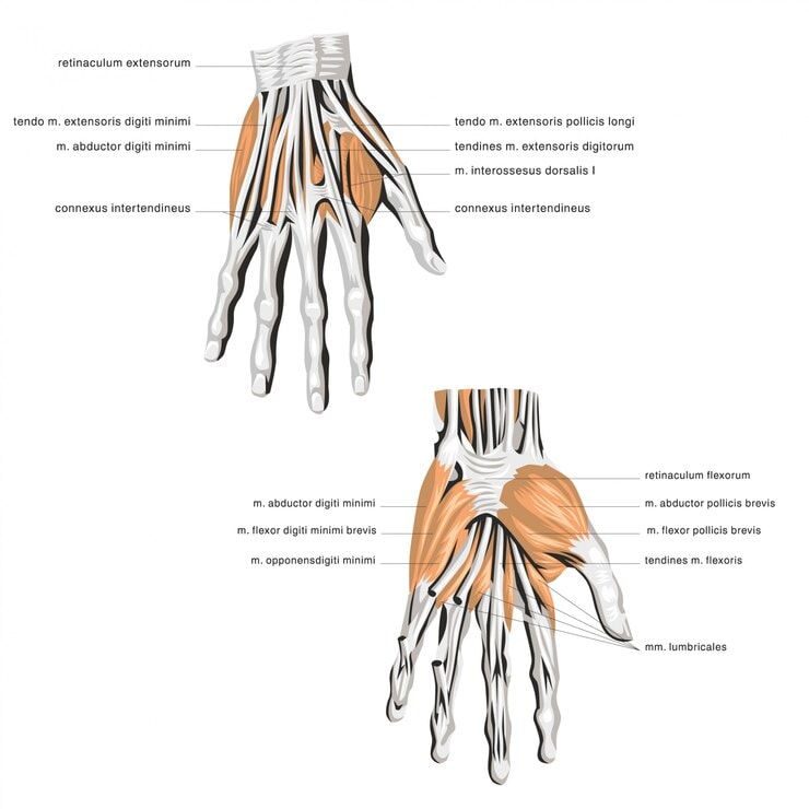
Muscles and Tendons of the Wrist
A complex network of intrinsic and extrinsic muscles and tendons maintains the wrist’s stability and mobility, facilitating various movements and supporting its function.
Flexor Tendons
Multiple muscle groups, categorized into flexor and extensor tendons, are responsible for wrist movement. The flexor carpi radialis attaches to the second and third metacarpal bones, aiding in wrist flexion. Its tendon helps bend the wrist toward the thumb side, while the flexor carpi ulnaris tendon bends it toward the little finger side.
The flexor carpi ulnaris connects to the pisiform bone and the fifth metacarpal, facilitating wrist flexion.
Extensor Tendons
The extensor carpi radialis longus, positioned on the thumb side, assists in lifting the wrist backward. The extensor carpi ulnaris straightens the wrist toward the pinky finger’s direction.
The extensor digitorum extends the fingers at the metacarpophalangeal joints, essential for finger movement. Extensor tendons are crucial for wrist movement, enabling activities involving hand positioning and finger actions.
Intrinsic and Extrinsic Muscles
Intrinsic muscles, originating within the hand, provide fine motor control for finger movements. Along with extrinsic muscles that start in the forearm, they control movements through their tendons.
Extrinsic muscles, which originate from the forearm, facilitate broader movements of the wrist and fingers.
Nerves and Blood Vessels in the Wrist
The wrist houses critical nerves and blood vessels essential for hand functionality and mobility, playing a significant role in maintaining wrist health.
Nerves and Blood Vessels in the Wrist Diagram
Median Nerve and Ulnar Nerve
The median nerve runs down the forearm and through the carpal tunnel, facilitating movement and sensation in the thumb, index finger, and part of the middle finger. It controls most flexor muscles in the forearm and provides sensation to the thumb, index, middle, and part of the ring finger.
The ulnar nerve travels along the ulna, primarily responsible for sensation in the ring and little fingers, and controls some hand muscles. It is crucial for the sensation of the small finger and half of the ring finger.
Radial and Ulnar Arteries
The ulnar and radial arteries, branching from the brachial artery, play a vital role in maintaining blood flow to the hand and wrist. The radial artery, on the thumb side, supplies blood to the hand’s lateral aspect, while the ulnar artery, nearer the pinkie, supplies the medial aspect.
Together, these arteries ensure adequate blood flow to the hand and wrist for proper function.
Movements of the Wrist
The wrist exhibits three main types of motion: flexion-extension, radial-ulnar deviation, and circumduction. These movements are essential for wrist functionality, allowing a wide range of hand activities.
Flexion and Extension
Daily activities require wrist flexion of approximately 54 degrees and extension of about 60 degrees. Normal wrist flexion can reach up to 80 degrees, while extension can extend to about 70 degrees, showcasing the wrist’s impressive range of motion.
Movements like flexion and extension are facilitated by muscles such as the flexor carpi radialis and extensor carpi ulnaris, enabling the wrist to bend forward and backward. These motions are crucial for many daily tasks, from typing to lifting objects.
Radial and Ulnar Deviation
Radial deviation allows for approximately 20 degrees of movement, while ulnar deviation enables about 30 degrees. These movements are essential for tasks requiring side-to-side wrist motion, such as waving or turning a doorknob.
The extensor carpi radialis longus and flexor carpi ulnaris control the wrist’s lateral movements. Ligaments support this range of motion, providing stability while allowing flexibility.
Circumduction
Circumduction involves coordinated wrist movement combining flexion, extension, and radial and ulnar deviation. This circular motion is crucial for activities requiring a combination of these movements, such as stirring a pot or drawing a circle.
The wrist joint plays a significant role in enabling this complex motion, relying on the coordinated efforts of various muscles and ligaments. This movement showcases the wrist’s versatility and adaptability in performing diverse tasks.
Common Conditions Affecting the Wrist
Wrist conditions can include issues like carpal tunnel syndrome, fractures, and tendonitis, all of which can significantly impede hand and wrist pain function. These conditions often arise from repetitive stress, trauma, or underlying health issues.
Diagram of Wrist Injury
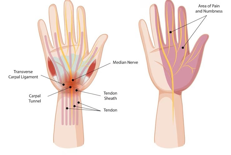
Carpal Tunnel Syndrome
Carpal tunnel syndrome primarily occurs due to pressure on the median nerve, often influenced by repetitive wrist movements or conditions like arthritis. Symptoms often include tingling and numbness, particularly in the thumb and fingers.
Treatments for carpal tunnel syndrome can include wrist splints, physical therapy, and, if necessary, surgical intervention. These options aim to relieve pressure on the median nerve and restore normal function.
Wrist Pain and Injuries
Fractures are a common source of wrist pain, often occurring due to falls or trauma. These injuries can significantly impact daily activities and require prompt medical attention.
Sprains can result from overstretching ligaments in the wrist, leading to pain and instability. Tendonitis, characterized by the inflammation of tendons near the wrist, is another common condition caused by repetitive motions or overuse.
Wrist Sprains and Strains
Wrist sprains and strains often result from sudden impacts or overuse, leading to damage in the ligaments and tendons.
Proper treatment and rehabilitation are crucial for recovery and preventing further injury.
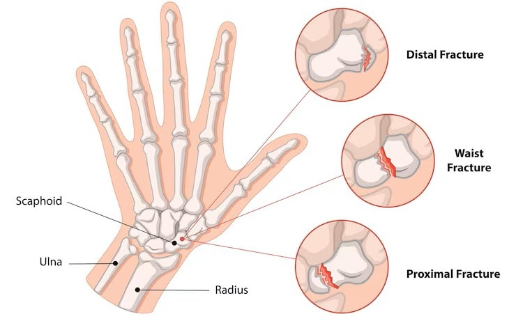
9.3. Fractures of the Wrist
Fractures of the wrist can vary in type and severity. The scaphoid bone is particularly prone to fractures, which may not be immediately visible on X-rays. These fractures require careful diagnosis and treatment to ensure proper healing.
Treatment options for wrist fractures can include casting, physical therapy, and, in some cases, surgical intervention. Recovery often involves a period of immobilization followed by rehabilitation to restore function.
9.4. Arthritis of the Wrist
Arthritis in the wrist can be either osteoarthritis or rheumatoid arthritis. Osteoarthritis is a degenerative condition characterized by the breakdown of cartilage, leading to pain and stiffness. Rheumatoid arthritis, an autoimmune disorder, causes inflammation and damage to the wrist joints.
Both types of arthritis can significantly impact wrist function and quality of life. Treatment options may include medication, physical therapy, and lifestyle modifications to manage symptoms and improve joint health.
Hand and Wrist Functionality
The wrist joint acts as a crucial link between the forearm and the hand, allowing for a wide range of movements essential for daily activities. Its unique structure enables simultaneous movement in two planes, facilitating complex motions like flexion, extension, abduction, and adduction.
Precision Grip and Fine Motor Movements
The wrist is a complex structure that plays a crucial role in allowing for precision grip and fine motor movements. Muscles such as the flexor digitorum superficialis are essential for bending the fingers, which is vital for fine motor control.
The intrinsic muscles of the hand contribute significantly to the precision grip by allowing for refined movements. Proper nerve function, particularly from the median and ulnar nerves, ensures that the small muscles necessary for precision grip can effectively respond to stimuli.
Stability and Flexibility
The wrist offers a unique combination of stability and flexibility, essential for various hand movements. Stability refers to the ability of the wrist to maintain its structure and function under load, while flexibility allows for a range of motions, adjusting to various tasks.
Ligaments such as the collateral ligaments and volar radiocarpal ligaments are crucial for maintaining the wrist’s stability during movement. Muscles like the flexor carpi radialis and extensor carpi ulnaris aid in providing the flexibility necessary for diverse motions of the wrist.


