The radius is a key bone in the forearm that supports arm movement and stability. This article explains its anatomy, including the proximal and distal regions, shaft, joint articulations, and common conditions affecting the radius anatomy.
Key Takeaways
The radius has key anatomical structures like the radial head and tuberosity that enable forearm rotation and muscle attachment, essential for arm movements.
The shaft of the radius serves as a sturdy anchor point for various muscles, contributing to wrist flexion and forearm stability through its connections with the ulna.
Common conditions like Colles fractures and osteoporosis can significantly affect the radius, impacting wrist function and stability.
Proximal Radius Anatomy
The proximal radius features important landmarks like the radial head, neck, and radial tuberosity. These structures facilitate the rotation and stability of the forearm, enabling diverse arm movements.
The radial head connects with the capitellum of the humerus and the radial notch of the ulna, enabling crucial rotational movements for forearm mobility.
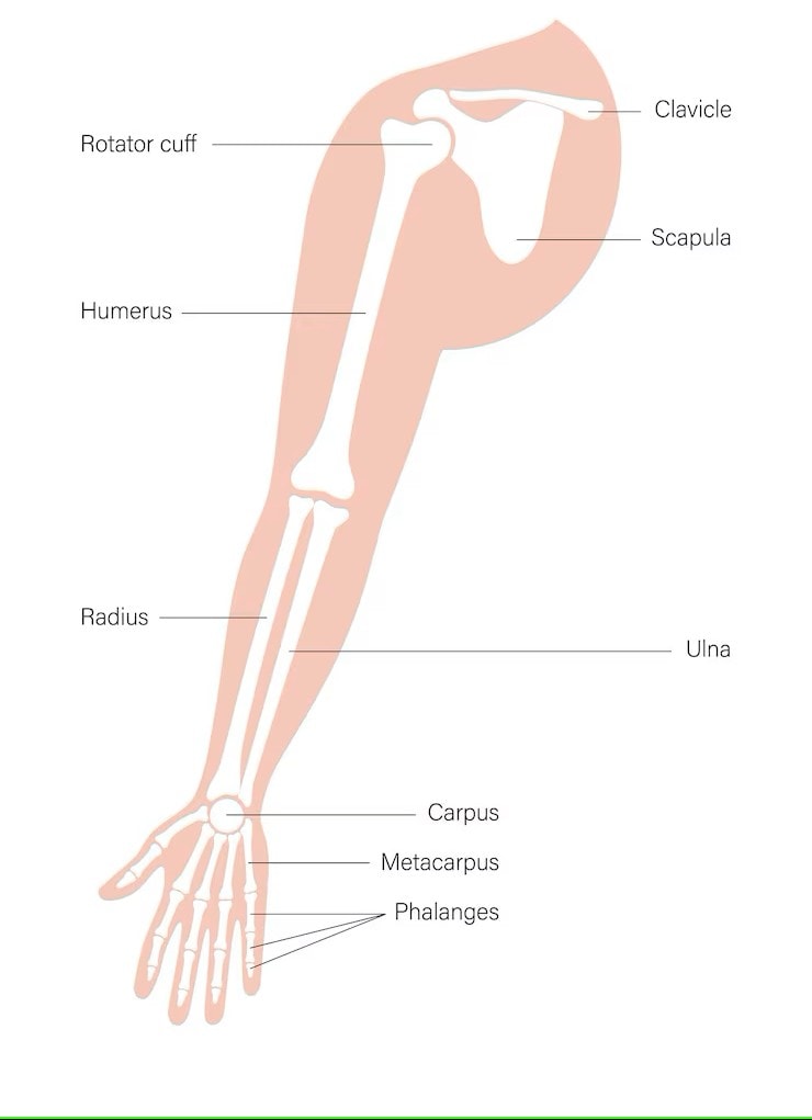
Diagram of the Radius
Radial Head
The radial head is a disc-shaped structure integral to the elbow and proximal radioulnar joints. It interacts with the capitellum of the humerus and the radial notch of the ulna, facilitating forearm rotation.
This joint’s flexibility and functionality allow the radius to bend and straighten in harmony with the humerus and ulna.
Neck of Radius
The neck of the radius, a narrowed region just below the radial head, stabilizes the elbow joint through its ligament attachments. This stability maintains proper arm movements and ensures joint integrity during activities.
Radial Tuberosity
Situated just below the radial head, the radial tuberosity is a bony projection and key attachment point for the biceps brachii muscle, enabling powerful elbow flexion.
A synovial bursa covers the anterior part of the radial tuberosity, offering a smooth surface for muscle attachment.
Shaft of the Radius
Extending from the neck to the distal end, the shaft of the radius features a triangular cross-section with three distinct borders. Its diameter increases distally, offering a sturdy structure for muscle attachments.
It serves as an anchor point for several muscles, including the pronator teres, pronator quadratus, supinator, and various extrinsic hand muscles.
Anterior Surface
The anterior surface of the radial shaft provides a flat area for muscle attachment, facilitating wrist flexion and movements necessary for gripping and manipulating objects.
Posterior Surface
The posterior surface of the radial shaft is generally smooth with minimal muscle attachments, making it less involved in muscle function compared to the anterior surface.
Interosseous Border
The interosseous border of the radius connects to the ulna through the interosseous membrane, enhancing stability between the two bones and ensuring coordinated forearm activities.
Distal Radius Anatomy
The distal end of the radius expands into a rectangular shape and consists of five surfaces, each contributing to wrist movements and interactions with the carpal bones.
The distal radioulnar joint, where the ulnar notch of the radius meets the head of the ulna, is crucial for forearm rotation.
Styloid Process
The styloid process, a bony projection extending distally from the lateral side of the distal radius, stabilizes and supports the wrist joint by providing attachment points for ligaments involved in wrist movements.
Ulnar Notch
Located at the distal end of the radius, the ulnar notch articulates with the head of the ulna to form the distal radioulnar joint, allowing the radius to rotate around the ulna and enabling pronation and supination of the forearm.
Carpal Articular Surface
The carpal articular surface of the distal radius interacts with the carpal bones, allowing for a range of fluid and coordinated wrist movements.
Joint Articulations Involving the Radius
The radius forms key joint articulations, including the elbow, proximal radioulnar, and distal radioulnar joints. These joints are essential for forearm functionality, enabling a wide range of movements and providing stability during activities.
Elbow Joint
The elbow joint, a compound structure formed by the humerus, radius, and ulna, enables arm flexion and extension, allowing for coordinated upper limb movements.
Proximal Radioulnar Joint
The proximal radioulnar joint, where the radial head articulates with the ulnar notch, allows for forearm rotation, essential for actions like turning the wrist.
Distal Radioulnar Joint
The distal radioulnar joint enables the radius and ulna to move in opposite directions for supination and pronation, maintaining wrist stability during forearm rotation.
Common Conditions Affecting the Radius
The radius is prone to conditions like distal radius fractures and osteoporosis, which can significantly impact forearm and wrist functionality and stability.
Colles Fracture
Colles fractures often result from trauma, like falling on an outstretched wrist. Symptoms include wrist pain, swelling, and a potential ‘dinner fork’ appearance.
Osteoporosis
Osteoporosis weakens bones, including the radius bone, increasing fracture risk. Bone density tests help with early detection, and higher-risk individuals include females and adults over 65.
Blood Supply and Innervation of the Radius
The radius receives blood mainly from the radial and interosseous arteries, which is crucial for maintaining bone health and supporting its functions.
Radial Artery
The radial artery supplies blood to the radius and neighboring structures, providing essential oxygen and nutrients that support the health and function of the radius.
Nerve Supply
The radial and median nerves innervate the muscles attached to the radius, playing a crucial role in motor control and proper function of these muscles.