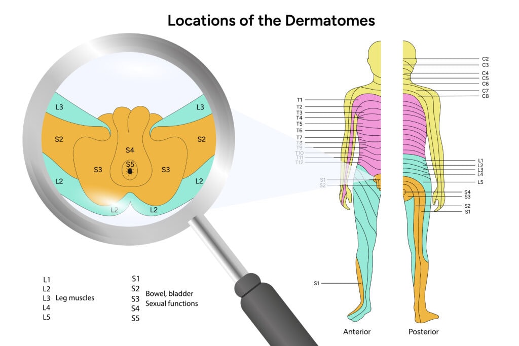
A dermatome map highlights specific skin areas linked to spinal nerves. These maps are key for diagnosing nerve issues and understanding sensory pathways. This guide will explore their anatomy, variations, and medical significance.
Key Takeaways
- Dermatomes are skin areas innervated by specific spinal nerves, crucial for sensory information relay to the brain.
- Dermatome maps vary among individuals and different types; they play a key role in clinical assessments and diagnosing conditions.
- Recognizing dermatome patterns helps healthcare professionals evaluate nerve issues and provide effective treatments, especially for conditions like shingles and radiculopathy.
What is Dermatomes
Dermatomes are areas of skin that are innervated by a single spinal nerve’s posterior root. Imagine these as neatly organized segments, each corresponding to a specific spinal nerve, except for the face, which is innervated by the trigeminal nerve. This segmental organization plays a crucial role in the sensory neurons distribution throughout the body, allowing us to feel touch, pain, and temperature sensations.
The primary function of dermatomes is to relay sensory information from the skin to the central nervous system. Each dermatome is like a messenger, carrying vital information to the brain about what’s happening on the skin’s surface. However, the exact pattern of these dermatomes can vary significantly between individuals, and often, for a dermatome to be completely numb, two or three neighboring posterior roots need to be affected.
Understanding dermatomes is not only fascinating but also crucial in clinical practice. Knowing the corresponding dermatomes allows healthcare professionals to diagnose and treat various conditions more effectively. This intricate map of sensory distribution is a vital tool in the world of neurology and beyond.
Development and Anatomy of Dermatomes
Dermatomes originate from the somitic mesoderm, a layer of cells adjacent to the developing neural tube in the embryo. As the embryo grows, these cells organize in a segmental pattern, forming the basis of our dermatome map. However, some overlap occurs between adjacent dermatomes, adding complexity to their distribution.
The arrangement of dermatomes in the limbs is particularly interesting due to embryonic limb rotation. This rotation causes a spiral pattern in the dermatomes of the arms and legs, leading to a unique and intricate sensory map. Each spinal nerve, except for the first cervical nerve (C1), which is primarily involved in motor innervation, contributes to the formation of these dermatomes.
Knowing the development and anatomy of dermatomes provides deeper insight into their functional significance and variability. This understanding is essential for accurately interpreting sensory information and diagnosing neurological conditions. The journey from embryonic development to the complex maps we use today showcases the intricate design and functionality of the human body.
Dermatome Maps and Their Variations
Dermatome maps are visual representations of the sensory nerve distribution in the body. They are essential tools for assessing sensation, localizing lesions, and determining the extent of injuries. However, not all dermatome maps are identical. Some, like the Keegan and Garrett map and the Foerster map, show significant variations in certain areas, such as the C4 dermatome.
Despite these variations, certain regions, like the thorax and abdomen, exhibit consistency across different maps. Recognizing these variations and their clinical significance is essential for accurate diagnosis and treatment.
Dermatome maps are more than just anatomical charts; they are vital instruments in clinical practice, guiding healthcare professionals in their assessments and interventions.
Major Dermatome Maps
The Keegan and Garrett map and the Foerster map are two major dermatome maps that provide a visual representation of sensory nerve distribution in the body. In the upper limb, these maps differ significantly. The Keegan and Garrett map shows a continuous representation of dermatomes, while the Foerster map presents them in a more discontinuous fashion.
In the lower limbs, the differences are even more pronounced. The Keegan and Garrett map displays a spiral arrangement of dermatomes. In contrast, the Foerster map presents a more segmental distribution. Recognizing these variations is essential for accurate clinical assessment and diagnosis, as the exact dermatome pattern influences the interpretation of sensory information.
Face, Head, and Neck Dermatomes
The face is primarily innervated by the trigeminal nerve, which has three main branches responsible for sensory innervation. This nerve covers the entire face, while spinal nerves C2-C4 provide sensory innervation to the neck and shoulder regions.
Recognizing the dermatomes of the face, head, and neck is essential for diagnosing and treating conditions affecting these areas. The overlapping and specific innervation of these regions highlight the complexity and precision of our nervous system.
Upper Limb Dermatomes
Spinal nerves C5 to T2 innervate the upper limb dermatomes. This means they are responsible for the sensory distribution in that area. These dermatomes cover the arms, from the shoulders down to the fingers. In the Keegan and Garrett map, these dermatomes appear continuous, whereas the Foerster map shows a more discontinuous representation.
These variations highlight the importance of understanding conflicting dermatome maps for accurate clinical assessments. By comparing these maps, healthcare professionals can better diagnose and treat conditions affecting the upper limbs.
Thorax and Abdomen Dermatomes
Thoracic dermatomes exhibit a distinct pattern of distribution that is essential for understanding referred pain. These dermatomes are arranged in nearly horizontal lines, with T10-T12 showing an inferior dip in their distribution. This arrangement is consistent across different dermatome maps.
Conditions like shingles commonly affect these dermatomes, causing significant discomfort and pain. Recognizing the thoracic and abdominal dermatomes is essential for diagnosing and treating such conditions.
Lower Limb and Genitalia Dermatomes
The lower limbs are innervated by specific spinal nerves L1 to S5, with S4 and S5 being located only on the posterior aspect. The Keegan and Garrett map illustrates a spiral arrangement of dermatomes. In contrast, the Foerster map displays a more segmental distribution.
Recognizing these variations and specific dermatome patterns is essential for diagnosing and treating conditions affecting the lower limbs and genitalia. These regions’ complex innervation highlights the importance of accurate dermatome maps in clinical practice.
Clinical Implications of Dermatome Maps
Dermatome maps are invaluable tools in clinical practice. They help healthcare professionals identify potential issues with the spine or associated nerves by evaluating sensory distribution and locating spinal cord injuries. These maps also assist in diagnosing conditions like disc herniation by pinpointing specific nerve root issues.
Disruptions in nerve connections can result in symptoms like numbness or pain. Dermatome maps enable clinicians to accurately assess these symptoms and determine the underlying cause, enhancing treatment effectiveness.
Herpes Zoster and Dermatomes
Herpes Zoster, commonly known as shingles, affects specific dermatomes where the varicella-zoster virus reactivates. This reactivation leads to pain and a characteristic rash in the affected dermatomes. Identifying the affected dermatomes can help in diagnosing and treating the condition effectively.
Treatment options for managing Herpes Zoster include physical therapy, medications, and spinal procedures to alleviate pain. By targeting the specific dermatomes involved, these treatments can provide significant relief to patients.
Radicular Pain and Radiculopathy
Radiculopathy is characterized by the objective loss of sensory symptoms and/or motor innervation. This occurs due to impaired conduction of impulses along a spinal nerve or its dorsal root, including the spinal nerve roots. Radicular pain, often caused by damage or irritation to a single posterior root or spinal ganglion, typically results from spinal stenosis or a herniated disc, including issues related to the dorsal root ganglion.
This condition can lead to numbness or weakness in specific areas, depending on the affected nerve root. Surgical options such as discectomy or laminectomy may be necessary if other treatments are ineffective.
Identifying the affected dermatomes is essential for accurate diagnosis and treatment of radicular pain.
Key Dermatome Landmarks
Recognizing key dermatome landmarks is vital for neurological examinations and clinical assessments. These landmarks represent uniform areas, but variations and overlaps among individuals can complicate their use in assessments.
For instance, the dermatomes of the head and neck are primarily innervated by spinal nerves C2-C4, while the face is innervated by branches of the trigeminal nerve (CN V). Knowing these landmarks helps clinicians accurately assess and diagnose sensory issues.
Dermatomes vs. Myotomes
Dermatomes are areas of skin linked to specific nerve roots in the spinal cord, facilitating the transmission of nerve signals. In contrast, myotomes are groups of skeletal muscles innervated by a single spinal nerve root. These maps are crucial in physical therapy and pain management, guiding treatment strategies based on the affected dermatomes.
Recognizing the differences between dermatomes and myotomes aids in diagnosing and treating various neuromuscular conditions. While dermatomes focus on sensory distribution, myotomes are concerned with motor function, making them both essential in clinical practice.
Educational Resources for Learning Dermatomes
Kenhub offers engaging videos and interactive quizzes specifically designed for mastering dermatome anatomy. Users can choose their preferred learning method, whether it be videos, quizzes, or a combination of both.
Additionally, Kenhub features a free ultimate anatomy study guide and in-depth articles that provide detailed insights into dermatomes, enhancing the learning experience. These resources are invaluable for students and healthcare professionals aiming to deepen their understanding of dermatomes.