

Lungs are vital organs that help us breathe by taking in oxygen and expelling carbon dioxide. They are crucial for maintaining life and overall health. In this article, we will explore how lungs work, their anatomy, and tips for keeping them healthy.
Key Takeaways
The lungs are essential organs comprised of lobes and segments that facilitate gas exchange, with the right lung containing three lobes and the left lung containing two.
The tracheobronchial tree is a critical component of the respiratory system, providing an airway structure supported by cartilage and ensuring the efficient movement of air to the alveoli for gas exchange.
Proactive measures such as quitting smoking, regular physical activity, and monitoring air quality are vital for maintaining lung health and preventing various respiratory diseases.
Lung Anatomy and Structure
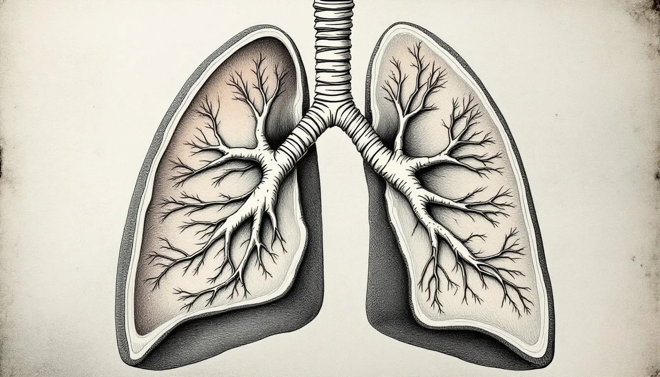
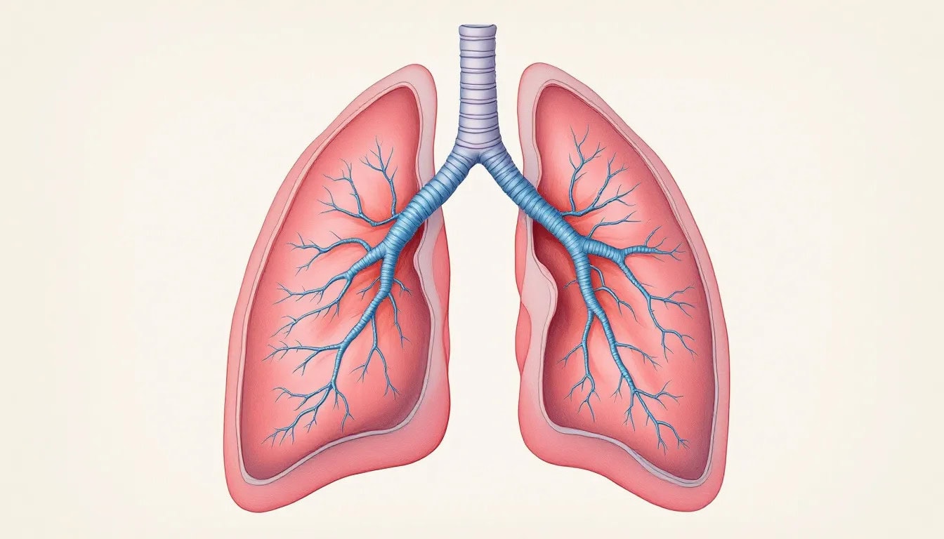
The lungs are cone-shaped organs nestled within the ribcage, a marvel of biological engineering that ensures we breathe effortlessly. Humans are endowed with a right lung and a left lung, with the left lung being slightly smaller to make room for the heart. Healthy lungs exhibit a spongy texture and a pinkish-gray hue, indicative of their vital role in gas exchange.
Each lung, encased in a serous pleural sac, is divided into lobes by fissures. The right lung has three lobes, while the left lung consists of two. These lobes are further segmented into bronchopulmonary segments, with the right lung having ten and the left lung with eight to ten segments.
The various lung surfaces and borders, including the mediastinal, diaphragmatic, and costal surfaces, play crucial roles in lung function and stability. Understanding the lung’s anatomy is fundamental to appreciating how these organs facilitate the essential process of gas exchange.
Diagram of Lung Anatomy
Lobes and Fissures
The right lung contains three lobes: superior, middle, and inferior, each crucial for lung function. The left lung, slightly smaller due to the heart’s position, has two: superior and inferior. These lobes, separated by fissures, enhance the lung’s flexibility and capability.
The right lung’s lobes are divided by horizontal and oblique fissures, while the left lung’s lobes are separated by the oblique fissure. Interestingly, the left lung features a cardiac notch, a concave space accommodating the heart. Understanding these lobes and fissures provides insight into the lung’s structural efficiency and adaptability.
Surfaces and Borders
The lungs feature three surfaces: costal (in contact with the rib cage, forming the anterior and lateral aspects), mediastinal (facing the mediastinum and housing vital structures like the heart and great vessels), and diaphragmatic (resting on the diaphragm, crucial for breathing).
The lungs’ borders-anterior (aligns with the midline and contours to the sternum), posterior (rounded and continuous with the thoracic wall, adding stability), and inferior (separates the lung from the diaphragm)-are equally important.
These surfaces and borders collectively contribute to the lungs’ dynamic functionality.
The Tracheobronchial Tree
Starting with the trachea, the tracheobronchial tree is crucial for the respiratory system. This sturdy tube, supported by 16 to 20 C-shaped cartilage rings, maintains an open airway. The trachea’s four layers-mucosal, submucosa, hyaline cartilage, and adventitia-protect and support the airway.
At its lower end, the trachea splits into the left and right mainstem bronchi at the carina. These primary bronchi, with incomplete tracheal rings and smaller cartilage plates, enter each lung. The left lung has two lobar bronchi, while the right lung has three, corresponding to their lobes. Terminal bronchioles, the smallest branches, mark the end of the respiratory system’s conducting zone.
The trachealis muscle, found at the back of the trachea, plays a vital role in expelling foreign materials through coughing. Lined with ciliated pseudostratified columnar cells and mucus-secreting goblet cells, the respiratory epithelium traps particles and debris, which cilia then move out of the respiratory tract. This system ensures clean air reaches the alveoli.
External Anatomy of the Lungs
Pyramid-shaped structures within the thoracic cavity, the lungs are marvels of biological design. Their external anatomy enables efficient gas exchange.
Adjacent to the heart and bordered by the diaphragm, the lungs are ideally situated for breathing.
Lung Position and Location
Situated in the thoracic cavity and bordered by the heart, ribs, and diaphragm, the lungs are strategically positioned. The left lung has a cardiac notch for the heart, while both rest on the diaphragm, a dome-shaped muscle essential for breathing.
Lung Shape and Size
The right lung is wider and shorter, while the left lung is longer and narrower to accommodate the heart. These shape and size differences ensure efficient space utilization within the thoracic cavity, including the right and left lungs.
Lung Lobes
The right lung has three lobes: superior, middle, and inferior, while the left has two: superior and inferior. Each lobe serves a specific function and is separated by fissures, enabling efficient lung expansion and contraction.
Understanding the significance of each lobe helps appreciate the lung’s complex functionality.
Internal Anatomy of the Lungs
The lungs, with a sponge-like texture internally, are covered by a double-layered pleura that lubricates them during breathing, essential for their function.
The Bronchial Tree
The bronchial tree starts with primary bronchi branching from the trachea, supported by cartilage. These primary bronchi divide into secondary lobar bronchi-three in the right lung and two in the left. Segmental bronchi branch from the lobar bronchi, forming distinct bronchopulmonary segments in each lung.
Bronchioles, the smaller branches of the bronchi, lack cartilage and have muscular walls that can adjust airflow. This network ensures that air reaches every part of the lung for optimal gas exchange.
Alveoli
Alveoli, tiny air sacs where primary gas exchange occurs, allow oxygen to enter the bloodstream and carbon dioxide to exit. Connected to alveolar ducts, they are crucial for the respiratory system’s function.
Type II pneumocytes within alveoli produce surfactant, reducing surface tension and preventing alveolar collapse during exhalation, ensuring the alveoli remain open and functional.
Blood Supply to the Lungs
The pulmonary artery transports deoxygenated blood from the heart to the lungs for oxygenation. Bronchial arteries supply oxygen-rich blood to the lung tissue itself. This dual blood supply system is crucial for lung function.
Lung Tissue Structure
Lung tissue, primarily consisting of parenchyma, plays a crucial role in respiratory function, facilitating gas exchange and supporting lung tissues function.
Pleura
The pleura, a protective covering for the lungs, consists of two layers: the visceral pleura covering the lungs and the parietal pleura adhering to the chest wall. The space between these layers, filled with pleural fluid in the pleural cavity, reduces friction during breathing.
Lung Parenchyma
Lung parenchyma includes structures like alveoli and bronchioles, essential for gas exchange, ensuring the efficient exchange of oxygen and carbon dioxide during breathing.
Physiology of the Lungs
The lungs’ primary function is to facilitate the exchange of oxygen and carbon dioxide between the atmosphere and the bloodstream, vital for sustaining life.
Ventilation Process
The mechanics of breathing involve inspiration and expiration. During inhalation, the diaphragm contracts, enlarging the thoracic cavity and reducing internal pressure.
Expiration occurs when the diaphragm relaxes, allowing the lungs to recoil passively.
Gas Exchange Mechanism
Gas exchange mainly occurs in the alveoli, where oxygen diffuses into the blood and carbon dioxide diffuses out based on pressure gradients, crucial for maintaining healthy lung function.
Lung Volumes and Capacities
Tidal volume refers to the amount of inhaled air or exhaled during normal breathing.
Vital capacity is the maximum amount of air that can be expelled from the lungs after a maximum inhalation.
Blood Supply and Circulation
The pulmonary artery carries deoxygenated blood from the heart to the lungs for oxygenation. Pulmonary veins transport oxygen-rich blood from the lungs to the heart’s left atrium. Bronchial arteries supply oxygenated blood to the lung tissue itself.
Bronchial veins are responsible for collecting deoxygenated blood. They then drain this blood into the azygos vein. This comprehensive blood supply system ensures that the lungs receive the necessary oxygen and nutrients to function effectively.
Nervous System Connections
The pulmonary plexus, formed from autonomic nerves and ganglia at the hilum of each lung, controls bronchial smooth muscle tone and blood flow. Sympathetic innervation from cardiac nerves linked to the cervical sympathetic ganglia and T2 to T6 spinal cord segments causes bronchodilation and pulmonary vessel constriction.
Parasympathetic fibers from the vagus nerve lead to bronchoconstriction and indirect vasodilation. Sensory nerves within the pulmonary plexus provide innervation to the visceral pleura surrounding the lungs.
Gas Exchange Process
Gas exchange occurs in the lungs between the alveoli and the surrounding capillaries, facilitating the transfer of oxygen into the blood and carbon dioxide out of it. During inhalation, alveoli inflate, creating a large surface area for efficient gas exchange with the bloodstream.
The walls of alveoli and capillaries share a thin membrane, allowing gases to diffuse easily. Oxygen binds to red blood cells after crossing the alveolar walls, and carbon dioxide diffuses from the blood into the alveoli to be expelled during exhalation. This process is fundamental to maintaining the body’s oxygen levels and expelling metabolic waste.
Lung Development and Growth
Lung development is a complex process that begins early in pregnancy. Vitamin A plays a critical role in proper lung development during this period. A deficiency in maternal vitamin A can lead to malformations in fetal lung morphology and altered expressions of developmental genes. Inadequate vitamin A during gestation may result in reduced air space size in the developing lungs of neonates.
Ensuring adequate vitamin A intake during pregnancy is essential for healthy lung development and function in newborns.
lungs diagram
A detailed diagram of the lungs can significantly enhance understanding of their structure and function. The lungs are depicted as cone-shaped organs located in the thoracic cavity, with their base resting on the diaphragm and an apex extending above the clavicle.
Each lung consists of multiple lobes separated by fissures, with the right lung having three lobes and the left lung two. The surface of the lungs includes a broad costal area that presses against the rib cage and a smaller mediastinal surface facing inward.
The hilum serves as the entry and exit point for airways, blood vessels, and nerves. The pleura, double-layered membranes surrounding the lungs, consist of the visceral and parietal pleura.
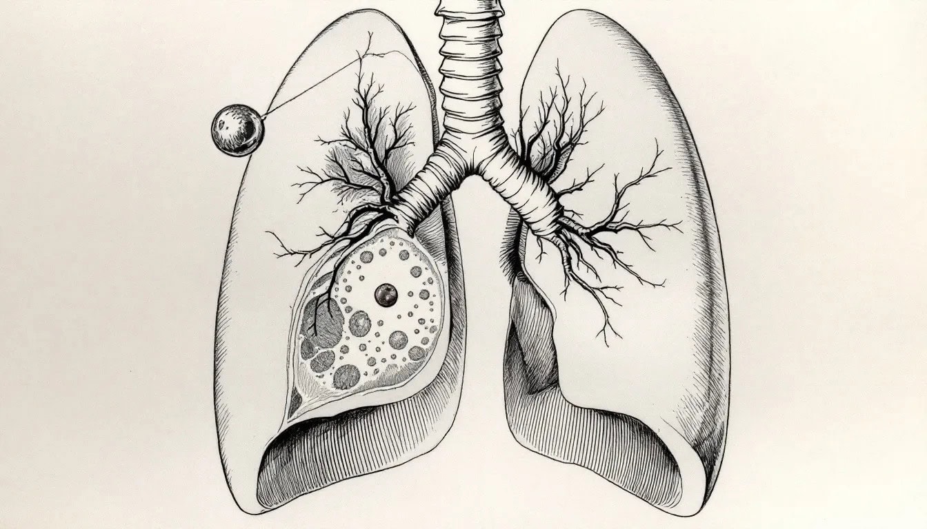
Common Lung Conditions and Diseases
Pulmonology is a specialized field of medicine. It focuses on diagnosing and treating respiratory diseases related to the lungs. Common lung conditions include asthma, bronchiectasis, and chronic obstructive pulmonary disease (COPD), which encompasses chronic bronchitis and emphysema. Pulmonary embolism, often caused by deep vein thrombosis in the legs, is another critical condition affecting lung function.
Vaccination against flu and pneumonia is crucial for lung protection.
Chronic Obstructive Pulmonary Disease (COPD)
Chronic obstructive pulmonary disease (COPD) is a progressive lung disease that causes breathing difficulties, often due to chronic bronchitis and emphysema. Symptoms include chronic cough, shortness of breath, and frequent respiratory infections.
Treatments include bronchodilators, steroids, and pulmonary rehabilitation to improve lung function and quality of life.
Lung Cancer
Lung cancer can be classified into two main categories: small-cell lung carcinoma and non-small-cell lung carcinoma, based on cell size. Smoking, particularly cigarette smoke, is the major risk factor for lung cancer.
Early detection and treatment are vital for improving survival rates.
Respiratory Infections
Respiratory infections, such as pneumonia, significantly impair lung function and overall health. Pneumonia, characterized by inflammation of lung tissue, can be caused by bacteria, viruses, or fungi, with tuberculosis being a major cause of bacterial respiratory infection.
Diagnostic and Surgical Procedures
Diagnosing and treating lung conditions often involve various procedures. Pulmonary embolism, defined as a blood clot lodged in the pulmonary arteries, requires prompt diagnosis and intervention.
Venous drainage from the lungs is primarily carried out by pulmonary veins.
Lung Function Tests
Pulmonary function tests (PFTs) are essential for diagnosing and monitoring lung diseases and assessing treatment responses. Spirometry, lung volume tests, lung diffusion capacity tests, and pulmonary exercise tests are crucial for evaluating lung capacity and functionality.
These tests guide treatment approaches and assess lung functionality.
Surgical Interventions
Surgical interventions for lung conditions include:
Lobectomy
Segmentectomy
Lung volume reduction surgery
Pneumonectomy
Lung transplantation is considered for patients with end-stage lung diseases who have not responded to other treatments.
Maintaining Healthy Lungs
Maintaining healthy lungs involves several proactive measures. Quitting smoking and avoiding secondhand smoke are crucial for lung health. Testing for radon gas in homes is essential, as it can lead to lung cancer. Regular physical activity enhances lung efficiency and reduces the risk of lung-related diseases. Monitoring the Air Quality Index can help limit exposure to outdoor air pollution. Keeping living and workspaces well-ventilated helps reduce indoor air pollution.
Using protective gear is advised for individuals working in environments with dust or chemical exposure. These measures collectively contribute to maintaining healthy lungs and preventing lung disease.


