

Thigh muscle anatomy involves the study of muscles in the anterior, medial, and posterior compartments of the thigh. These muscles are crucial for movements like walking, running, and squatting. Key muscles include the quadriceps, hamstrings, and adductors, each playing vital roles in lower body function. This article dives into the functions, locations, and importance of these muscles.
Key Takeaways
Thigh muscles are organized into three compartments: anterior, medial, and posterior, each responsible for unique movements such as flexion, extension, and adduction.
The femoral artery supplies blood to the thigh muscles, while the femoral, obturator, and sciatic nerves provide essential innervation for muscle function and coordination.
Common thigh injuries include strains, tendonitis, and muscle imbalances, necessitating proper management and rehabilitation to maintain mobility and prevent long-term complications.
Thigh Muscles Anatomy
Thigh muscles are skeletal muscles, controlled voluntarily and responsible for various movements. They are grouped into three primary compartments: anterior, medial, and posterior, based on location and function. Each compartment has muscles performing distinct functions like flexion, extension, adduction, and stabilization of the lower body.
The major muscle groups in the thigh include the quadriceps, hamstrings, adductors, pectineus, and sartorius. These muscles are crucial for walking, squatting, and maintaining balance. They support body weight and proper alignment, vital for daily activities and athletic performance.
Thigh Muscle Diagram
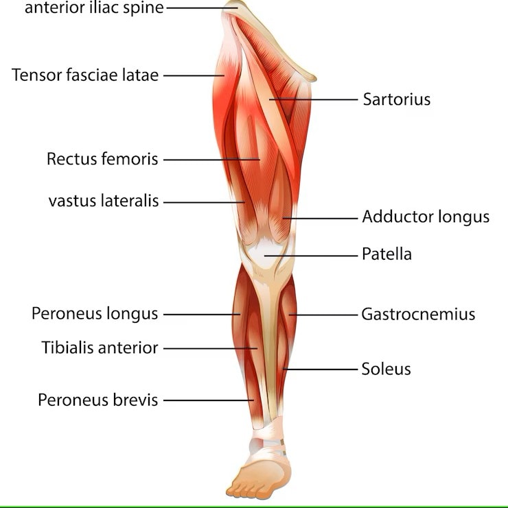
Anterior Compartment Muscles
The anterior compartment primarily consists of muscles responsible for knee extension and hip flexion. It includes the quadriceps femoris group, crucial for powerful leg movements like running and jumping. It also contains hip flexors such as the iliacus, psoas major, sartorius, and pectineus.
The femoral artery supplies the anterior compartment, ensuring these muscles receive adequate oxygen and nutrients for optimal performance. Muscles like the rectus femoris, vastus muscles, and sartorius each play unique roles in movement and stability.
Rectus Femoris
The rectus femoris is one of the four muscles in the quadriceps femoris group. It has two heads: one originating from the anterior inferior iliac spine and another from above the acetabulum. It extends down the thigh, inserting into the quadriceps femoris tendon, which attaches to the patella and continues as the patellar tendon to the tibia.
The rectus femoris extends the knee and flexes the hip, crucial for activities like kicking a ball or sprinting. Its innervation by the femoral nerve and blood supply from the femoral artery ensure it functions effectively.
Vastus Muscles
The vastus muscles, part of the quadriceps femoris, include the vastus lateralis, vastus medialis, and vastus intermedius. These muscles primarily extend the knee, critical for activities like standing up or climbing stairs.
Each muscle has a specific location and function. The vastus lateralis is on the lateral thigh, the vastus medialis on the medial side, and the vastus intermedius lies deep to the rectus femoris. Together, they stabilize and extend the knee joint.
Sartorius Muscle
The sartorius is the longest muscle in the human body. It extends from the anterior superior iliac spine to the medial tibia. It flexes both the knee and hip joints, essential for crossing the legs or performing a high kick.
Besides flexion, the sartorius assists in hip abduction and lateral rotation. Its unique path allows it to act on multiple joints, providing versatility in movement.
Medial Compartment Muscles
The medial compartment contains muscles responsible for hip adduction. It includes the gracilis, obturator externus, adductor brevis, adductor longus, and adductor magnus. These muscles stabilize the pelvis and aid in movements like walking and running.
Each muscle has a specific role and origin, contributing to the medial thigh’s overall function. The obturator nerve provides motor innervation, ensuring proper function during movement.
Adductor Longus
The adductor longus is a flat muscle originating from the pubis and inserting on the linea aspera of the femur. It plays a key role in thigh adduction, pelvis stabilization, and assists in thigh flexion and external rotation.
Innervated by the obturator nerve, it is essential for movements like bringing the legs together, riding a horse, or certain dance moves. Its fan-shaped structure allows it to exert significant force during these actions.
Adductor Magnus
The adductor magnus is the largest muscle in the medial compartment. It comprises both adductor and hamstring components. It adducts the thigh and assists in hip extension, providing additional stability during movement.
It receives dual innervation from the obturator nerve and the tibial part of the sciatic nerve, enabling it to perform complex functions effectively. Its broad origin and insertion points support a wide range of movements and stabilize the pelvis.
Gracilis
The gracilis is the most superficial muscle in the medial compartment, running from the pubis to the medial tibia. It uniquely crosses both the hip and knee joints, assisting in thigh adduction and knee flexion.
The gracilis plays a critical role in bending the knee and bringing the legs together, crucial for athletic activities or daily tasks like sitting down. Its superficial position makes it easily palpable and a key muscle in clinical examinations.
Posterior Compartment Muscles
The posterior compartment is primarily involved in knee flexion and hip extension, essential for activities like walking, running, and jumping. It includes the hamstring muscles, crucial for these movements.
These muscles are innervated by the sciatic nerve, essential for their function and coordination. Understanding these muscles is vital for addressing mobility and athletic performance issues.
Hamstring Group
The hamstring group consists of the biceps femoris, semitendinosus, and semimembranosus. These muscles originate from the ischial tuberosity and are responsible for knee flexion and hip extension.
The biceps femoris has a long head for hip extension and a short head for knee flexion. The semitendinosus and semimembranosus also play crucial roles, with the semimembranosus providing additional knee stability during flexion.
Sciatic Nerve
The sciatic nerve is the largest and longest nerve, innervating most posterior thigh muscles, including the semimembranosus, semitendinosus, and adductor magnus. This nerve is crucial for lower limb mobility and coordination.
The sciatic nerve branches into the tibial and common fibular nerves, each innervating different muscles and contributing to various lower limb movements. Proper sciatic nerve function is essential for activities involving knee bending and hip extension.
Blood Supply and Lymphatics
The blood supply and lymphatic drainage are critical for thigh muscle function and health. The femoral artery is the main vessel supplying the thigh, ensuring muscles receive necessary oxygen and nutrients. The lymphatic system, including the external iliac lymphatic plexus, is vital for immune response and fluid balance.
Understanding the thigh’s vascular network is essential for diagnosing and treating poor circulation or lymphatic drainage conditions. The next sections will delve into the specifics of the femoral artery and the veins and lymph nodes in the thigh.
Femoral Artery
The femoral artery is the primary blood supply to the thigh. Originating from the external iliac artery, it enters the thigh beneath the inguinal ligament. It branches into the deep femoral artery (profunda femoris) and the superficial femoral artery, each supplying different muscle groups.
The profunda femoris is the largest femoral artery branch, supplying deep thigh structures, including medial and lateral circumflex femoral arteries.
The superficial femoral artery continues down the thigh, becoming the popliteal artery after passing through the adductor hiatus.
Veins and Lymph Nodes
The thigh’s vascular network includes major veins like the femoral vein, essential for effective blood circulation. These veins return deoxygenated blood to the heart for reoxygenation.
The lymphatic system also plays a crucial role, with lymph nodes aiding in immune response and fluid balance. For example, the external iliac lymphatic plexus drains lymph from the anterior thigh muscles, merging into the common iliac plexus.
Nerve Supply
The nerve supply is crucial for sensory perception and motor control of thigh muscles. Primary nerves innervating the thigh muscles include the femoral, obturator, and sciatic nerves. Each nerve plays a specific role in muscle function and coordination.
Understanding the nerve supply is crucial for diagnosing and treating nerve damage or dysfunction conditions. The following sections will provide detailed information about the femoral and obturator nerves.
Femoral Nerve
The femoral nerve originates from lumbar spinal roots L2 to L4, providing sensory and motor innervation to anterior thigh muscles. It is essential for hip flexion and knee extension, playing a critical role in walking and running.
The femoral nerve also supplies sensation to the anterior thigh and medial leg, ensuring proper sensory feedback during movement. Its importance in both motor and sensory functions makes it a key nerve for maintaining lower limb mobility.
Obturator Nerve
The obturator nerve originates from spinal roots L2 to L4 and is responsible for providing motor function to the adductor muscles of the medial thigh. These muscles are crucial for thigh adduction, a movement essential for activities such as walking and running.
The obturator nerve supplies sensory innervation to the medial side of the thigh. This innervation plays a role in overall sensory perception. The dual innervation of the adductor magnus by both the obturator and sciatic nerves enhances its role in stabilizing the thigh during movement.
Muscles of the Anterior Thigh
The muscles of the anterior thigh are primarily responsible for extending the knee and flexing the hip. This compartment includes the quadriceps femoris group, the iliopsoas, and the sartorius. These muscles are essential for powerful leg movements and maintaining stability during activities such as walking and running.
The femoral nerve innervates these muscles, ensuring proper motor function and coordination. The following sections will provide detailed information about the iliopsoas, quadriceps femoris, and sartorius muscles.
Iliopsoas
The iliopsoas muscle is a composite structure comprising the iliacus, psoas major, and often the psoas minor. This muscle group originates from the lumbar vertebrae and iliac fossa, inserting at the lesser trochanter of the femur. It plays a crucial role in hip flexion and contributes to the stabilization of the lumbar spine.
The iliopsoas is essential for movements that require lifting the thigh, such as climbing stairs or performing high-intensity athletic activities. Its importance in both stability and mobility makes it a key muscle in the anterior thigh.
Quadriceps Femoris
The quadriceps femoris consists of four distinct muscles: rectus femoris, vastus lateralis, vastus medialis, and vastus intermedius. This muscle group is primarily responsible for extending the knee joint, playing a significant role in activities such as walking, running, and jumping.
Each muscle in the quadriceps group has a specific function. The rectus femoris also assists in hip flexion, while the vastus muscles focus on knee extension. The femoral nerve innervates this muscle group, ensuring proper motor function and coordination.
Sartorius
The sartorius muscle, the longest muscle in the human body, originates from the anterior superior iliac spine and inserts on the medial aspect of the tibia. It facilitates flexion, abduction, and lateral rotation of the hip, as well as flexion of the knee.
This muscle’s unique path across the thigh allows it to act on multiple joints, providing significant versatility in movement. The sartorius is essential for activities such as crossing the legs or performing high kicks.
Muscles of the Posterior Thigh
The posterior thigh is primarily composed of the hamstring group, which includes the biceps femoris, semitendinosus, and semimembranosus. These muscles are essential for knee flexion and hip extension, playing key roles in activities such as running and jumping.
The sciatic nerve innervates these muscles, ensuring their proper function and coordination during movement. The following sections will provide detailed information about the hamstrings and the posterior portion of the adductor magnus.
Hamstrings
The hamstrings consist of the biceps femoris, semitendinosus, and semimembranosus. These muscles originate from the ischial tuberosity and are responsible for knee flexion and hip extension.
The biceps femoris has a long head for hip extension and a short head for knee flexion. The semitendinosus and semimembranosus also play crucial roles, with the semimembranosus providing additional knee stability during flexion.
Adductor Magnus (Posterior Portion)
The posterior portion of the adductor magnus helps in both hip extension and adduction. This muscle is crucial for movements such as squatting, where it provides additional power and stability.
It receives dual innervation from the obturator nerve and the tibial part of the sciatic nerve, enabling it to perform complex functions effectively. Its broad origin and insertion points support a wide range of movements and stabilize the pelvis.
Muscles of the Medial Thigh
The medial thigh muscles are primarily responsible for hip adduction, a movement essential for activities such as walking and running. This compartment includes the adductor longus, adductor brevis, adductor magnus, gracilis, and obturator externus.
The obturator nerve provides motor innervation, ensuring proper function during movement. The following sections will provide detailed information about the adductor group and the obturator externus.
Adductors Group
The adductor group includes the adductor longus, adductor brevis, and the anterior portion of the adductor magnus. These muscles are primarily responsible for thigh adduction, bringing the legs together during movement.
The gracilis muscle is the most superficial of the adductors and crosses both the hip and knee joints, playing a role in knee flexion as well. The obturator nerve innervates these muscles, ensuring their proper function and coordination.
Obturator Externus
The obturator externus muscle contributes to both thigh adduction and lateral rotation. It originates from the obturator membrane and surrounding bones, inserting into the trochanteric fossa of the femur.
This muscle plays a crucial role in stabilizing the hip joint during movement, ensuring proper alignment and coordination. Its function is essential for activities that require precise control of hip movements, such as dancing or martial arts.
Muscles of the Lateral Thigh
The lateral thigh compartment primarily includes the tensor fasciae latae (TFL) and the iliotibial band (IT band), which play critical roles in lower limb function. These structures are essential for hip stabilization and movement.
Understanding the muscles of the lateral thigh is vital for addressing issues related to hip and knee stability. The following sections will provide detailed information about the TFL and IT band.
Tensor Fasciae Latae (TFL)
The tensor fasciae latae (TFL) originates from the anterior superior iliac spine and the iliac crest, inserting into the fascia of the IT band. This muscle is crucial for stabilizing the hip and knee during movements and assists in hip abduction, internal rotation, and weak hip flexion.
The TFL plays a significant role in facilitating hip flexion and abduction, making it essential for activities such as running and walking. Its importance in both stability and movement makes it a key muscle in the lateral thigh.
Iliotibial Band (IT Band)
The iliotibial band (IT band) forms a connection with the TFL, aiding in its functions. This band serves as a connective structure that provides stability to the knee joint during activities such as running and walking. The IT band’s primary role is to maintain knee stability by resisting lateral forces, ensuring proper alignment and coordination during movement.
Its function is essential for athletes and individuals engaged in high-impact activities.
Innervation of Thigh Muscles
The innervation of thigh muscles is crucial for their proper function and coordination. The primary nerves responsible for innervating these muscles include the femoral nerve, sciatic nerve, obturator nerve, and superior gluteal nerve.
Understanding the nerve supply is crucial for diagnosing and treating nerve damage or dysfunction conditions. The following sections will provide detailed information about the femoral, sciatic, obturator, and superior gluteal nerves.
Femoral Nerve
The femoral nerve provides motor innervation to key muscles in the anterior compartment, including the quadriceps and sartorius. It is essential for hip flexion and knee extension, playing a critical role in walking and running.
The femoral nerve also supplies sensation to the anterior thigh and medial leg, ensuring proper sensory feedback during movement. Its importance in both motor and sensory functions makes it a key nerve for maintaining lower limb mobility.
Sciatic Nerve
The sciatic nerve innervates the hamstring muscles located in the posterior compartment of the thigh. This nerve is crucial for lower limb mobility and coordination, as it provides motor and sensory innervation to these muscles.
The sciatic nerve branches into the tibial and common fibular nerves, each innervating different muscles and contributing to various lower limb movements. Proper sciatic nerve function is essential for activities involving knee bending and hip extension.
Obturator Nerve
The obturator nerve is responsible for motor innervation. It specifically targets the adductor muscles located in the medial thigh. These muscles are crucial for thigh adduction, a movement essential for activities such as walking and running.
The obturator nerve supplies sensory innervation to the medial side of the thigh. This innervation plays a role in overall sensory perception. The dual innervation of the adductor magnus by both the obturator and sciatic nerves enhances its role in stabilizing the thigh during movement.
Superior Gluteal Nerve
The superior gluteal nerve is responsible for innervating the tensor fasciae latae, a muscle involved in hip stabilization. This nerve plays a crucial role in ensuring the proper function and coordination of the TFL during movements such as walking and running.
Proper innervation by the superior gluteal nerve ensures that the TFL can perform its functions effectively, contributing to overall hip and knee stability. Understanding this nerve’s role is essential for addressing issues related to hip stabilization and movement.
Blood Supply to the Thigh Muscles
The blood supply to the thigh muscles is essential for their function and health. The primary artery supplying blood to these muscles is the femoral artery, which ensures that the muscles receive the oxygen and nutrients they need.
Understanding the vascular network in the thigh is crucial for diagnosing and treating conditions related to poor circulation. The following sections will provide detailed information about the femoral artery, deep femoral artery, and obturator artery.
Femoral Artery
The femoral artery is the main artery supplying blood to the thigh muscles. It enters the thigh beneath the inguinal ligament at the midpoint between the anterior superior iliac spine (ASIS) and the pubic symphysis. From this point, it extends downward, branching into several arteries that provide blood to the surrounding tissues.
The profunda femoris artery, the largest branch of the femoral artery, supplies the deep structures of the thigh, including both medial and lateral circumflex femoral arteries. These branches ensure that all parts of the thigh receive adequate blood supply, maintaining muscle health and function.
Deep Femoral Artery (Profunda Femoris)
The deep femoral artery, also known as the profunda femoris, branches off the femoral artery and provides blood to both the posterior and medial muscles of the thigh. This artery is crucial for delivering oxygen and nutrients to the deeper structures of the thigh, ensuring their proper function.
By supplying the deeper muscles, the profunda femoris artery plays a vital role in maintaining the health and performance of the thigh muscles, particularly those involved in powerful movements like running and jumping.
Understanding this artery’s function is essential for diagnosing and treating conditions related to poor circulation in the thigh.
Obturator Artery
The obturator artery primarily nourishes the muscles located in the medial compartment of the thigh. This artery branches from the internal iliac artery and provides blood to the adductor muscles, ensuring their proper function during activities like walking and running.
By supplying the adductor muscles, the obturator artery plays a crucial role in maintaining hip stability and thigh adduction. Proper blood flow through this artery is essential for the health and performance of the medial thigh muscles.
Muscle Actions and Movements
The thigh muscles are involved in various actions and movements that are essential for daily activities and athletic performance. These actions include hip flexion, hip extension, hip abduction and adduction, knee flexion, and knee extension.
Understanding these movements and the primary muscles involved helps in diagnosing and treating injuries, improving performance, and maintaining overall muscle health. The following sections will provide detailed information about each movement and the muscles responsible.
Hip Flexion
Hip flexion is primarily facilitated by the iliopsoas, rectus femoris, and sartorius muscles. These muscles work together to lift the thigh towards the abdomen, a movement essential for walking, running, and climbing stairs.
Proper function of these muscles is crucial for maintaining stability and mobility in the lower limb. Any impairment in hip flexion can significantly impact daily activities and athletic performance.
Hip Extension
Hip extension involves the gluteus maximus and the hamstring muscles, including the biceps femoris, semitendinosus, and semimembranosus. These muscles work together to move the thigh backward, a movement essential for jumping, sprinting, and maintaining an upright posture.
The ability to extend the hip properly is crucial for explosive movements and overall lower limb strength. Any impairment in hip extension can hinder athletic performance and daily activities.
Hip Abduction and Adduction
Hip abduction is primarily facilitated by the gluteus medius, gluteus minimus, and tensor fasciae latae (TFL), while hip adduction involves the adductor longus, adductor brevis, and adductor magnus muscles. These movements are essential for maintaining balance and performing lateral movements, such as side-stepping or pivoting.
Proper function of these muscles ensures stability and coordination during movement, reducing the risk of falls and improving overall performance in activities that require lateral motion.
Knee Flexion
Knee flexion is primarily facilitated by the hamstring muscles, including the biceps femoris, semitendinosus, and semimembranosus. These muscles work together to bend the knee, a movement essential for walking, running, and squatting. Proper function of these muscles is crucial for maintaining lower limb mobility and stability.
Any impairment in knee flexion can significantly impact daily activities and athletic performance.
Knee Extension
Knee extension involves the quadriceps femoris group, which includes:
Rectus femoris
Vastus lateralis
Vastus medialis
Vastus intermedius
These muscles work together to straighten the knee, a movement essential for standing up, walking, and running.
The ability to extend the knee properly is crucial for maintaining lower limb stability and performing various activities. Any impairment in knee extension can hinder daily activities and athletic performance.
Common Disorders and Injuries of the Thigh Muscles
Thigh muscles are susceptible to various disorders and injuries, particularly in athletes and individuals engaged in high-impact activities. Common issues include muscle strains, tears, tendinopathies, and muscle imbalances, which can lead to significant pain and impact daily activities.
Understanding these common disorders and injuries is crucial for prevention, diagnosis, and treatment. The following sections will provide detailed information about specific injuries and their management.
Strains and Tears
Muscle strains in the thigh often occur due to sudden movements or overexertion during physical activities. These strains can lead to tears in muscle fibers, causing pain, swelling, and impaired function.
The severity of muscle strains can vary, with grades ranging from mild (grade 1) to severe (grade 3). Treatment typically involves rest, ice, compression, and elevation (RICE), along with physical therapy to restore strength and flexibility.
Muscle Imbalances
Muscle imbalances in the thighs can arise from discrepancies in strength between opposing muscle groups, which may increase the risk of injuries. These imbalances can result in decreased performance and increased susceptibility to strains and other injuries.
Physical therapy is often recommended to address muscle imbalances, focusing on strengthening weaker muscles and improving overall muscle balance. Proper training and exercise can help prevent imbalances and maintain muscle health.
Tendinopathies (e.g., Iliopsoas Tendinopathy)
Iliopsoas tendinopathy is a common condition caused by repetitive hip flexion or overuse in sporting activities. This condition leads to pain in the groin area, affecting hip flexion and overall mobility.
Management of tendinopathies often involves rest, anti-inflammatory medications, and gradual return to activity. Physical therapy can also help strengthen the affected muscles and improve flexibility, reducing the risk of recurrence.
Groin Injuries
Groin injuries often involve the adductor muscles and can occur during sports that require sudden direction changes, such as soccer or hockey. These injuries can cause significant pain and impair movement, requiring proper treatment and rehabilitation.
Rehabilitation for groin injuries focuses on restoring strength, flexibility, and ensuring a safe return to activity. Proper warm-up and stretching exercises can help prevent these injuries and maintain muscle health.
Common Thigh Muscle Injuries
Muscle strains, pulls, and tears are common conditions that affect the thigh muscles. These injuries can cause discomfort and limit mobility. These injuries can result in symptoms such as pain, numbness, weakness, and trouble putting weight on the leg, which should prompt a call to a healthcare provider.
Taking precautions during exercise, such as proper warm-up and stretching, can help maintain thigh muscle health and prevent injuries. Understanding these common injuries is crucial for prevention and effective treatment.
Muscle Strains
Muscle strains commonly affect the hamstrings and quadriceps, resulting in a tearing sensation and severe, sudden pain. These strains can also cause localized pain and swelling in the affected area.
Treatment typically involves rest, ice, compression, and elevation (RICE), along with physical therapy to restore strength and flexibility. Understanding the causes and symptoms of muscle strains can help in their prevention and management.
Tendonitis
Tendonitis in the thigh often arises from repetitive motion, resulting in pain and swelling in the affected tendon. This condition is commonly caused by overuse and can lead to joint pain and stiffness.
Management of tendonitis typically involves rest, anti-inflammatory medications, and physical therapy to strengthen the affected muscles and tendons. Proper warm-up and stretching exercises can help prevent tendonitis and maintain muscle health.
Clinical Significance
Understanding thigh muscle anatomy is crucial for effective diagnosis and treatment of injuries and conditions affecting the lower limb. This knowledge aids physicians in identifying pathology during physical exams and determining appropriate treatments.
Thigh muscle injuries can lead to chronic conditions affecting overall mobility. Proper management and treatment of these injuries are essential for maintaining muscle health and preventing long-term complications.
Physical Examination
Specific examination techniques can effectively assess the functionality of thigh muscles during clinical evaluations. These techniques include palpation, strength testing, and range of motion assessments, which help identify any abnormalities or injuries.
Understanding the anatomy and function of thigh muscles is essential for accurate diagnosis and effective treatment planning. Proper physical examination can help identify the underlying causes of pain and dysfunction, leading to better patient outcomes.
Surgical Considerations
Understanding thigh anatomy is crucial in surgery as it minimizes neurovascular injury, aids in identifying pathology, and determines appropriate muscle portions for grafts. Knowledge of thigh anatomy helps surgeons avoid damaging significant nerves and blood vessels during surgical procedures, significantly reducing the risk of complications.
Peripheral nerve blocks are used in thigh surgery to minimize postoperative pain by ensuring the correct identification and anesthesia of the nerve. Overall, understanding thigh anatomy contributes to safer surgeries, improved healing, and enhanced functional outcomes for patients.


