

Foot anatomy is fascinating and vital to our daily functions. This article explains the bones, muscles, ligaments, and common issues of the foot. You’ll learn how these elements work together to support weight, enable movement, and maintain balance.
Key Takeaways
The foot is a complex structure with 26 bones, 33 joints, and over 100 muscles, ligaments, and tendons, all working together to support mobility and absorb stress.
Key components include the tarsal, metatarsal, and phalangeal bones, which each play critical roles in foot stability and function during activities like walking and running.
Common foot issues, such as plantar fasciitis and stress fractures, can significantly impact mobility, but proper care and preventive measures can help maintain foot health.
Foot Anatomy
The foot is a remarkable structure, supporting the entire body weight, especially during activities like walking or running. It contains more than 100 muscles, tendons, and ligaments that enable complex movements and contribute to mobility.
The foot absorbs a tremendous amount of force and stress with each step, making it vital for our daily activities and overall mobility. Understanding the anatomy of the foot can help in appreciating its role in our day-to-day lives and recognizing the importance of maintaining its health.
The human foot is composed of 26 bones, including tarsal, metatarsal, and phalangeal bones that provide structure and support. These bones are connected by 33 joints, facilitating various movements and contributing to stability during ambulation. Each component, from the bones to the muscles and ligaments, plays a critical role in the foot’s functionality.
Examining each aspect allows us to understand how our feet function and how to keep them healthy.
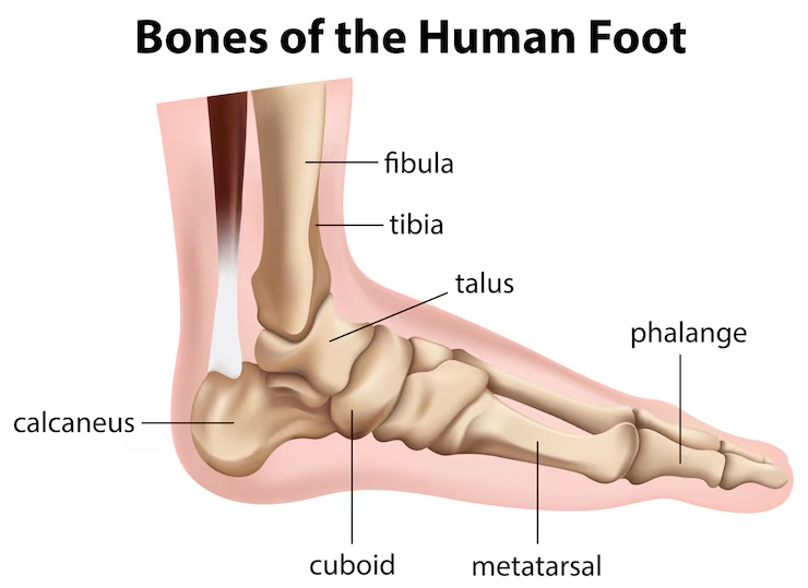
Foot Anatomy Diagram
Bones of the Foot
The bones of the foot are categorized into three main groups: tarsal bones, metatarsal bones, and phalanges. These foot bones form the structural framework of the foot, enabling it to support body weight and facilitate movement. Each group of bones has a specific role, and together they contribute to the foot’s overall function and stability.
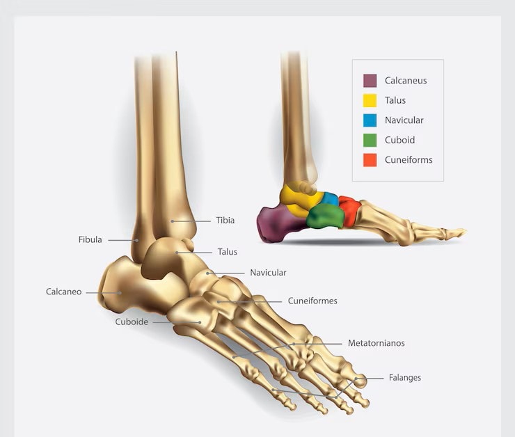
Bones of the Foot Diagram
1.1 Tarsal Bones
The tarsal bones are located in the rear and midfoot and include seven tarsal bones: the talus, calcaneus, navicular, cuboid, and three cuneiform bones. The calcaneus, or heel bone, is the largest tarsal bone and plays a crucial role in weight support. The talus sits above the calcaneus and forms the lower part of the ankle joint, enabling dorsiflexion and plantar flexion.
The navicular bone, cuboid bone, and cuneiform bones form the midfoot and connect to the metatarsal bones. These bones are integral to the foot’s arch structure, providing stability and flexibility. These bones must function properly to absorb shock and distribute forces during activities like walking and running.
1.2 Metatarsal Bones
The metatarsal bones are the long bones in the middle part of the foot, numbering from one to five, starting from the big toe. These bones connect the tarsal bones to the phalanges and play a critical role in weight-bearing and balance, including the fifth metatarsal bone.
Each metatarsal bone has a base, shaft, and head, contributing to the foot’s arch and overall stability during movement.
1.3 Phalanges
The phalanges are the toe bones, with each toe (except the big toe) having three phalanges: proximal, middle, and distal. The big toe, also known as the hallux, consists of only two phalanges. These are the proximal and distal phalanges. These bones are crucial for balance and propulsion during walking and running, allowing for fine motor movements and stability.
2. Joints of the Foot
The foot consists of 33 joints that facilitate movement and flexibility. These joints, including the major ones like the ankle joint, subtalar joint, and midtarsal joint, are essential for both movement and stability. The joints of the foot enable various types of movements, including dorsiflexion, plantar flexion, inversion, and eversion, contributing to the overall functionality of the foot.
2.1 Ankle Joint (Talocrural Joint)
The ankle joint, or talocrural joint, is formed by the tibia, fibula, and talus bones. This joint allows for dorsiflexion and plantar flexion, playing a vital role in walking, running, and other activities.
Ankle joint stability helps prevent injuries and ensures smooth movement.
2.2 Subtalar Joint
The subtalar joint is formed between the talus and calcaneus bones. This joint allows for inversion and eversion of the foot, contributing to the foot’s ability to adapt to uneven surfaces and maintain balance.
2.3 Midtarsal Joint (Chopart’s Joint)
The midtarsal joint, also known as Chopart’s joint, consists of the talonavicular and calcaneocuboid joints. This joint plays a crucial role in foot flexibility and stability, allowing for smooth transitions during walking and running.
2.4 Tarsometatarsal Joints (Lisfranc Joint)
The tarsometatarsal joints, or Lisfranc joints, are formed between the tarsal and metatarsal bones. These joints provide stability to the midfoot and are essential for weight-bearing and movement.
2.5 Metatarsophalangeal Joints (MTP Joints)
The metatarsophalangeal joints (MTP joints) connect the metatarsal bones to the phalanges. These joints play a significant role in toe movement, crucial for walking, running, and maintaining balance. The MTP joints allow for flexion, extension, and limited rotation of the toes, contributing to the overall flexibility and functionality of the foot.
2.6 Interphalangeal Joints
The interphalangeal joints are the joints between the phalanges of the toes. Each toe has proximal and, in the case of all but the big toe, distal interphalangeal joints. These joints enable flexion and extension of the toes, which are essential for gripping surfaces and maintaining balance.
3. Muscles of the Foot
The muscles of the foot are categorized into two primary groups: intrinsic and extrinsic. Together, these muscles contribute to the foot’s stability, movement, and fine motor control. Intrinsic muscles are located within the foot, while extrinsic muscles originate in the leg and extend into the foot.
3.1 Intrinsic Muscles
The intrinsic muscles of the foot have both their origin and insertion within the foot itself. Primarily responsible for fine motor control of the toes, these muscles include the extensor digitorum brevis and extensor hallucis brevis. They play a crucial role in stabilizing the foot and providing proprioceptive feedback, which is essential for maintaining balance.
3.2 Extrinsic Muscles
Originating in the leg, extrinsic muscles mainly control movements like dorsiflexion and plantarflexion. Key extrinsic muscles include the tibialis anterior, tibialis posterior, peroneus longus and brevis, gastrocnemius and soleus (via the Achilles tendon), flexor digitorum longus, and flexor hallucis longus.
These muscles control larger movements of the foot and ankle, contributing to activities such as walking and running.
4. Ligaments of the Foot
Foot ligaments connect bones and stabilize the joints, maintaining stability and support. They are crucial in preventing injuries and ensuring smooth and coordinated movements.
4.1 Medial Ligaments (Deltoid Ligament)
The medial ligaments, including the deltoid ligament, play a crucial role in stabilizing the ankle joint. The deltoid ligament comprises several components, such as the tibionavicular and tibiocalcaneal ligaments, which collectively stabilize the inner ankle and prevent excessive inward rolling of the foot.
4.2 Lateral Ligaments
Lateral ligaments, such as the anterior talofibular ligament (ATFL), calcaneofibular ligament (CFL), and posterior talofibular ligament (PTFL), primarily support the outer ankle and are commonly susceptible to sprains. These ligaments are often involved in ankle sprains due to their location and their role in supporting the lateral aspect of the foot.
4.3 Plantar Ligaments
Before:
The plantar ligaments, including the plantar fascia, long plantar ligament, and short plantar ligament, are crucial for maintaining the arch of the foot and absorbing shock during weight-bearing activities. The plantar fascia, in particular, is vital for arch support and helps distribute forces across the foot during movement.
After:
The plantar ligaments include:
The plantar fascia, which is vital for arch support and helps distribute forces across the foot during movement
The long plantar ligament
The short plantar ligament
These ligaments are crucial for maintaining the arch of the foot and absorbing shock during weight-bearing activities.
4.4 Other Ligaments
Other ligaments, such as the spring ligament (plantar calcaneonavicular ligament) and the Lisfranc ligament, play a significant role in supporting the foot’s structure and maintaining the arch. The spring ligament is particularly important for maintaining the foot’s arch structure.
5. Arches of the Foot
The foot comprises three main arches: the medial longitudinal arch, lateral longitudinal arch, and transverse arch, each serving to support weight and absorb shock. These arches provide structural support and shock absorption during movement.
5.1 Medial Longitudinal Arch
The medial longitudinal arch, the tallest foot arch, is formed by the calcaneus, talus, navicular, three cuneiforms, and the first three metatarsal bones. This arch is primarily composed of these bones and plays a crucial role in foot mechanics and weight distribution.
5.2 Lateral Longitudinal Arch
The lateral longitudinal arch is flatter compared to the medial arch and consists of the calcaneus, cuboid, and the fourth and fifth metatarsal bones. This arch provides stability and support to the lateral aspect of the foot.
5.3 Transverse Arch
The transverse arch runs across the foot at the midtarsal and tarsometatarsal regions, formed by the bases of the metatarsals and the three cuneiform bones. This arch provides structural support and helps maintain the foot’s shape.
6. Nerves of the Foot
The nerves in the foot originate from the sciatic nerve, specifically from the L4 to S3 nerve roots. These nerves provide both motor and sensory functions to the foot, playing a crucial role in its movement and sensation.
6.1 Tibial Nerve
Branching from the sciatic nerve, the tibial nerve supplies muscles in the leg’s posterior compartment and divides into the medial and lateral plantar nerves at the tarsal tunnel, providing motor and sensory functions to the foot.
6.2 Common Peroneal Nerve
The common peroneal nerve branches into the superficial and deep peroneal nerves, which innervate different compartments of the leg and foot.
The deep peroneal nerve has a primary function of innervating the muscles located in the anterior compartment of the leg. It plays a crucial role in motor control for this area.
6.3 Sural Nerve
The sural nerve is formed from branches of both the common peroneal nerve and tibial nerve, providing sensory innervation to the posterior and lateral aspects of the foot.
6.4 Saphenous Nerve
The saphenous nerve is a sensory branch of the femoral nerve, supplying sensation to the medial side of the foot. It provides sensory input to the skin along the medial aspect of the leg and ankle.
7. Blood Supply of the Foot
The dorsalis pedis and posterior tibial arteries primarily supply blood to the foot, providing oxygenated blood to ensure proper function and health.
7.1 Arteries
The dorsalis pedis artery arises from the anterior tibial artery and supplies the dorsal aspect of the foot.
The posterior tibial artery enters the foot through the tarsal tunnel and divides into the medial and lateral plantar arteries, supplying the plantar side of the foot.
7.2 Veins
Venous drainage of the foot is mainly through the dorsal venous arch, which collects blood from the toes and dorsal foot. The great saphenous vein runs along the medial side of the leg and foot, while the small saphenous vein runs along the posterior aspect of the leg, draining into the popliteal vein.
8. Fascia and Retinacula
Fascia provides structural support and protection to the foot’s underlying tissues, playing a crucial role in foot biomechanics. The retinacula hold tendons in place as they cross the ankle, preventing bowstringing during movement.
8.1 Plantar Fascia
Divided into medial, central, and lateral sections, the plantar fascia helps maintain the foot’s arch and distribute forces during walking and running.
8.2 Retinacula
The extensor retinaculum (superior and inferior) and the flexor retinaculum are crucial in securing muscle tendons and the tibial nerve. They prevent bowstringing and aid in smooth foot movement.
Key Foot Structures
The key structures of the foot, including the plantar fascia, Achilles tendon, and sesamoid bones, are critical for movement and stability. These structures work in unison to facilitate walking, running, and other activities.
Plantar Fascia
The plantar fascia is a thick band of tissue that runs along the bottom of the foot, connecting the heel to the front of the foot. Its primary role is to support the arches of the foot, allowing for proper weight distribution and stability during movement.
Issues with the plantar fascia can cause plantar fasciitis, leading to pain and inflammation in the heel and arch.
Achilles Tendon
The Achilles tendon connects the calf muscle to the heel bone and is vital for walking, running, and jumping. Achilles tendinitis is common among athletes, with nearly 24% likely to experience issues with this tendon in their lifetime. Symptoms include pain and stiffness, especially after physical activity, and treatment often includes rest, physical therapy, and in severe cases, surgery.
Sesamoid Bones
Located in the tendons of the big toe, sesamoid bones provide mechanical advantages during toe movements. These bones help absorb shock and reduce friction in the big toe’s flexor tendon during activities like walking and running. They act as pulleys for tendons, improving their mechanical efficiency and helping with weight distribution during movement.
Muscles and Tendons of the Foot
The muscles and tendons of the foot are crucial for both movement and stability, enabling actions like walking and running. These muscles are classified into intrinsic and extrinsic groups, each with distinct roles in foot movement.
Intrinsic Muscles
Located within the foot, intrinsic muscles contribute to its shape and stability, allowing for fine motor control. The plantar aspect of the foot contains ten intrinsic muscles that stabilize the foot’s arches and control digit movements.
Extrinsic Muscles
Originating from the leg, extrinsic muscles control larger movements of the foot and ankle, such as dorsiflexion and plantarflexion. The tibialis anterior muscle is responsible for dorsiflexing the foot and is located in the anterior compartment of the leg.
These muscles facilitate broader movements like eversion and dorsiflexion of the foot.
Flexor Hallucis Brevis Tendon
The flexor hallucis brevis tendon aids in flexing the great toe and is innervated by the medial plantar nerve. This tendon has two heads of origin, contributing to its unique structure and function in toe movement. The largest sesamoid bone is located in the flexor hallucis brevis tendon beneath the big toe.
Common Foot Conditions
Foot pain can arise from various injuries or diseases affecting the bones, tendons, ligaments, and soft tissues. These conditions can significantly impact daily life and mobility.
Common foot conditions include plantar fasciitis, stress fractures, and heel spurs.
Plantar Fasciitis
Plantar fasciitis often results in discomfort at the bottom of the foot, particularly near the heel. It is a common foot condition that causes pain in the heel and bottom of the foot. The main causes can include excessive walking or standing, obesity, and high-impact sports.
Treatment options include stretching exercises, orthotics, physical therapy, and in severe cases, surgery.
Stress Fractures
Stress fractures are small cracks in a bone, often caused by repetitive force or overuse, particularly prevalent in the metatarsal bones of the foot. Symptoms include localized pain, swelling, and tenderness, especially during activities such as walking or running.
Preventive measures include proper footwear, gradual training increases, and strengthening exercises, while treatment may involve rest and, in severe cases, a walking boot or crutches.
Heel Spurs
Heel spurs are bony growths that form on the underside of the heel bone. They often occur due to strain or injury. The pain associated with heel spurs can arise from the inflammation of surrounding tissues due to the strain or injury incurred.
Treatment options may include rest, ice, anti-inflammatory medications, and physical therapy.


