

The diaphragm is a crucial muscle that separates the chest cavity from the abdominal cavity and plays a key role in breathing. When you inhale, the diaphragm contracts and flattens, allowing your lungs to expand. When you exhale, it relaxes and moves upwards, helping push air out of your lungs. This article will explore the diaphragm’s anatomy, functions, common issues, and its importance in overall health.
Key Takeaways
The diaphragm is a crucial dome-shaped muscle that separates the thoracic and abdominal cavities and plays a vital role in respiration.
It features three main parts (sternal, costal, lumbar) and various attachments, ensuring effective contraction and relaxation for breathing.
Common issues like diaphragmatic hernia and paralysis can significantly affect respiratory function, highlighting the importance of the diaphragm in overall health.
Diaphragm Anatomy
The diaphragm is a dome-shaped, musculotendinous barrier that separates the thoracic cavity from the abdominal cavity. This unique structure plays a crucial role in respiration and various bodily functions by altering the volumes of the thoracic and abdominal cavities. It comprises three main muscular parts: the sternal, costal, and lumbar segments, each contributing to its overall function and structure, including the thoracic diaphragm.
The diaphragm is well-anchored within the body, connecting to the sternum via the xiphoid process, to the ribs through costal cartilages, and to the upper lumbar vertebrae through its lumbar portion. This anchorage allows it to function efficiently during the breathing cycle, contracting and relaxing rhythmically to facilitate airflow into and out of the lungs.
The diaphragm also features three significant openings allowing vital structures to pass through. These include the inferior vena cava at the T8 level, the esophageal hiatus at the T10 level, and the aortic hiatus at the T12 level. Each opening serves a specific purpose, such as allowing the passage of the inferior vena cava through the vena caval foramen within the central tendon, the esophagus near the right crus of the diaphragm, and the aorta, thoracic duct, and azygos vein between the crura of the diaphragm.
Understanding the diaphragm’s anatomy is crucial for appreciating its vital functions and the complex interplay of its parts.
Diaphragm Diagram

The diaphragm is a complex musculotendinous structure that functions as a partition between the thoracic and abdominal cavities. This unique positioning makes it a crucial anatomical division, separating the thoracic cavity above from the abdominal cavity below. Examining its structure reveals how it supports various physiological processes, particularly respiration.
The diaphragm’s intricate structure can be further understood by examining its general anatomy and the specific muscle fibers that contribute to its function. This detailed exploration highlights how each part plays a role in the overall movement and function of the diaphragm.
General Anatomy
The diaphragm’s general anatomy is both fascinating and functional. It features a unique double-domed shape, with a convex upper surface and a concave lower surface, which is essential for its role in respiration. This dome shape allows the diaphragm to effectively change the volume of the thoracic cavity during breathing, facilitating the inhalation and exhalation processes.
Positioned within the body, the diaphragm’s left side is slightly lower than the right due to the presence of the liver underneath it. This slight asymmetry is a perfect example of how human anatomy adapts to accommodate various internal organs. The divisions of the diaphragm into sternal, costal, and lumbar parts further illustrate its complex structure and functionality.
Understanding the diaphragm’s anatomical position and its divisions helps appreciate its role in respiration and other bodily functions. These structural details form the basis for how it operates in both normal and stressed conditions.
Diaphragmatic Muscle Fibers
The diaphragm’s muscle fibers are integral to its function as a primary respiratory muscles. These fibers converge towards a central tendon, which acts as an anchor point and facilitates the diaphragm’s movement during inhalation and exhalation. This tendon is crucial for the diaphragm’s ability to flatten and expand the thoracic cavity when it contracts.
The muscular portion of the diaphragm comprises different fiber types, each contributing to its function during breathing. These muscle fibers are categorized based on their roles in contracting and relaxing the diaphragm, highlighting their importance in sustaining the breathing process. The central tendon’s role as a key attachment point for these fibers underscores its importance in the diaphragm’s overall structure and function.
Understanding the arrangement of diaphragmatic muscle fibers around the central tendon provides insight into how the diaphragm achieves its rhythmic contractions and relaxations. This intricate design is crucial for its role as a primary muscle in respiration.
Attachments of the Diaphragm
The diaphragm’s effectiveness as a respiratory muscle is largely due to its firm attachments within the body. These attachments can be categorized into anterior, lateral, and posterior connections, each playing a vital role in anchoring the diaphragm. The diaphragm is primarily anchored to the lower six ribs and the sternum, ensuring its stability during the breathing cycle.
These attachments reveal how the diaphragm is securely fixed within the thoracic and abdominal cavities, enabling it to perform its functions efficiently. Each attachment point allows the diaphragm to contract and relax, facilitating normal breathing and other essential bodily processes.
Anterior Attachment
The anterior attachment of the diaphragm includes connections to the posterior aspect of the xiphoid process and the internal surfaces of the 7th to 12th ribs. These connections are crucial for the diaphragm’s structural integrity and its ability to anchor firmly within the thoracic cavity.
The anterior connections extend from the lower ribs and converge at the xiphoid process, forming a strong foundation for the diaphragm’s function. This robust anchorage is essential for the diaphragm’s movement during respiration, ensuring that it can contract and relax effectively.
Lateral Attachment
The lateral attachment of the diaphragm involves connections to the lower 11th and 12th ribs. These lateral connections play a vital role in stabilizing the diaphragm, particularly during the contraction and relaxation phases of the breathing cycle. By anchoring to the lower ribs, the diaphragm can maintain its structural integrity and perform its functions efficiently.
These lateral attachments complement the anterior and posterior connections, providing a comprehensive support system for the diaphragm within the thoracic and abdominal cavities.
Posterior Attachment
The posterior attachment of the diaphragm is primarily to the lumbar vertebrae, with the crura of the diaphragm serving as musculotendinous bands connecting to these vertebrae. These connections are crucial for providing posterior support and stability to the diaphragm.
The firm attachment to the lumbar vertebrae ensures that the diaphragm remains securely in place, allowing it to perform its respiratory and other functions effectively. This posterior anchorage complements the anterior and lateral attachments, forming a robust support system for the diaphragm.
Diaphragmatic Openings
The diaphragm features several openings that allow essential structures to pass between the thoracic and abdominal cavities. These include:
The caval hiatus, which facilitates the passage of the inferior vena cava
The esophageal hiatus, which allows the esophagus and vagus nerve to pass through
The aortic hiatus, which permits the aorta and thoracic duct to enter the abdominal cavity
Each of these openings plays a crucial role in the anatomy and function of the diaphragm.
Understanding these openings and their roles is crucial for appreciating how the diaphragm accommodates various physiological processes, such as respiration, digestion, and circulation. Each opening is strategically positioned to ensure efficient functioning of the body’s systems.
Diaphragmatic Openings Diagram
Caval Hiatus
The caval hiatus is located at the T8 vertebral level and allows the passage of the inferior vena cava and the terminal branches of the right phrenic nerve. This opening is essential for the return of venous blood to the heart, playing a crucial role in maintaining circulation.
Positioned within the central tendon of the diaphragm, the caval hiatus ensures that the inferior vena cava can traverse the diaphragm without being compressed during respiratory movements. This strategic positioning highlights the diaphragm’s role in facilitating efficient blood flow.
Esophageal Hiatus
The esophageal hiatus, found at the T10 vertebral level, permits the passage of the esophagus and the right and left vagus nerves. This opening is crucial for the digestive system, allowing food to pass from the esophagus into the stomach.
Positioned near the right crus of the diaphragm, the esophageal hiatus also helps prevent acid reflux by maintaining the position of the lower esophageal sphincter. This strategic placement ensures that the diaphragm can support both respiratory and digestive functions efficiently.
Aortic Hiatus
The aortic hiatus, located at the T12 vertebral level, serves as a passage for the aorta, thoracic duct, and the azygos vein. This opening is crucial for transporting blood from the thoracic to the abdominal cavity, facilitating circulation.
Positioned between the crura of the diaphragm, the aortic hiatus ensures that these essential structures can traverse the diaphragm without being compressed. This strategic location enhances the diaphragm’s role in maintaining efficient blood flow and lymphatic drainage.
Muscle Groups of the Diaphragm
The diaphragm consists of three main muscular parts: the sternal, costal, and lumbar sections. Each part has distinct origins but all converge at the central tendon, working together to facilitate the diaphragm’s movement during respiration.
These muscle groups are crucial for the diaphragm’s function, providing the necessary force for contraction and relaxation during breathing. Understanding these muscle groups and their roles is essential for appreciating the diaphragm’s overall function.
Sternal Part
The sternal part of the diaphragm originates from the posterior side of the xiphoid process. This segment contributes to the primary action of the diaphragm by aiding in inspiration through its contraction. The sternal portion lowers the costal cartilages, facilitating inhalation by expanding the chest cavity.
This action is essential for drawing air into the lungs, highlighting the importance of the sternal part in the respiratory process.
Costal Part
The costal part of the diaphragm attaches to the internal surfaces of the lower six ribs and their corresponding costal cartilages. This part plays a crucial role in respiration by helping to expand the thoracic cavity during contraction.
By attaching to the lower ribs, the costal part of the diaphragm ensures that the thoracic cavity can expand and contract efficiently, facilitating normal breathing. This segment’s role in respiration is vital for maintaining adequate airflow into and out of the lungs.
Lumbar Part
The lumbar part of the diaphragm is connected to the bodies of vertebrae L1-L3 and their intervertebral discs. This section assists in stabilizing the diaphragm during activities that increase abdominal pressure, such as heavy lifting.
Anchored to the medial and lateral arcuate ligaments and the lumbar vertebrae, the lumbar part plays a crucial role in maintaining intra-abdominal pressure. This stabilizing function is essential for various bodily processes, including respiration and digestion.
Nerve Supply of the Diaphragm
The diaphragm receives its primary motor innervation from the phrenic nerve, which is crucial for its contraction during breathing. This innervation allows the diaphragm to perform its vital functions, particularly in the respiratory process.
Other nerves, such as the intercostal and vagus nerves, also contribute to the diaphragm’s function by providing sensory and autonomic control. Knowing the diaphragm’s nerve supply is crucial for understanding how it operates within the body’s nervous system.
Phrenic Nerve
The phrenic nerve originates from the cervical spinal roots C3, C4, and C5, and it is essential for initiating the breathing process. This nerve serves both motor functions for diaphragm contraction and sensory functions for the pericardium and mediastinal pleura, including the phrenic nerves.
In addition to motor functions, the phrenic nerve carries sensory information from the diaphragm and surrounding structures, highlighting its importance in the nervous supply of the diaphragm. This dual role ensures that the diaphragm can perform its functions effectively.
Other Nerves Contributing to Diaphragm Function
Intercostal nerves provide sensory innervation to the peripheral parts of the diaphragm, assisting in its overall control and function. These nerves play a role in modulating the diaphragm’s activity by contributing to thoracic movements.
The vagus nerve also influences the diaphragm’s function indirectly by regulating autonomic control of breathing. This nerve’s role in autonomic regulation underscores the complexity of the diaphragm’s nervous supply and its importance in maintaining respiratory function.
Blood Supply to the Diaphragm
The diaphragm receives blood from several arteries, including the superior phrenic, inferior phrenic, and pericardiacophrenic arteries. This extensive blood supply ensures that the diaphragm can perform its functions efficiently by providing the necessary oxygen and nutrients.
Knowing the diaphragm’s blood supply is vital for understanding how it maintains its metabolic needs during various activities, particularly respiration.
Superior Phrenic Arteries
The superior phrenic arteries originate from the thoracic aorta or the 10th intercostal artery. These arteries provide blood to the upper surface of the diaphragm, forming connections with the musculophrenic and pericardiacophrenic arteries. This arterial supply is essential for maintaining the diaphragm’s function, ensuring that it receives adequate oxygen and nutrients to perform its respiratory and other roles efficiently.
Inferior Phrenic Arteries
The inferior phrenic arteries typically emerge from the anterior trunk of the aorta, just above the celiac artery. These arteries supply blood to the lower surface of the diaphragm, ensuring that it can perform its functions efficiently. This blood supply is crucial for maintaining the diaphragm’s metabolic needs, particularly during activities that require increased respiratory effort.
Pericardiacophrenic Arteries
The pericardiacophrenic arteries branch off from the internal thoracic artery and supply the diaphragm. These arteries play a significant role in providing the diaphragm with the necessary blood supply to maintain its function.
Understanding the role of the pericardiacophrenic arteries in the diaphragm’s blood supply highlights the complexity of its vascular network and its importance in maintaining respiratory function.
Lymphatic Drainage of the Diaphragm
The diaphragm’s lymphatic drainage predominantly occurs through the thoracic duct. This drainage system is essential for maintaining fluid balance and immune function within the diaphragm and surrounding structures.
Knowing the diaphragm’s lymphatic drainage is vital for understanding how it maintains its metabolic and immune functions, especially during activities that increase abdominal pressure.
Lymphatic Vessels
Lymphatic vessels from the diaphragm collect lymph and transport it to the regional lymph nodes. These vessels primarily drain from the pars costalis and pars sternalis to the sternal lymph node and other abdominal lymph nodes.
This drainage system ensures that the diaphragm can maintain its fluid balance and immune function, highlighting the importance of its lymphatic vessels in overall health.
Regional Lymph Nodes
Lymph from the diaphragm is primarily drained into the ilioinguinal and lumbar lymph nodes. The mediastinal lymph nodes also play a role in draining lymph from the diaphragm.
Understanding the role of regional lymph nodes in the diaphragm’s lymphatic drainage is essential for appreciating how it maintains its immune function and fluid balance.
Function of the Diaphragm
The diaphragm serves as the primary muscle involved in the breathing process, regulating airflow into the lungs by contracting and relaxing rhythmically. This function is crucial for maintaining adequate oxygen levels in the body and facilitating normal breathing.
In addition to its respiratory role, the diaphragm also performs various non-respiratory functions, such as regulating abdominal pressure and aiding in blood circulation. Understanding these functions is essential for appreciating the diaphragm’s overall role in maintaining health and well-being.
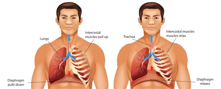
Respiratory Function
During inhalation, the diaphragm contracts and flattens, increasing the volume of the thoracic cavity and drawing air into the lungs. This action creates a negative pressure that facilitates airflow, highlighting the diaphragm’s primary role in respiration.
Exhalation occurs when the diaphragm relaxes, returning to its dome shape and pushing air out of the lungs. This rhythmic contraction and relaxation are essential for maintaining normal breathing and ensuring adequate oxygen supply to the body.
The diaphragm’s role in the breathing cycle underscores its importance as a primary respiratory muscle, facilitating the exchange of air and maintaining respiratory system function.
Non-Respiratory Functions
The diaphragm plays a key role in maintaining abdominal pressure, which is essential for bodily functions like urination and defecation. This pressure regulation is achieved through the diaphragm’s contraction and relaxation, highlighting its importance in various physiological processes.
The diaphragm also influences blood circulation by aiding the return of venous blood to the heart during its contraction. This function underscores the diaphragm’s role in maintaining cardiovascular health and facilitating efficient blood flow.
Knowing the diaphragm’s non-respiratory functions helps appreciate its overall role in maintaining health and well-being, beyond its primary role in respiration.
Clinical Relevance
The diaphragm is crucial for separating the thoracic cavity from the abdominal cavity and plays a significant role in respiration. Its importance is underscored by the various clinical conditions that can affect it, such as diaphragmatic hernia, phrenic nerve injury, and diaphragmatic paralysis.
Knowing the clinical relevance of the diaphragm helps appreciate its role in maintaining health and the potential impact of related disorders.
Diaphragmatic Hernia
Diaphragmatic hernias can be congenital or acquired, with congenital ones often requiring emergency surgical intervention. These hernias can lead to severe respiratory issues due to the abnormal positioning of organs within the thoracic cavity.
Congenital diaphragmatic hernia (CDH) is a defect where an abnormal opening in the diaphragm allows abdominal organs to move into the chest cavity, leading to underdeveloped lungs and respiratory distress in newborns. Acquired hernias, often resulting from trauma or surgery, can also cause significant health issues and require prompt treatment.
Knowing the causes, symptoms, and treatment options for diaphragmatic hernias helps appreciate the potential impact of this condition on respiratory health.
Phrenic Nerve Injury
Injury to the phrenic nerve can significantly impair diaphragmatic movement, leading to respiratory difficulties. This injury can result from trauma, surgery, or diseases affecting the central nervous system, highlighting the importance of the phrenic nerve in diaphragm function.
Diagnosis of phrenic nerve injury may involve imaging techniques such as ultrasound or MRI, along with nerve conduction tests to assess diaphragm function. These diagnostic tools are essential for identifying the extent of the injury and planning appropriate treatment.
Management of phrenic nerve injury may include supportive therapies, respiratory exercises, or surgical options in severe cases, underscoring the importance of early diagnosis and intervention.
Diaphragmatic Paralysis
Diaphragmatic paralysis often results from nerve injuries, trauma, or diseases affecting the central nervous system. This condition can lead to symptoms like shortness of breath, especially noticeable when lying down or engaging in physical activity.
Management of diaphragmatic paralysis may involve breathing exercises, respiratory therapy, or surgical options in severe cases. These interventions aim to improve respiratory function and quality of life for affected individuals.
Knowing the causes, symptoms, and management options for diaphragmatic paralysis helps appreciate its impact on respiratory health and the importance of early intervention.
Development and Structure
Diaphragm development involves contributions from the septum transversum, pleuroperitoneal folds, and somites. These structures play a crucial role in the formation of the diaphragm, ensuring its proper function within the body.
The septum transversum acts as an initial partition between the thoracic and abdominal cavities during diaphragm development, highlighting its importance in the early stages of formation. Muscle progenitors for the diaphragm originate from the cervical somites, specifically from segments C3 to C5, underscoring the complexity of its development.
Knowing the development and structure of the diaphragm provides insights into its formation and growth, highlighting the intricate processes involved in its morphogenesis.
Motor Innervation and Control
The phrenic nerve is crucial for diaphragm movement, originating from spinal nerves C3 to C5, and is responsible for its complete motor innervation. This innervation allows the diaphragm to perform its vital functions, particularly in the respiratory process.
Activation of the phrenic nerve leads to diaphragm contraction during inhalation, allowing for an increase in thoracic volume. This process is regulated by a complex neural network involving phrenic motor neurons, which are the final output for diaphragm muscle force generation.
Knowing motor innervation and control of the diaphragm helps appreciate how it operates within the body’s nervous system, highlighting the importance of the phrenic nerve in maintaining respiratory function.
Sensory Innervation and Proprioception
The distribution of muscle spindles in the diaphragm is sparse, with only 10 spindles identified, all located within the crural diaphragm. This sparse distribution highlights the minimal influence of diaphragmatic proprioception on breathing regulation.
Some afferents in the phrenic nerve originate from structures outside the diaphragm, such as the pericardium and liver, underscoring the complexity of its sensory innervation. Knowing sensory innervation and proprioception helps appreciate the diaphragm’s function and its role in maintaining respiratory health.
Diaphragm Function in Respiration
The diaphragm is primarily responsible for breathing by contracting rhythmically and involuntarily. During inhalation, the diaphragm flattens, resulting in an increase in chest cavity volume. When the diaphragm relaxes, it returns to its dome shape, which helps expel air from the lungs.
During inspiratory actions, diaphragm motor unit recruitment follows a size principle, where smaller, more excitable motor neurons are activated before larger ones. This recruitment pattern ensures efficient force generation during breathing, highlighting the diaphragm’s role as a primary respiratory muscle.
Knowing the diaphragm’s function in respiration helps appreciate its role in maintaining adequate oxygen levels in the body and facilitating normal breathing.
Diaphragm’s Role in Gastrointestinal Functions
During swallowing, the crural diaphragm temporarily relaxes to allow food passage, indicating a functional divergence from respiratory activity. This relaxation allows the esophagus to expand and accommodate food, highlighting the diaphragm’s role in digestion.
The crural diaphragm’s selective inhibition during esophageal distension may involve reflex pathways that operate independently of the brainstem.
Proper function of the diaphragm is critical in maintaining the pressure needed for the lower esophageal sphincter to prevent reflux.
Dysfunction of the diaphragm can lead to disruptions in esophageal motility and contribute to gastroesophageal reflux disease (GERD).
Increased abdominal pressure during diaphragm contraction aids in the effective propulsion of food through the digestive tract. Knowing the diaphragm’s role in gastrointestinal functions helps appreciate its overall contribution to increasing intra abdominal pressure and digestive health.
Common Diaphragm Issues and Disorders
Diaphragmatic paralysis occurs when the phrenic nerve is damaged, leading to an elevated diaphragm visible on imaging. This condition often leads to shortness of breath, especially noticeable when the individual is lying down or engaging in physical activity. Understanding this condition is crucial for diagnosing and treating respiratory difficulties effectively.
Cervical spinal cord injuries can lead to diaphragmatic paralysis, although the diaphragm can still function if the phrenic nerve remains intact. Congenital diaphragmatic hernias result from insufficient diaphragm development, allowing abdominal contents to enter the thoracic cavity. Assessing diaphragm function through ultrasound is beneficial for diagnosing conditions like diaphragm paralysis or phrenic nerve injury post-surgery.
Mutations affecting the extracellular matrix can lead to congenital diaphragmatic hernias by compromising structural integrity. Knowing these common issues and disorders helps appreciate the potential impact on respiratory health and the importance of early intervention.
Evolutionary Perspective of the Diaphragm
The diaphragm’s evolution reflects an adaptation to both respiratory needs and the demands of internal pressure regulation in larger vertebrates. In reptiles, the diaphragm’s partitioning function emerged to prevent internal organs from interfering with lung expansion during breathing. This adaptation highlights the diaphragm’s role in enhancing respiratory efficiency.
In some reptiles, adaptations evolved to stabilize lungs against the movements of abdominal organs, further enhancing respiratory efficiency. The physical partitioning created by the diaphragm enhances the efficiency of pressure generation in both thoracic and abdominal cavities.
Symmorphosis, a principle of biological design, suggests that the diaphragm’s structure is matched to its multiple physiological functions. Knowing the evolutionary perspective of the diaphragm provides insights into its development across species and its importance in maintaining respiratory and internal pressure functions.
Diaphragm in Medical Imaging and Surgery
Computed tomography (CT) and magnetic resonance imaging (MRI) are considered reference tests for diaphragm exploration. These imaging techniques allow for detailed assessment of diaphragm anatomy and function, making them crucial for diagnosing various conditions.
Ultrasound imaging is a key non-invasive technique that can be performed at the bedside, making it particularly useful in critical care settings. Fluoroscopy was traditionally used to analyze diaphragm function but poses risks due to radiation exposure. Diaphragmatic thickness measurements can indicate muscle weakness or paralysis, useful for various neuromuscular disorders.
Knowing diaphragm anatomy is crucial for accurate diagnosis and treatment in medical imaging and surgical interventions. Surgical repair options for diaphragm-related conditions often depend on accurate imaging to assess the degree of diaphragmatic dysfunction.
Recordings and Studies on Diaphragm Activity
Recent studies highlight the significant activity of motor neurons in diaphragm function. Motor units in the diaphragm exhibit homogeneous properties, meaning all muscle fibres within a unit share similar contractile characteristics, which is essential for consistent force output. These studies provide insights into the diaphragm’s role in various physiological processes.
During swallowing and esophageal distension, there is a dramatic divergence of activity between the crural and costal diaphragm, demonstrating complex functional responses. This divergence highlights the diaphragm’s ability to perform multiple roles simultaneously, such as aiding in digestion and maintaining respiratory function.
During the retching phase of vomiting, the diaphragm contracts strongly as a single muscle with abdominal muscles, increasing gastric pressure. Knowing these recordings and studies on diaphragm activity provides valuable insights into its complex functions and importance in maintaining overall health.


