
The extensor digitorum muscle enables finger extension, essential for tasks like typing and grasping. Located in the posterior forearm, it also aids in wrist movements. This article explores its anatomy, function, and clinical relevance.
Key Takeaways
The extensor digitorum muscle originates from the lateral epicondyle of the humerus and inserts on the dorsal surfaces of fingers 2-5, allowing for essential finger extension.
It is innervated by the posterior interosseous nerve, a branch of the radial nerve, which plays a crucial role in finger and wrist extension.
Injuries to the extensor digitorum, such as tendonitis or mallet finger, can severely affect hand function, highlighting the importance of rehabilitation and proper medical intervention.
Muscle Origin and Insertion
The extensor digitorum muscle originates from the lateral epicondyle of the humerus, which is a bony prominence on the outer side of the elbow. From there, its tendons extend down the forearm and insert on the dorsal surface of digits 2-5 via the extensor hood.
This configuration allows the muscle to control finger extension effectively, crucial for various hand movements.
Extensor Digitorum Muscle Diagram
Anatomical Relations
Positioned as the most superficial muscle of the posterior forearm, the extensor digitorum sits prominently under the skin. Lateral to this muscle are the extensor digiti minimi and extensor carpi ulnaris, while medial to it lies the extensor carpi radialis brevis.
Additionally, it is located superficially relative to deeper muscles such as the abductor pollicis longus and extensor pollicis longus. The tendons of the extensor digitorum occupy the fourth extensor compartment, ensuring its pivotal role in finger extension.
Innervation Details
The extensor digitorum muscle receives innervation from the posterior interosseous nerve, a branch of the radial nerve. This nerve travels through the forearm, providing motor impulses essential for muscle function.
Blood Supply Overview
The anterior tibial artery primarily supplies blood to the extensor digitorum muscle on the anterior surface, ensuring it receives the necessary oxygen and nutrients to maintain its function and health, supporting finger and wrist extension, along with the anterior tibial vessels.
Extensor Digitorum Overview
Located in the posterior forearm compartment, the extensor digitorum muscle extends the fingers. Originating from the lateral epicondyle of the humerus, it extends tendons to the medial four fingers, facilitating metacarpophalangeal and interphalangeal joint extension. This function is vital for daily activities like grasping and typing.
Beyond finger extension, the extensor digitorum also aids in wrist extension. This dual role highlights its importance in hand movements and dexterity. Innervated by the posterior interosseous nerve, a branch of the radial nerve, it ensures precise movement control. The posterior interosseous and radial recurrent arteries supply blood, maintaining muscle health and function.
Interestingly, the extensor digitorum has a lower limb counterpart, the extensor digitorum longus. This muscle extends the toes, crucial for walking and running. It is innervated by the deep peroneal nerve and supplied by the anterior tibial artery, emphasizing its importance in both upper and lower limb functionality.
Muscle Structure and Composition
The extensor digitorum muscle comprises multiple fibers organized into fascicles. These fibers generate the force required for muscle contraction.
Tendons, connective tissues that attach the muscle to bones, transmit force during muscle contractions, enabling finger and toe extension.
Tendons
Tendons are crucial for the extensor digitorum muscle’s function, attaching to the dorsal surfaces of the middle and distal phalanges, essential for digit extension. They pass through the inferior extensor retinaculum, a fibrous band that secures them, ensuring smooth movement.
The extensor digitorum longus in the lower limb also relies on tendons for toe extension, inserting into the distal phalanges to enable walking and running.
The seamless integration of tendons with the muscle fibers allows for efficient force transmission, highlighting the importance of tendons in both hand and foot movements.
Muscle Belly and Fascicles
The extensor digitorum’s muscle belly is dense with fibers organized into fascicles, crucial for generating contraction force. The orientation of these fibers affects the muscle’s range of motion and strength, enabling effective finger and toe extension.
The extensor digitorum’s fiber arrangement is optimized for function, allowing efficient contraction and facilitating digit extension. The muscle belly’s composition and fiber alignment ensure high efficiency in performing tasks.
Innervation of Extensor Digitorum
The posterior interosseous nerve, a branch of the radial nerve, primarily innervates the extensor digitorum muscle. This pathway is crucial for delivering motor impulses required for effective finger and toe extension.
Nerve Supply: Radial Nerve
The radial nerve is vital for the extensor digitorum muscle’s innervation. Originating from the brachial plexus’s posterior cord, it travels down the arm, entering the forearm deep to the radial artery. The posterior interosseous nerve, a deep branch, specifically innervates the extensor digitorum, providing necessary motor signals.
As the radial nerve progresses through the forearm, it ensures the extensor digitorum receives precise motor impulses for finger and wrist extension. This pathway highlights the radial nerve’s importance in upper limb extensor muscle functionality.
Motor and Sensory Innervation
The posterior interosseous nerve facilitates finger extension by innervating the extensor digitorum muscle, essential for various hand movements. Additionally, radial nerve sensory fibers provide sensation to the dorsal hand, allowing us to feel touch and pressure.
In the lower limb, the extensor digitorum longus receives motor innervation from the deep peroneal nerve, a branch of the common fibular nerve, supplying motor impulses for toe extension. Sensory fibers of the lumbar nerve L5 provide sensation to the skin over the muscle, highlighting the deep fibular nerve’s dual role in lower limb functionality.
Blood Supply to the Extensor Digitorum
The anterior and posterior interosseous arteries primarily supply blood to the extensor digitorum muscle, ensuring it receives the necessary oxygen and nutrients to maintain function and health, supporting finger and wrist extension.
Arterial Supply
The anterior and posterior interosseous arteries, branching from the common interosseous artery originating from the ulnar artery, primarily supply blood to the extensor digitorum muscle. The radial recurrent artery also contributes, ensuring adequate oxygen and nutrient supply.
The anterior and posterior interosseous arteries are crucial for maintaining the extensor digitorum muscle’s health and function. By providing a consistent blood supply, they ensure the muscle can perform effectively, supporting various hand movements.
Venous Drainage
Venous blood from the extensor digitorum muscle is primarily drained by deep veins accompanying the arteries, particularly the posterior interosseous vein. These veins efficiently return deoxygenated blood from the muscle to the heart, maintaining proper circulation.
In the lower limb, venous drainage of the extensor digitorum longus muscle is primarily through the anterior tibial vein, which accompanies the anterior tibial artery. Additional pathways include the fibular vein, collecting blood from the leg’s lateral compartment.
This comprehensive venous drainage system integrates with the superficial venous system of the foot, ensuring effective removal of deoxygenated blood from the muscle.
Functional Anatomy of the Extensor Digitorum
The extensor digitorum muscle extends the fingers and toes, significantly contributing to hand and foot movements. It works in synergy with other extensor muscles, ensuring coordinated and smooth movements for various activities.
Extension of the Digits
The extensor digitorum muscle extends the fingers at the metacarpophalangeal (MCP), proximal interphalangeal (PIP), and distal interphalangeal (DIP) joints. This extension is vital for functions like opening the hand, typing, and playing musical instruments.
In the lower limb, the extensor digitorum longus extends the toes at the metatarsophalangeal and interphalangeal joints, vital for walking and running. This allows the toes to lift off the ground, contributing to a smooth gait.
Coordinated Movements with Other Extensor Muscles
The extensor digitorum muscle collaborates with other extensor muscles to facilitate coordinated finger and wrist movements. It synergizes with the extensor indicis and extensor pollicis longus, ensuring smooth finger and thumb extension, crucial for activities requiring fine motor skills like writing and playing instruments.
In the lower limb, the extensor digitorum longus collaborates with the tibialis anterior and other muscles for efficient movement. This synergy is essential for walking, running, and balancing, ensuring stability and fluidity of movement.
Clinical Relevance and Disorders
Understanding the extensor digitorum muscle’s clinical relevance is vital for diagnosing and treating disorders. Issues like tendonitis, dysfunction, and weakness can significantly impact hand and foot function, hindering everyday tasks.
Tendon Injuries
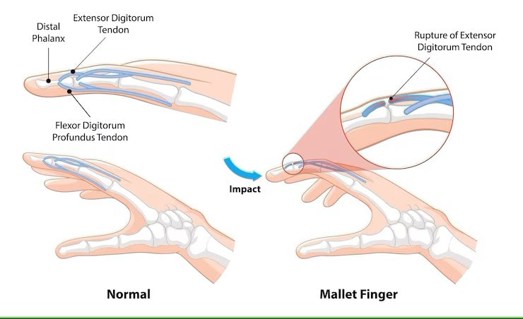
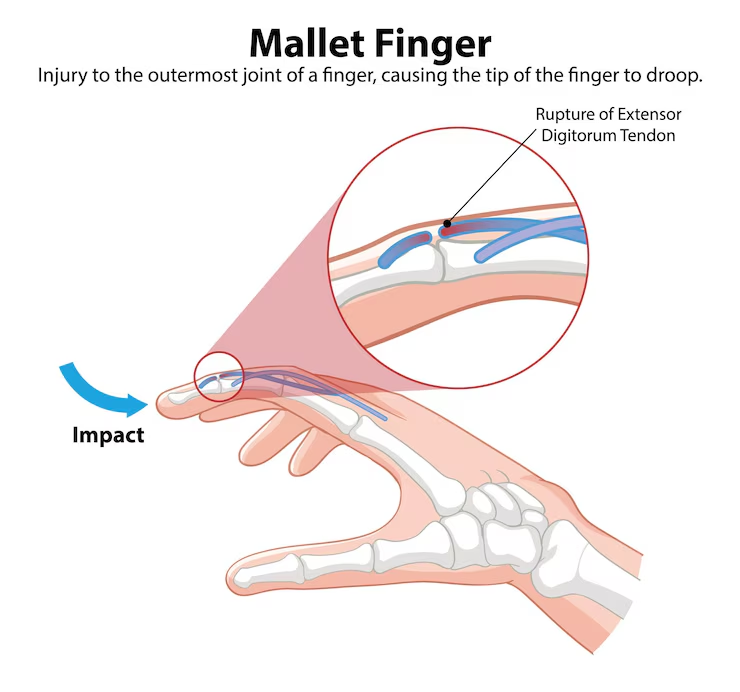
Tendon injuries can severely impact the extensor digitorum muscle. Conditions like mallet finger, where the tendon is injured or torn, can prevent full finger extension. Such injuries often result from deep cuts, overstretching, or blunt trauma. Non-surgical treatments, including splinting and hand therapy, can aid recovery, though they may require prolonged periods.
In the lower limb, injuries to the extensor digitorum longus tendon can lead to conditions like ‘drop foot,’ where the foot cannot dorsiflex. This condition severely hampers mobility and requires immediate medical attention to restore function and prevent further complications.
Dysfunction and Weakness
Dysfunction and weakness in the extensor digitorum muscle can stem from causes like radial nerve injury, leading to reduced digit extension or an inability to extend the fingers, significantly impacting hand function. Weakness in the extensor digitorum longus can be linked to conditions affecting the deep peroneal nerve, highlighting nerve health’s importance for muscle function.
Radial nerve injury significantly causes extensor digitorum dysfunction, leading to a diminished ability to extend the digits. This dysfunction can profoundly affect daily activities, necessitating timely medical intervention to restore normal function.
Extensor Tendonitis
Extensor tendonitis, often presenting with pain during active extension of the toes, is a common issue affecting the extensor digitorum muscle. Symptoms include inflammation, pain, and swelling around the affected tendon, especially after physical activity. This condition can significantly hinder hand and foot movements, making daily tasks challenging.
Management of extensor tendonitis typically involves rest, ice application, and anti-inflammatory medications. These treatments aim to reduce inflammation and alleviate pain, promoting healing and restoring normal function.
In severe cases, physical therapy may be necessary to strengthen the muscle and prevent recurrence.
Functional Role
The extensor digitorum muscle is crucial for extending the four medial fingers at both the metacarpophalangeal and interphalangeal joints. During the opening of the hand, it plays a vital role in counteracting the actions of finger flexors.
Its contraction also contributes to wrist extension while allowing the fingers to extend, highlighting its importance in hand and wrist movements.
Palpation Techniques
To palpate the extensor digitorum muscle, locate the dorsal surface of the forearm and feel for muscle contraction while the fingers extend. Positioned between the extensor carpi radialis brevis and extensor digiti minimi, the tendons of the extensor digitorum pass deep to the extensor retinaculum.
Assessing for abnormalities involves checking for tenderness or asymmetry during active finger extension.
Clinical Significance
Injuries to the extensor digitorum, such as mallet finger, can lead to an inability to straighten fingers or extend the wrist, severely impacting hand function. Treatment often involves splinting and may include surgical repair to restore function.
These injuries underscore the clinical importance of the extensor digitorum in maintaining hand and wrist mobility.
Injury on Digitorum Muscle Diagram
Strengthening and Rehabilitation
Physical therapy is crucial after extensor tendon injuries to improve motion and enhance functional recovery. Rehabilitation techniques focus on exercises that strengthen the extensor digitorum muscle, helping to restore hand and finger function and prevent future injuries.
Comparative Anatomy
The extensor digitorum muscle in humans originates from the lateral epicondyle of the humerus and inserts on the distal phalanges of digits 2-5 via the extensor hood. Its tendons extend towards the fingers, enabling finger extension and playing a key role in hand movements, particularly involving the musculus extensor digitorum.
In contrast, the extensor digitorum longus, originating from the lateral condyle of the tibia and the medial surface of the fibula, inserts onto the dorsal surfaces of the lateral four toes, facilitating toe extension. Both muscles work together to enhance functional mobility in the upper and lower limbs.
While the extensor digitorum longus has a long tendon structure, the extensor digitorum brevis consists of short muscular slips, highlighting the anatomical differences that contribute to their respective functions. This comparative anatomy underscores the evolutionary adaptations that optimize muscle function for specific tasks


