The humerus is the long bone in your upper arm, running from shoulder to elbow. It’s crucial for arm movement and muscle attachment. In this guide on humerus anatomy, you’ll learn about its location, structure, and the key features that enable a wide range of arm movements.
Key Takeaways
The humerus is the largest bone in the upper arm, crucial for shoulder and arm movement, connecting the shoulder and elbow joints.
Key features of the humerus, like the head, anatomical neck, and tuberosities, are vital for shoulder stability and muscle attachment.
Common humerus fractures occur in three areas: proximal, mid-shaft, and distal, with treatment often involving immobilization or surgery.
Overview of the Humerus
The humerus is a long bone found in the upper arm. It extends from the shoulder to the elbow. Measuring about one foot in length, it is the largest bone in the upper extremity. This single bone is responsible for supporting numerous muscles, tendons, and ligaments that are essential for arm movement. Its primary function is to facilitate the movement of the shoulder and arm, playing a crucial role in stabilizing the elbow and hand.
More than just a support structure, the humerus is a central player in the complex ballet of arm movements. It connects with the shoulder joint at its proximal end and the elbow joint at its distal end, making it indispensable for a wide range of motions.
The humerus consists of the proximal humerus, the shaft, and the distal humerus, each with its own distinct features and functions.
Diagram of Humerus
Proximal Humerus Anatomy
The proximal humerus is the upper part of this long bone and is packed with features that are vital for its function. It consists of:
the rounded head
narrow anatomical neck
surgical neck
two tuberosities-the greater and lesser tuberosities
These features play a significant role in forming the shoulder joint and providing attachment points for muscles.
The head of the humerus connects with the glenoid fossa of the scapula. This connection creates the shoulder joint. Let’s dive deeper into each of these features.
Head of the Humerus
The head of the humerus is shaped like an irregular hemisphere, perfectly designed to fit into the glenoid fossa of the scapula. This ball-and-socket joint, known as the glenohumeral joint, allows for a wide range of shoulder movements. The head of the humerus serves as the proximal articular surface, playing a crucial role in shoulder joint movement.
This articulation is not just about movement; it’s also about stability. The head of the humerus, along with the glenoid fossa, ensures that the shoulder joint remains stable while allowing for flexibility. This balance between stability and mobility is what makes the shoulder one of the most versatile joints in the human body.
Anatomical Neck
Located just below the head of the humerus, the anatomical neck serves as a critical site for attaching the joint capsule of the shoulder joint. This narrow region plays a significant role in maintaining the stability of the shoulder joint by anchoring the joint capsule.
Greater and Lesser Tuberosities
The greater and lesser tuberosities are bony projections situated below the humeral head. The greater tuberosity is positioned laterally and serves as an attachment point for three rotator cuff muscles. In contrast, the lesser tuberosity is located anteriorly and provides attachment for the subscapularis muscle.
These tuberosities are crucial for shoulder stability and movement, facilitating muscle attachment and action.
Shaft of the Humerus
The shaft of the humerus is cylindrical in its upper portion, gradually transitioning to a prismatic shape as it descends. This section of the bone also becomes triangular as it approaches the distal end. The shaft supports the attachment of various muscles and acts as a conduit for nerves and blood vessels.
Mid-shaft humerus fractures, typically caused by direct impacts, are a common type of injury in this region.
Deltoid Tuberosity
The deltoid tuberosity is a rough triangular area located on the anterolateral surface of the humeral shaft. This feature serves as the attachment point for the deltoid muscle, one of the primary muscles responsible for shoulder abduction. The deltoid muscle plays a pivotal role in lifting the arm away from the body, making the deltoid tuberosity an essential feature for upper limb movement.
In addition to the deltoid muscle, the humeral shaft features three distinct surfaces: anterolateral, anteromedial, and posterior surface. These surfaces, along with the anterior surface, medial, and lateral borders, provide attachment points for various muscles and ligaments, contributing to the overall functionality and stability of the upper arm.
Radial Groove
The radial groove is a shallow groove that runs obliquely down the posterior aspect of the humeral shaft. This groove is important due to its role as a passageway. It accommodates both the radial nerve and the profunda brachii artery. The radial nerve, in particular, is crucial for the innervation of the posterior compartment of the arm and forearm, impacting wrist and finger extension.
Given its importance, the radial groove is a critical landmark in the anatomy of the upper arm. Injuries to this area, such as radial nerve palsy, can have significant implications, often resulting from fractures or trauma to the mid-shaft of the humerus.
Distal Humerus Anatomy
The distal humerus is characterized by its two prominent epicondyles, the trochlea, and the capitulum, along with three fossae. These features are crucial for the articulation and movement of the elbow joint. The medial and lateral epicondyles serve as attachment points for muscles involved in forearm movement, while the trochlea and capitulum facilitate elbow flexion and extension.
Medial and Lateral Epicondyles
The medial and lateral epicondyles are prominent features of the distal humerus. The medial epicondyle is larger and serves as a key attachment site for several forearm flexor muscles. This epicondyle extends more distally compared to its lateral counterpart, making it a notable landmark in the anatomy of the elbow.
The lateral epicondyle, while smaller, is equally important. It serves as an attachment point for muscles that extend the forearm. Together, these epicondyles play a crucial role in the movement and stability of the elbow joint, facilitating various forearm actions.
Trochlea and Capitulum
The trochlea and capitulum are central features of the distal humerus’ condyle. The trochlea is positioned medially and articulates with the ulna, facilitating elbow flexion and extension. On the other hand, the capitulum connects with the radius, contributing to the elbow joint’s overall function.
Fossae
The distal humerus features three fossae: the olecranon fossa, coronoid fossa, and radial fossa. These fossae accommodate the corresponding processes of the ulna and radius during elbow movements, enabling smooth flexion and extension.
The olecranon fossa, in particular, accommodates the ulna’s olecranon process during elbow extension.
Blood Supply and Innervation
The humerus receives its blood supply primarily from the brachial artery, which branches out to nourish the bone. The humeral head primarily receives its blood supply from the posterior humeral circumflex artery. This artery plays a significant role in vascularization of the area. Additionally, the profunda brachii artery runs alongside the radial nerve within the radial groove, providing critical nourishment to this region.
In terms of innervation, the axillary nerve is responsible for the sensory innervation of the lateral aspect of the shoulder. The musculocutaneous nerve primarily innervates the biceps brachii and other flexor muscles in the arm.
The radial nerve, which innervates the posterior compartment of the arm and forearm, is crucial for wrist and finger extension. Other important nerves include the ulnar nerve and median nerves, each providing motor and sensory innervation to various parts of the arm and hand.
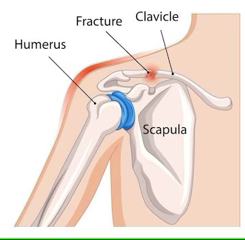
Common Humerus Fractures
Humerus fractures are commonly classified by their location-proximal, mid-shaft, or distal. The proximal humerus is particularly prone to fractures, often resulting from trauma such as falls, car accidents, or sports injuries. Treatment can vary from immobilization with a splint or cast to surgical intervention for realignment.
Let’s explore these fractures in more detail.
Proximal Humerus Fractures
Proximal humerus fractures typically occur near the shoulder joint, affecting the upper part of the bone. These fractures often require surgical intervention to realign the bone fragments, especially if they are displaced.
The radial nerve, which routes through the spiral groove of the humerus, is susceptible to injury in these cases.
Mid-Shaft Humerus Fractures
Mid-shaft humerus fractures are usually caused by a direct blow to the upper arm. The initial treatment involves using a splint or sling to realign the bones and reduce movement.
These fractures typically take up to four months to heal, depending on the severity and the individual’s overall health.
Distal Humerus Fractures
Distal humerus fractures often result from direct impacts or falls, affecting the lower part of the bone near the elbow joint. These fractures can be particularly challenging to treat due to the complex anatomy of the distal humerus. Surgical treatment often involves open reduction and internal fixation to realign the bone fragments.
Recovery from distal humerus fractures can be lengthy, sometimes taking up to a year to fully heal. The severity of the fracture and the individual’s overall health can significantly impact the recovery time.
Clinical Relevance
Understanding the clinically oriented anatomy of the humerus is crucial for diagnosing and treating injuries and conditions related to this bone. Common issues include fractures, osteoporosis, and nerve or muscle damage. For instance, humerus fractures can occur due to significant trauma such as falls or car accidents, often requiring several months for recovery.
Radial nerve injury is a common complication associated with humeral fractures, especially in the distal third of the humerus. Metastatic disease can also lead to pathologic fractures, necessitating careful management. Understanding these clinical aspects can help in providing better treatment and care for patients.