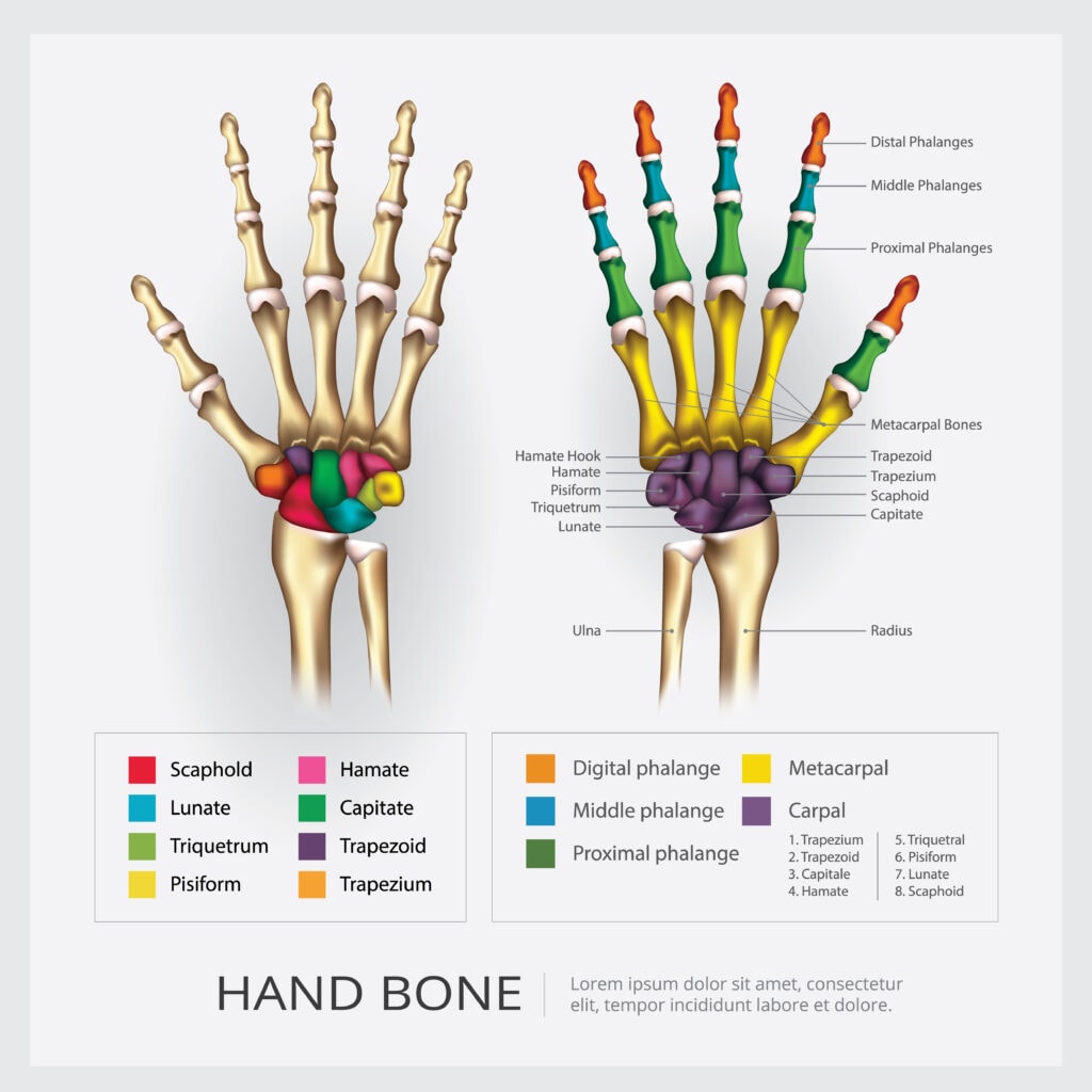
The hand has 27 bones, known as the hand bones, crucial for movement and dexterity. Understanding these bones helps in comprehending hand function. This guide explores each bone diagram, from carpal bones in the wrist to the phalanges in the fingers, detailing their roles and significance.
Key Takeaways
- The human hand consists of 27 bones categorized into carpal, metacarpal, and phalangeal groups, all contributing to its functionality and dexterity.
- Injuries like fractures commonly affect specific bones such as the scaphoid and fifth metacarpal, emphasizing the importance of prompt treatment and rehabilitation.
- Post-surgical care and rehabilitation, including physical therapy, are crucial for restoring hand function after injuries or surgeries, highlighting the need for comprehensive recovery plans.
Overview of Hand Bones
The human hand is an intricate structure composed of 27 bones, each serving a distinct function. These bones are integral to the hand’s functionality, allowing for a range of movements including flexion, rotation, and extension.
They provide structural support and shape, anchoring other anatomical structures such as muscles, tendons, and ligaments. Grouped by location and function, these bones collectively contribute to the hand’s impressive range of motion and dexterity.
From the robust carpal bones forming the foundation of the wrist to the slender phalanges enabling fine motor skills, each bone is a vital component in the symphony of hand movements.
This overview sets the stage for a deeper exploration of the individual groups of bones and their unique contributions to hand function.
Carpal Bones: The Foundation of the Wrist
The carpal bones form a crucial part of the wrist anatomy, consisting of eight small bones arranged in two rows.
These wrist bones are essential for wrist flexibility and stability, allowing for a wide range of movements.
The proximal row of carpal bones consists of the scaphoid, lunate, triquetrum, and pisiform. In contrast, the distal row includes the trapezium, trapezoid, capitate, and hamate bones.
These eight carpal bones play a pivotal role in the overall function of the hand and wrist. They facilitate the complex sliding movements among the bones, which are essential for wrist motion and stability.
Scaphoid Bone
The scaphoid bone is a unique carpal bone that serves as a link between the proximal and distal rows, making it a crucial element in wrist biomechanics.
The most commonly fractured carpal bone is often injured. This typically occurs due to a fall on an outstretched hand (FOOSH). Scaphoid fractures are characterized by pain and tenderness in the anatomical snuffbox and are generally treated with cast immobilization.
Due to its unique blood supply, a scaphoid fracture can lead to avascular necrosis, particularly affecting the proximal pole. This complication can have long-term consequences, including the risk of developing osteoarthritis of the wrist.
Lunate Bone
The lunate bone is centrally positioned in the proximal row of carpal bones and plays a critical role in wrist movement.
Its central location makes it pivotal for the overall function of the wrist, contributing to the smooth and coordinated movements necessary for various hand activities.
However, the lunate bone is susceptible to conditions such as Kienböck’s disease, where its blood supply is compromised, leading to bone deterioration.
Metacarpal Bones: Connecting the Wrist to the Fingers
The metacarpal bones are the skeletal connectors between the wrist and the fingers, comprising five long bones that contribute to the hand’s structure and function. Each metacarpal bone has a base, shaft, and head, and corresponds to the digits: thumb, index, long, ring, and small fingers.
These bones articulate proximally with the carpal bones and distally with the proximal phalanges, facilitating a range of hand movements.
These five metacarpal bones are crucial for hand function, providing the framework for finger mobility and strength.
First Metacarpal Bone
The first metacarpal bone is essential for the thumb’s mobility, playing a vital role in hand function. Its saddle-shaped articulation allows for a wide range of thumb movements, crucial for grasping and pinching actions.
Common issues related to the first metacarpal bone include Bennett’s fracture, which impacts thumb function.
Fifth Metacarpal Bone
The fifth metacarpal bone, often referred to as the bone of the little finger, plays a significant role in grip strength and is frequently involved in injuries sustained during contact sports. Boxer’s fractures predominantly occur at the neck of the fifth metacarpal and are common among individuals engaged in activities involving punching.
Phalanges: The Finger Bones
The phalanges are the finger bones of the hand, comprising a total of 14 bones organized into three types: proximal, middle, and distal. Each finger contains three phalanges, except for the thumb, which has two. These bones are critical for finger movement, dexterity, and fine motor skills.
Proximal Phalanges
The proximal phalanges are critical in enabling finger movement as they connect to the metacarpals to facilitate articulation. They form metacarpophalangeal (MCP) joints with their respective metacarpals, allowing for a range of movements including flexion and extension.
Common injuries to proximal phalanges include fractures and dislocations, often resulting from sports or accidents. Proper treatment and rehabilitation are essential to restore full functionality and prevent long-term complications.
Distal Phalanges
The distal phalanges feature flat surfaces that support the fingertips, including the nail beds. They are critical for fine motor skills and tactile sensation at the fingertips, enabling precise movements and sensory feedback.
These bones play a vital role in daily activities, from typing on a keyboard to playing musical instruments.
Joints of the Hand
The hand consists of three main types of joints: Carpometacarpal (CMC), Metacarpophalangeal (MCP), and Interphalangeal (IP) joints, each serving distinct functions.
The carpometacarpal joints are formed by the connection of the metacarpal bones to the carpal bones, facilitating thumb mobility and grasping movements.
Proximal phalanges are crucial for forming the metacarpophalangeal joints, enabling finger flexibility and allowing for flexion and extension. Ligaments are fibrous tissues that stabilize and connect the joints in the hand, ensuring proper function and preventing dislocations.
Carpometacarpal Joint
The carpometacarpal joint of the thumb is unique as it allows a greater range of motion compared to other CMC joints. This joint is crucial for thumb opposition, enabling a wide range of hand movements essential for grasping and manipulating objects.
The range of motion at the carpometacarpal joint varies between fingers, with the small finger allowing for more movement than the index finger. Understanding this joint’s anatomy and function is key to appreciating the thumb’s versatility.
Proximal Interphalangeal Joint
The proximal interphalangeal (PIP) joint plays a key role in flexing and extending fingers during hand functions. Common injuries to the PIP joint include dislocations or fractures, impacting finger mobility.
The PIP joint enables the bending and straightening of the fingers and can become stiff after an injury. Proper treatment and rehabilitation are essential to restore full functionality and prevent long-term stiffness.
Tendons and Ligaments
Tendons and ligaments are crucial for hand and wrist movement and stability. The hand contains 34 muscles that contribute to various movements, including the intrinsic and extrinsic muscles. The intrinsic muscles found in the hand include the thenar, hypothenar, lumbrical, and interossei groups.
Ligaments are fibrous tissues that stabilize and connect the joints in the hand, ensuring proper function and preventing dislocations.
Flexor Tendons
Flexor tendons play a crucial role in bending the fingers and wrist. Injuries to these tendons can lead to significant functional impairment in hand movement. Tendon repair is critical for restoring function after avulsion injuries, particularly of the flexor tendon digitorum profundus.
Surgical intervention is necessary when tendons are completely ruptured or cut, impacting hand movement. Proper treatment and rehabilitation are essential to restore full functionality and prevent long-term complications.
Extensor Tendons
Extensor tendons are essential for straightening the fingers and wrist. Common injuries to extensor tendons can occur from overstretching or trauma, affecting hand function. Understanding the role of extensor tendons helps in recognizing their importance in hand function and the impact of injuries.
Proper treatment and rehabilitation are essential to restore full functionality and prevent long-term complications.
Hand Bones Diagram
Blood Supply to the Hand
The blood supply to the hand is primarily provided by the ulnar and radial arteries. These arteries ensure that the hand receives the necessary nutrients and oxygen to function properly.
The radial artery primarily supplies the lateral aspect of the hand, while the ulnar artery primarily supplies the medial aspect.
Radial Artery
The radial artery plays a crucial role in supplying blood to the lateral aspect of the hand and fingers. It enters the hand and contributes to both the superficial and deep palmar arches.
Ulnar Artery
The ulnar artery divides into deep and superficial palmar branches, forming palmar arches. It primarily supplies the rest of the digits and the medial side of the index finger, along with the radial and ulnar arteries and ulnar arteries.
The ulnar artery is essential for proper circulation in the hand. Recognizing its role underscores the need for proper treatment and rehabilitation to restore full functionality and prevent complications.
Nerve Supply to the Hand
The main nerves that provide sensation to the hand and wrist are the median, ulnar, and radial nerves. These nerves have both sensory and motor components, contributing to the hand’s overall function.
Median Nerve
The median nerve descends along the arm and enters the hand through the carpal tunnel. It is responsible for sensory input from the thumb, index finger, middle finger, and half of the ring finger.
The median nerve also innervates the thenar muscles, crucial for thumb movements and grip. Recognizing its role underscores the need for proper treatment and rehabilitation to restore full functionality and prevent complications.
Ulnar Nerve
The ulnar nerve is responsible for power grasp and fine motor control in the hand. It provides nerve supply to the dorsal and volar ulnar side of the fourth digit. Additionally, it also innervates the fifth digit.
The motor branch of the ulnar nerve innervates the flexor carpi ulnaris and flexor digitorum profundus muscles.
Common Injuries and Conditions
Common injuries and conditions affecting the hand include fractures and arthritis. The scaphoid bone is the most frequently fractured among the carpal bones, often due to falls on an outstretched hand. Injury to the scaphoid can lead to significant complications, including the risk of avascular necrosis due to its unique blood supply.
Conditions like arthritis can significantly limit the functionality of the carpometacarpal joint, leading to pain and decreased grip strength.
Fractures
A Bennett fracture occurs at the base of the first metacarpal and involves an intra-articular fracture resulting from forced abduction. Bennett’s fracture is a specific type of fracture affecting the base of the first metacarpal, often caused by axial loading.
A Boxer’s fracture typically refers to a fracture of the fifth metacarpal neck, common due to punching. Injuries to the fifth metacarpal often result in dorsal angulation and may lead to ‘pseudo-clawing’ if severe.
Arthritis
Rheumatoid arthritis in the hand can cause characteristic deformities such as swan neck and boutonniere deformities, as well as ulnar deviation of digits. Pain and stiffness in the joints of the hand are typical symptoms of arthritis that can worsen with activity.
Osteoarthritis in the hand typically leads to joint stiffness and pain, especially after periods of inactivity.
Surgical Considerations
Surgical interventions for severe arthritis may involve joint fusion or reconstruction to alleviate symptoms. Consideration of surgery is warranted for severe injuries or conditions that impact function significantly.
The anatomical relationship between nerves, tendons, ligaments, and blood vessels must be considered when designing an incision for hand and wrist surgery.
Fracture Fixation
Treatment for severe hand fractures may involve surgical intervention to realign broken bones. Surgical intervention for metacarpal fractures is recommended if there’s any malrotation affecting function.
Surgical fixation for hand fractures may involve the use of plates and screws or intramedullary rods.
Tendon Repair
Extensor tendons are generally easier to access and repair than flexor tendons during surgery. Tendon repair is a significant surgical procedure utilized to restore function and mobility after tendon injury. Surgical techniques for tendon repair involve precise methods that can vary depending on the tendon type and location.
Successful tendon repair can lead to improved hand function and reduced pain for patients.
Rehabilitation and Recovery
Post-surgery care for hand injuries typically emphasizes the importance of following a prescribed rehabilitation plan to ensure optimal healing. Proper post-surgical care includes maintaining a balanced diet enriched with calcium and vitamin D to support bone healing.
Physical Therapy
Physical therapy focuses on restoring strength and flexibility to enhance hand functionality post-injury. Recovery from tendon repair surgery may demand up to three months for the tendon to regain strength.
Exercises like wrist flexion and extension, thumb movements, and finger pinching are essential for regaining strength and dexterity in the hand after injury. Incorporating resistance training, such as ball squeezes and finger lifts, can enhance strength and dexterity during recovery.
Using therapy putty or balls can enhance grip strength and finger dexterity as part of a hand recovery exercise routine.
Exercises such as wrist flexion and extension help improve the range of motion and strength in the hand during rehabilitation. Exercises like wrist supination and finger tendon glides are essential for regaining hand mobility post-injury. Hand fractures may heal in about 6 to 8 weeks, depending on the severity and location of the fracture.
Post-Surgical Care
Post-surgical care is essential to monitor healing and prevent complications following hand surgeries. Complications such as inflammation or infection can arise post-surgery, necessitating further treatment.
Proper post-surgical care for hand injuries includes managing swelling, protecting the surgical site, and gradually increasing hand movement to aid healing. Post-surgical care for hand surgery typically includes managing swelling through elevation and ice application, alongside prescribed pain medications.
Final Thoughts
It will highlight the critical roles of bones, joints, tendons, ligaments, blood supply, and nerve supply in maintaining hand function and health.
The conclusion will inspire readers to appreciate the complexity of the hand and encourage them to take proactive steps in maintaining hand health, whether through proper care, rehabilitation, or seeking medical attention when necessary.
Frequently Asked Questions
What are the main bones in the human hand?
The main bones in the human hand are the carpal bones, metacarpal bones, and phalanges, totaling 27 bones. These structures are essential for the hand’s diverse movements and functionality.
What is the role of the scaphoid bone in the hand?
The scaphoid bone is vital for connecting the proximal and distal rows of carpal bones, playing a crucial role in wrist movement and stability. Its importance is underscored by the fact that it is the most commonly fractured carpal bone.
How do the radial and ulnar arteries supply blood to the hand?
The radial artery supplies blood to the lateral aspect of the hand, while the ulnar artery serves the medial aspect. Together, they ensure the hand receives essential nutrients and oxygen for proper function.
What are common injuries that affect the hand?
Common injuries that affect the hand include fractures, such as Bennett’s and Boxer’s fractures, along with conditions like arthritis. These injuries can severely impair hand functionality and necessitate appropriate treatment and rehabilitation.
What is the importance of post-surgical care for hand injuries?
Post-surgical care for hand injuries is crucial for monitoring healing, preventing complications, and ensuring a proper recovery. Effective management of swelling and protection of the surgical site, along with gradual movement, facilitates optimal healing.