
Cutaneous myiasis occurs when fly larvae burrow into the skin, creating painful, inflamed lesions. While unsettling, proper removal techniques minimize complications. Initial signs include itching, swelling, and a small punctum where the maggot breathes. Risk factors include poor wound care, tropical climates, and exposure to infected flies. Safe extraction methods range from suffocating larvae with petroleum jelly to careful surgical removal.
Cutaneous Myiasis and Its Symptoms
How does a tiny fly larva end up burrowing into human skin? Cutaneous myiasis occurs when flies, like Dermatobia hominis, lay eggs on the skin or nearby surfaces. Initially, the larvae hatch, they burrow into the skin, creating painful, itchy swellings.
These lesions often have a small opening, allowing the larva to breathe and move—visible signs of infestation.
Symptoms include red, inflamed bumps with a central punctum, sometimes accompanied by a crawling sensation or mild fever. The discomfort can range from mild irritation to sharp pain, depending on the larvae’s location.
While cutaneous myiasis is more common in tropical areas, travelers or those exposed to flies risk infection. Recognizing these signs promptly helps guarantee proper treatment and prevents complications like secondary infections. Immediate removal of the larvae is key to recovery.
Identifying Risk Factors for Myiasis Infestation
The humid, buzzing air of tropical regions isn’t just uncomfortable—it’s a breeding ground for flies that cause myiasis. Risk factors for infestation include residing in warm climates, where flies thrive, or rural areas with limited sanitation.
People with chronic conditions like diabetes or poor circulation face higher risks because open wounds attract flies. Poor hygiene, homelessness, or substance abuse can also increase vulnerability, as neglected skin or unclean environments invite infestation.
The head, neck, and lower limbs are common targets, especially if left exposed or injured. Comprehension of these risk factors helps prevent myiasis by addressing hygiene, wound care, and environmental controls. Awareness of personal and regional risks empowers individuals to take protective steps against this painful condition.
Types of Myiasis and Their Clinical Presentation
Different types of myiasis present with distinct symptoms and affect specific areas of the body. Cutaneous myiasis often appears as inflamed, boil-like lesions with small openings where larvae breathe.
Other forms, like wound or creeping myiasis, cause intense discomfort as maggots move through tissue or infest open sores.
Cutaneous Myiasis Symptoms
Painful, itchy, and often alarming, cutaneous myiasis occurs as fly larvae burrow into the skin, creating inflamed lesions that can resemble boils or infected wounds. The most common symptoms include swelling, redness, and a sensation of movement under the skin, sometimes accompanied by pus or discharge.
In wound myiasis, open sores attract flies, which lay eggs that hatch into maggots, worsening tissue damage and increasing infection risk. Creeping myiasis causes a distinctive crawling feeling as larvae migrate, leaving winding tracks under the skin. Patients may also notice small breathing holes where larvae surface for air.
While these symptoms can be distressing, proper identification and treatment typically prevent complications. Recognizing early signs facilitates timely medical intervention and reduces discomfort.
Common Myiasis Infestation Sites
Because myiasis can affect various parts of the body, the infestation sites often determine the type and severity of symptoms. Furuncular myiasis, for example, typically appears as raised, boil-like lesions on the skin, often with a visible opening where the larva breathes.
Wound myiasis targets open sores or decaying tissue, while creeping myiasis causes itchy, tunnel-like tracks as larvae move beneath the skin. Body cavity myiasis is more severe, involving the eyes, ears, or nasal passages, leading to pain and potential complications. Accidental myiasis occurs when larvae are ingested, usually through contaminated food.
Each type presents distinct challenges, making timely identification crucial. Recognizing these sites helps in choosing the right removal method and preventing further discomfort or infection.
Noninvasive Treatment Techniques for Maggot Removal
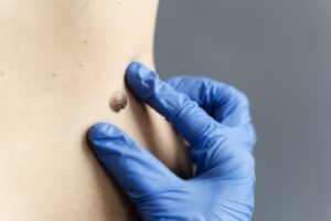
While maggots infest a wound, noninvasive removal methods can often resolve the issue without surgery. Treatments like petroleum jelly or beeswax, as noted by et al, cover the punctum, coaxing larvae out naturally over hours. A wide variety of substances, including liquid paraffin or bacon strips, can help without causing further harm. Gentle pressure or tweezers might assist if larvae resist. Covering the wound loosely avoids suffocating the larva, ensuring it emerges head-first.
| Method | Application | Timeframe |
|---|---|---|
| Petroleum jelly | Cover punctum | 3–24 hours |
| Bacon strips | Place on wound | 6–12 hours |
| Liquid paraffin | Apply lightly | 4–18 hours |
| Tweezers | Gently grasp | Immediate |
| Loose covering | Avoid restriction | Until emergence |
Enlarging the punctum slightly could ease removal for stubborn cases. These techniques minimize discomfort while encouraging safe extraction.
Surgical Removal of Larvae: When Local Anesthesia Is Needed
Some wounds hold stubborn larvae that refuse to loosen their grip despite gentle removal efforts. In these cases, emergency medicine might require surgical intervention under local anesthesia.
Lidocaine is often injected around the punctum to numb the area and help extrude deeply embedded larvae. A small 4-5 mm punch excision of the punctum and surrounding skin improves access for removal. Toothed forceps are then used carefully to extract the larvae without tearing or damaging them.
This method is reserved whenever non-invasive techniques fail, ensuring minimal discomfort and reduced risk of infection. The procedure is quick, precise, and typically performed in clinical settings to address severe or persistent myiasis. Proper wound care afterward promotes healing and prevents complications.
The Role of Ivermectin in Myiasis Treatment
As larvae burrow deep into the skin and resist removal, ivermectin offers a non-invasive alternative to surgical intervention. This antiparasitic medication targets the nervous system of larvae, making it effective for treating varieties of myiasis, including cutaneous and cavitary forms.
Oral or topical use: Depending on severity, ivermectin can be taken as a pill or applied directly to infested areas.
Reduces inflammation: It eases swelling and discomfort caused by larval activity.
Kills embedded larvae: Works systematically to eliminate parasites without manual extraction.
Prevents spread: Stops larvae from migrating deeper or causing secondary infestations.
Ivermectin is particularly useful for extensive or hard-to-access infestations, offering relief when physical removal isn’t viable. Always follow medical guidance to guarantee safe and effective treatment.
Managing Wound Myiasis and Secondary Infections
Effective maggot extraction techniques involve irrigation and topical agents like chloroform or ether, guaranteeing minimal trauma to the tissue. Controlling secondary infections requires antibiotics should bacterial complications arise, alongside proper wound care.
Consulting specialists verifies appropriate management based on the infection’s location and severity.
Maggot Extraction Techniques
Though maggots can appear unsettling in a wound, they frequently can be extracted securely with suitable techniques to avert additional difficulties. Fly larvae removal requires care to minimize discomfort and prevent further tissue damage.
- Manual Extraction: Gently use tweezers to remove visible maggots, ensuring the wound is cleaned afterward.
- Irrigation: Flushing the wound with saline or sterile water can dislodge larvae and cleanse the area.
- Occlusion Method: Applying petroleum jelly or adhesive tape over the wound smothers maggots, forcing them to surface for easier removal.
- Natural Remedies: A paste of turmeric or honey might help expel larvae while soothing the skin.
Each method should be performed with clean hands or sterile tools to reduce infection risk. Should maggots be deeply embedded, medical assistance is recommended to avoid complications.
Infection Control Measures
After safely removing maggots from a wound, the next step is confirming the area stays clean and free from infection. Proper disposal of maggots in sealed containers averts reinfestation.
Dilute hydrogen peroxide can help cleanse the wound, but thorough irrigation with saline is key to removing debris. Debridement removes dead tissue, reducing infection risks. Monitoring heart rate helps detect signs of systemic infection, like fever or increased pulse.
Antibiotic ointments or prescribed medications could be needed if bacteria enter the wound. Keeping the area dry and covered with sterile dressings prevents contamination. Follow hospital protocols for handling maggots to avoid cross-contamination.
Watch for redness, swelling, or pus, which signal infection. Prompt medical attention guarantees proper healing and prevents complications.
Safe Disposal of Removed Maggots and Biohazard Considerations
Since maggots removed from cutaneous myiasis can carry harmful bacteria, proper disposal is critical to prevent contamination. Scientific research confirms that mishandling maggots can spread infections, so following biohazard protocols is essential.
Seal maggots in a leak-proof container to avoid accidental exposure to bacteria.
Use biohazard bags or sharps containers for disposal, as they’re designed for infectious waste.
Wear gloves and protective gear whenever handling maggots to minimize direct contact.
Never flush maggots down drains—this can lead to environmental spread and reinfestation.
Healthcare facilities follow strict guidelines for biohazard waste, but even at home, these steps help reduce risks. Proper disposal protects both people and the environment, ensuring maggots don’t cause further harm. Following these measures keeps everyone safe while addressing the issue effectively.
Key Precautions to Prevent Reinfestation and Complications
Preventing reinfestation and complications after maggot removal requires proper wound care to avoid infection and promote healing.
Maintaining environmental hygiene, such as keeping abodes clean and free of decaying matter, reduces the risk of attracting flies.
Follow-up medical monitoring verifies any lingering issues are addressed promptly, safeguarding against further complications.
Proper Wound Care
While maggot removal is the initial step in treating cutaneous myiasis, proper wound care afterward is just as critical to guarantee, verify, and ascertain reinfestation and complications do not occur. Effective wound healing relies on meticulous attention to cleanliness and monitoring.
- Clean thoroughly: Remove all maggots and dead tissue to eliminate sources of reinfestation.
- Apply antimicrobials: Use topical treatments to prevent bacterial infections that slow recovery.
- Monitor closely: Watch for redness, swelling, or pus, which signal infection requiring medical attention.
- Educate the patient: Teach proper dressing changes and hygiene to avoid future issues.
Disposing of contaminated materials in sealed containers stops maggots from spreading. Keeping the wound clean and protected certifies, validates, and confirms faster healing and reduces risks. Consistent care and awareness are key to full recovery.
Environmental Hygiene Practices
To keep the wound protected after maggot removal, maintaining a clean environment is just as essential as treating the infestation itself. Proper disinfection procedures reduce the risk of reinfestation and secondary infections. All surfaces and tools used during treatment should be thoroughly cleaned with appropriate disinfectants.
Removed maggots and contaminated materials must be sealed in leak-proof containers before disposal to prevent spreading. Hands and equipment should be washed meticulously between procedures to avoid cross-contamination. Patients should be educated on keeping their habitats clean and monitoring for signs of reinfection. A coordinated hygiene plan with healthcare providers guarantees long-term prevention.
Simple steps like regular cleaning, proper waste disposal, and personal hygiene create a safer healing environment. These practices protect both the patient and others from further complications.
Follow-Up Medical Monitoring
Follow-up medical monitoring plays a critical role in guaranteeing a full recovery after maggot removal. Post-procedure patient education helps prevent reinfestation and complications by guiding patients on proper wound care and recognizing warning signs.
Key steps include:
- Regular wound checks – Inspect the site for redness, swelling, or unusual discharge, which could indicate infection.
- Scheduled follow-ups – Attend medical visits to track healing progress and address concerns early.
- Infection prevention – Keep the wound clean and covered until fully healed to avoid contamination.
- Lifestyle adjustments – Avoid unsanitary conditions and practice good hygiene to reduce future risks.
Patients should report fever, increased pain, or delayed healing promptly. Clear communication with healthcare providers guarantees timely intervention and long-term recovery.
When to Seek Specialist Consultation for Myiasis Cases
Some cases of myiasis—especially those affecting sensitive areas like the eyes, nose, or open wounds—require a specialist’s expertise for proper care. Prompt referral to specialists guarantees accurate diagnosis and stops complications.
Dermatologists handle wound and furuncular myiasis, while ophthalmologists manage eye infections (ophthalmomyiasis). Otorhinolaryngologists treat oral, nasal, or facial cases. Severe pain, deep tissue involvement, or signs of infection (redness, swelling, fever) also warrant professional help.
Delaying care risks worsening the condition, as larvae can burrow deeper or cause secondary infections. Specialists use precise tools and medications to remove maggots safely, especially in delicate areas. Whether home remedies fail or symptoms persist, consulting a specialist quickly improves the result. Timely intervention minimizes discomfort and prevents long-term damage.
Conclusion
The clinic walls smell of antiseptic as borrowed tweezers finally pull free the last wriggling intruder. A shallow breath escapes the patient—half relief, half disbelief—while the nurse seals the wound with clean gauze. Outside, flies circle discarded bandages, but for now, the skin is quiet. Healing begins where invasion ends, though the memory of movement under flesh could/might linger longer than scars.


