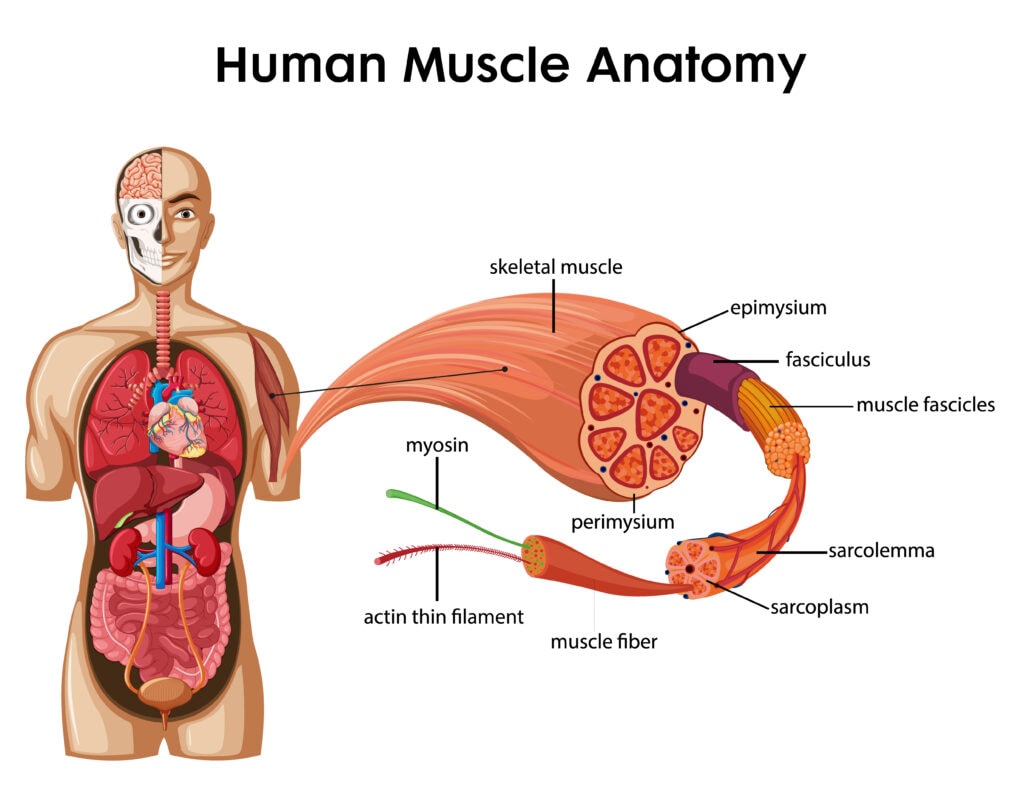
A muscle diagram illustrates the human muscle anatomy, providing clear visual guidance on muscle locations and functions.
There are three types of muscle tissue: skeletal, cardiac, and smooth, each with unique characteristics and involuntary or voluntary control.
Common muscle-related issues include strains, sprains, tension, and overuse injuries, which are especially prevalent in sports and daily activities. For example, muscle strains are a leading cause of discomfort for active individuals and can account for a significant portion of sports injuries.
Using our muscle diagram, you can gain insight into the specific muscle groups that may be causing pain or discomfort. In fact, research shows that visual tools like muscle diagrams can enhance understanding and treatment planning by up to 50%.
This article will explore various types of muscle diagrams and their usefulness in understanding muscle structure and jobs.
Overview of Muscle Diagrams
Muscle diagrams serve as essential tools for visualizing muscle structure, aiding students and professionals in anatomical education. These diagrams provide a detailed view of both superficial and deep muscle layers, offering a comprehensive understanding of muscular anatomy. Think of them as maps that guide us through the complex landscape of the human body.
Muscle diagrams help in understanding the relationships between different muscles and how they work together for movement and posture. This visual aid makes it easier to grasp complex anatomical concepts. For instance, observing how the biceps and triceps work together to move the arm can be more enlightening than reading about it in text alone.
Using muscle diagrams in your study routine can greatly enhance your understanding of muscle functions and their roles in the body. These diagrams show muscle locations and illustrate their connections to bones and other tissues, providing a comprehensive view of the muscular system.
Types of Muscle Tissue
The human body contains three primary types of muscle tissue. These are skeletal muscle, smooth muscle, and cardiac muscle. Each type has unique characteristics and functions, contributing to the body’s overall physiology.
Understanding these differences is key to appreciating how our muscles work.
Skeletal Muscle
Skeletal muscles are the most well-known type, making up about 40% of an individual’s body weight. These muscles are under voluntary control, meaning we consciously decide when to move them. Structurally, skeletal muscle cells are composed of muscle fibers wrapped in connective tissue sheaths, including the epimysium, perimysium, and endomysium. This organization allows for effective contraction and coordinated movements.
Each muscle fiber in skeletal muscles is surrounded by a connective tissue sheath called the endomysium. These fibers are grouped into compartments known as fasciculi, which are surrounded by perimysium. Fasciculi enhance the muscle’s ability to generate force by grouping muscle fibers together. This striated appearance is due to the arrangement of muscle fibers within the fasciculi.
Skeletal muscles are primarily responsible for movement and are attached to bones. The connective tissue covering these muscles provides support, protection, and pathways for blood vessels and nerves. These muscles work in pairs; for example, when the biceps contract to lift the arm, the triceps relax to allow the movement.
Grasping the structure and function of skeletal muscles is essential for understanding voluntary movements. Whether lifting weights or typing, these muscles are constantly at work, showcasing the human body’s remarkable capabilities.
Cardiac Muscle
Cardiac muscle is unique to the heart and operates involuntary muscle, meaning it functions without conscious control. These muscles are responsible for pumping blood throughout the body, a vital process for sustaining life. The cells in cardiac muscle are interconnected, allowing for synchronized contractions that are essential for heart function.
This muscle type has a striated appearance similar to skeletal muscle but is distinguished by its ability to contract rhythmically and continuously without fatigue. The heart’s ability to pump blood efficiently relies on the coordinated effort of cardiac muscle cells and muscle cell function, making it a marvel of biological engineering.
Smooth Muscle
Smooth muscles are found in the walls of hollow organs, such as the intestines, blood vessels, and bladder. Unlike skeletal muscles, smooth muscles operate involuntarily, controlling essential functions like digestion and circulation without conscious effort. These muscles facilitate involuntary contractions that are crucial for various bodily processes.
The structure of smooth muscles is different from that of skeletal and cardiac muscles. They lack the striations seen in the other two types and are more spindle-shaped. This unique structure allows them to contract in a more sustained and controlled manner, performing precise movements necessary for bodily functions.
Muscle Physiology and Function
Muscle physiology encompasses how muscles contract, the role of ATP, and the neuromuscular junction in muscle function. Understanding these processes is essential for appreciating how our muscles enable movement and maintain posture.
Let’s dive into the fascinating mechanics behind muscle contraction.
How Muscles Contract
Muscle contraction begins with an electrical signal from a motor neuron, which triggers the release of calcium ions within the muscle fibers.
This process is initiated by action potentials that travel down the sarcolemma and activate calcium release from the sarcoplasmic reticulum. The interaction between actin and myosin filaments within the sarcomeres causes the muscle fiber to shorten, resulting in contraction.
At a microscopic level, muscle contraction occurs through the sliding filament model. In this model, actin and myosin filaments slide past each other, shortening the muscle fiber. The presence of ATP is crucial during this process, as it provides the energy needed for the myosin heads to bind to actin and perform the power stroke.
The neuromuscular junction plays a vital role in this process. It is the site where the motor neuron and muscle fiber communicate, allowing for the transmission of impulses necessary for contraction. Understanding this intricate process highlights the complexity and efficiency of our muscular system.
ATP in Muscle Contraction
Adenosine triphosphate (ATP) is crucial for muscle contraction as it detaches myosin heads from actin, enabling relaxation. ATP provides the energy required for muscle contraction by facilitating the movement of myosin heads along actin filaments.
During muscle contraction, ATP is hydrolyzed to ADP and inorganic phosphate, releasing energy that powers the process.
The power stroke during muscle contraction occurs when myosin heads pivot and pull actin filaments, driven by the energy released from ATP hydrolysis. Regulatory proteins like tropomyosin and troponin help control the contraction process by managing the binding sites on actin. This coordinated effort ensures smooth and efficient muscle function.
Neuromuscular Junction
The neuromuscular junction is the site where motor neurons communicate with skeletal muscle fibers, facilitating muscle contraction.
This communication occurs at a chemical synapse, where synaptic vesicles release acetylcholine (ACh) into the synaptic cleft. ACh binds to receptors on the muscle membrane, triggering the electrical signals necessary for contraction.
To terminate the signal and prevent continuous muscle contraction, acetylcholinesterase breaks down ACh in the synaptic cleft. Diseases affecting the neuromuscular junction, like Myasthenia Gravis, can lead to muscle weakness by reducing the effectiveness of ACh at the receptors.
Major Muscle Groups and Their Functions
Muscle health is crucial for overall well-being, as muscle dysfunction can lead to various medical conditions. Understanding muscle anatomy and the functions of different muscle groups helps us appreciate their roles in movement and stability.
Let’s explore the major muscle groups: upper limb, lower limb, and trunk muscles.
Upper Limb Muscles
The upper limb muscles include the biceps, which primarily facilitates arm flexion. Another important muscle in this group is the rhomboid major, which retracts and stabilizes the scapula during upper limb movements. These arm muscles work together to allow for a wide range of motions, from lifting objects to performing intricate tasks.
Functions of upper limb muscles are crucial for activities requiring precise movements and coordination. Whether throwing a ball or typing, these muscles are constantly at work, highlighting the body’s remarkable capabilities.
Lower Limb Muscles
The lower limb muscles, including the quadriceps, are essential for extending the knee joint. These muscles play a crucial role in facilitating movement and maintaining posture throughout various activities. The strength and function of lower limb muscles significantly impact daily activities and athletic performance.
Muscles in the lower limb work together to support major movements such as walking, running, and jumping. Knowing their roles helps in appreciating how these muscles contribute to mobility and overall physical health.
Trunk Muscles
Trunk muscles are categorized into three groups: superficial, intermediate, and intrinsic back muscles. These muscles attach to bones across the body and start just under the skull, extending down to the pelvis. The erector spinae muscles extend the back. They also facilitate side bending. These muscles are crucial for maintaining posture and allowing a range of trunk movements.
The intrinsic back muscles consist of superficial, intermediate, and deep subgroups, working together in multiple directions. The latissimus dorsi, a wide and flat muscle located in the lower part of the trunk, provides stability for back movements. The trapezius muscle, located across the upper part of the trunk, helps move the scapula. These muscles facilitate movement by both stretching and pressing together.
Intermediate back muscles are located above and below the ribcage, aiding in trunk movements. The serratus anterior muscle, shaped like a fan, provides stability and mobility to the shoulder blade.
Human Muscle Diagram Labeled
Anatomy of Specific Muscles
Muscle anatomy charts include detailed information on the origin, insertion, innervation, and function of specific muscles. This knowledge is essential for understanding how muscles coordinate to perform movements and maintain stability.
Let’s take a closer look at the sternocleidomastoid muscle, latissimus dorsi, and rhomboid major.
Sternocleidomastoid Muscle
The sternocleidomastoid muscle has two heads: one originating from the manubrium of the sternum, and the other from the medial third of the clavicle. It inserts on the lateral surface of the mastoid process of the temporal bone and the superior nuchal line of the occipital bone. When contracted unilaterally, this muscle flexes the neck to the same side while rotating the head to the opposite side.
Bilateral contraction of the sternocleidomastoid muscle assists in neck flexion and extension of the head. Its primary nerve supply comes from the accessory nerve and branches from the cervical plexus.
This muscle also plays a role in elevating the sternum and clavicle, assisting in breathing. Pathological conditions in the sternocleidomastoid can lead to wryneck, causing head tilt and rotation.
Latissimus Dorsi
The latissimus dorsi muscle is a large, flat muscle located in the back that contributes significantly to upper body movements. It plays a key role in various movements, including adduction and extension of the shoulder. Positioned in the lower back and extending to the sides, it forms a ‘V’ shape.
This muscle originates from the lower six thoracic vertebrae and the lumbar vertebrae, and inserts into the humerus. The latissimus dorsi is essential for movements like pulling and lifting, and it provides stability for the back during these activities.
Rhomboid Major
The rhomboid major is a muscle located in the upper back, connecting the shoulder blades to the spine. It originates from the spinous processes of the upper thoracic vertebrae (T2 to T5) and inserts onto the medial border of the scapula. This muscle is crucial for retracting the scapula, pulling it towards the spine and stabilizing the shoulder blade during arm movements.
The rhomboid major plays an important role in activities that involve shoulder elevation and retraction, such as rowing or pulling movements. Understanding this muscle’s function helps in recognizing its importance in upper back stability and movement.
Blood Supply to Muscles
Skeletal muscles receive blood through primary arteries that branch into smaller vessels, ensuring proper distribution of blood supply within the muscle tissue. This blood supply is crucial for delivering oxygen and nutrients necessary for muscle function and endurance.
Blood Vessels in Muscles
Each muscle fiber is surrounded by a dense network of capillaries, which are critical for delivering oxygen and nutrients directly to the fibers. The terminal arterioles, which are the last branches containing smooth muscle, lead to groups of capillaries known as the microvascular unit, regulating blood flow in skeletal muscles. Blood vessels are closely located with muscle fibers, facilitating efficient oxygen and nutrient exchange.
The skeletal muscle pump helps return blood to the heart by compressing veins during muscle contraction. This mechanism ensures that muscles receive an adequate blood supply to sustain their activities, especially during physical exertion.
Role of Oxygen and Nutrients in Muscle Function
Muscles utilize oxygen and nutrients primarily to generate ATP, which is essential for contraction and overall muscle function. Enhanced blood flow during muscle contraction leads to increased delivery of oxygen and nutrients necessary for energy production. Effective blood circulation is crucial for muscle endurance and aids in recovery after physical exertion.
Blood flow limitations can lead to muscle fatigue, as inadequate supply affects aerobic respiration. Knowing the importance of oxygen and nutrients helps appreciate how muscles sustain prolonged activities and recover efficiently.
Muscle Contraction Mechanism
Muscle fibers contain sarcomeres, the fundamental units of contraction. The two essential myofilaments in muscle fibers are actin and myosin. Regulatory proteins involved in the sliding mechanism leading to contraction include troponin C, I, T, and tropomyosin.
Let’s explore the roles of motor neurons and the neuromuscular junction in this process.
Role of Motor Neurons
Motor neurons transmit action potentials to muscle fibers, leading to their contraction through the release of neurotransmitters. The process begins when the neurotransmitter acetylcholine (ACh) is released from a motor neuron, which depolarizes the muscle fiber membrane. Motor neurons can branch out to connect with multiple muscle fibers, forming a unit that can control muscle contraction strength.
This connection between motor neurons and muscle fibers ensures precise control over muscle movements. Grasping the role of motor neurons helps appreciate how our nervous system coordinates complex activities like walking, running, and lifting.
Neuromuscular Junction
The neuromuscular junction is a chemical synapse where the motor neuron communicates with the muscle fiber at the motor end-plate. At this junction, the synaptic cleft, about 50 nm wide, serves as the space where neurotransmitters like acetylcholine are released. The motor end plate contains folds that increase the surface area for acetylcholine receptors, facilitating efficient neurotransmission.
Acetylcholine is released at the neuromuscular junction, binding to receptors that trigger the electrical signals necessary for muscle contraction. Acetylcholinesterase inactivates acetylcholine after its release, ensuring that muscle contractions are not prolonged unnecessarily. This precise mechanism highlights the importance of the neuromuscular junction in muscle function.
Clinical Significance of Muscles
Conditions such as myasthenia gravis are characterized by muscle fatigue and weakness, significantly impacting daily activities. Muscles form a crucial part of other body systems, contributing to overall physiological function.
Let’s explore some common muscle-related conditions and their clinical significance.
Muscle Cramps
Muscle cramps can be caused by idiopathic factors, known diseases, and exercise-associated factors. Skeletal muscle cramps commonly occur during sports or exercise. These cramps are sudden, involuntary contractions that can be quite painful.
Preventing muscle cramps involves staying hydrated, maintaining electrolyte balance, and performing proper warm-up exercises before engaging in physical activities. Understanding the causes and prevention of muscle cramps helps in managing and reducing their occurrence.
Myopathy
Myopathy encompasses a range of disorders that result in muscle weakness due to muscle fiber damage. These disorders can be genetic or acquired and often lead to significant functional impairments. Myopathy can result from genetic disorders, leading to muscle weakness.
Treatment for myopathy may involve physical therapy and medication. Early diagnosis and intervention are crucial for managing symptoms and improving the quality of life for individuals affected by myopathy.
Muscular Dystrophy
Muscular dystrophy is a group of genetic disorders that cause progressive weakness and loss of muscle mass. These disorders impact muscle tissue, leading to symptoms such as difficulty walking, muscle stiffness, and reduced mobility. Understanding muscular dystrophy helps in recognizing its impact on individuals and the importance of supportive care.
Treatment options for muscular dystrophy focus on managing symptoms and improving the quality of life. These may include physical therapy, medications, and assistive devices. Research continues to explore potential cures and advanced therapies for these conditions.
Myasthenia Gravis
Myasthenia gravis is an autoimmune disease that affects muscle strength and function. This condition impacts the neuromuscular junction, where it interferes with the communication between motor neurons and muscle fibers. The result is muscle weakness and fatigue, particularly in muscles responsible for:
eye movements
facial movements
chewing
swallowing
Treatment for myasthenia gravis may include medications that improve neuromuscular transmission and immune-suppressing drugs. Understanding this condition underscores the importance of the neuromuscular junction in maintaining muscle function and highlights the complexities of autoimmune disorders.
Strains and Sprains
Strains and sprains are common injuries that affect muscles and ligaments, respectively. Muscle strains occur when muscle fibers are overstretched or torn, often due to excessive force or improper use. Symptoms include pain, swelling, and limited movement.
Ligament sprains, on the other hand, involve the stretching or tearing of ligaments, usually due to sudden twists or impacts. Treatment for both strains and sprains typically involves rest, ice, compression, and elevation (RICE), along with physical therapy to restore function.
Bottom Line
From the types of muscle tissue to the detailed mechanisms of muscle contraction, this guide has explored the intricacies of the muscular system.
By learning about major muscle groups, their specific functions, and the clinical significance of muscle-related conditions, we gain valuable insights into maintaining muscle health and addressing potential disorders.