
The shoulder is a complex and versatile joint that allows for a wide range of motion. It consists of three main bones – the humerus (upper arm bone), scapula (shoulder blade), and clavicle (collarbone) – as well as multiple muscles, tendons, and ligaments.
The primary function of the shoulder is to provide mobility and stability to the upper body. This makes it an essential component in daily activities such as reaching, lifting, throwing, and pushing.
This guide explores these components, including the clavicle, scapula, and key muscles, to help you grasp how the shoulder functions and maintains stability.
Key Takeaways
The shoulder complex consists of bones, muscles, ligaments, and nerves that work together for mobility and stability.
Key components include the humerus, scapula, and clavicle, all essential for shoulder movement and function.
What is Shoulder Anatomy
The shoulder complex is a masterpiece of biological engineering, designed to provide both incredible mobility and stability. It’s made up of the shoulder girdle, which links the upper limb with the axial skeleton through the sternoclavicular joint. This intricate system includes bones, muscles, ligaments, and nerves, all working together to facilitate a wide range of movements while maintaining stability.
Shoulder muscles connect the upper limb to the trunk, supporting various shoulder movements. These muscles facilitate lifting, rotating, and stabilizing the arm, enabling everyday tasks with ease. Without this sophisticated structure, our ability to move our arms in almost any direction would be severely limited.
Key Bones in the Shoulder Complex
At the heart of the shoulder joint are three key bones: the humerus, scapula, and clavicle. These bones work together to provide the shoulder with its remarkable range of motion and stability.
The humerus forms the “ball” of the shoulder joint, fitting into the socket known as the glenoid on the scapula. This ball and socket arrangement allows for extensive movement, making the shoulder one of the most mobile joints in the body.
The scapula, or shoulder blade, is a flat, triangular bone that connects the upper arm to the clavicle and serves as an attachment point for various muscles. The clavicle, shaped like an ‘S’, links the shoulder girdle to the torso, enhancing both stability and mobility. Together, these bones form the foundation of the shoulder complex, enabling a wide range of functional movements.
1. Bones of the Shoulder
The shoulder complex is an intricate structure consisting of three main bones: the clavicle, scapula, and humerus. Each of these bones plays a crucial role in the overall function and stability of the shoulder joint. These bones not only provide the necessary support and framework but also serve as attachment points for muscles, ligaments, and tendons that facilitate movement.
The clavicle, scapula, and humerus work in unison to ensure that the shoulder can perform a wide range of movements, from simple tasks like lifting an object to complex athletic maneuvers. Understanding the structure and function of these bones is essential for appreciating the complexity and versatility of the shoulder joint.
1.1 Clavicle (Collarbone)
The clavicle, commonly known as the collarbone, serves as a strut that connects the arm to the body, facilitating movement and stability of the shoulder. It has a distinctive ‘S’ shape and connects the upper limb to the trunk, playing multiple critical roles such as force transmission. At one end, the clavicle articulates with the sternum at the sternoclavicular joint, while at the other end, it connects to the acromion of the scapula, forming part of the shoulder girdle.
This bone is essential for maintaining the structural integrity of the shoulder and providing a stable base for the upper limb. It helps to protect neurovascular structures that pass from the neck to the arm and acts as a shock absorber during falls, distributing forces across the shoulder to prevent injuries.
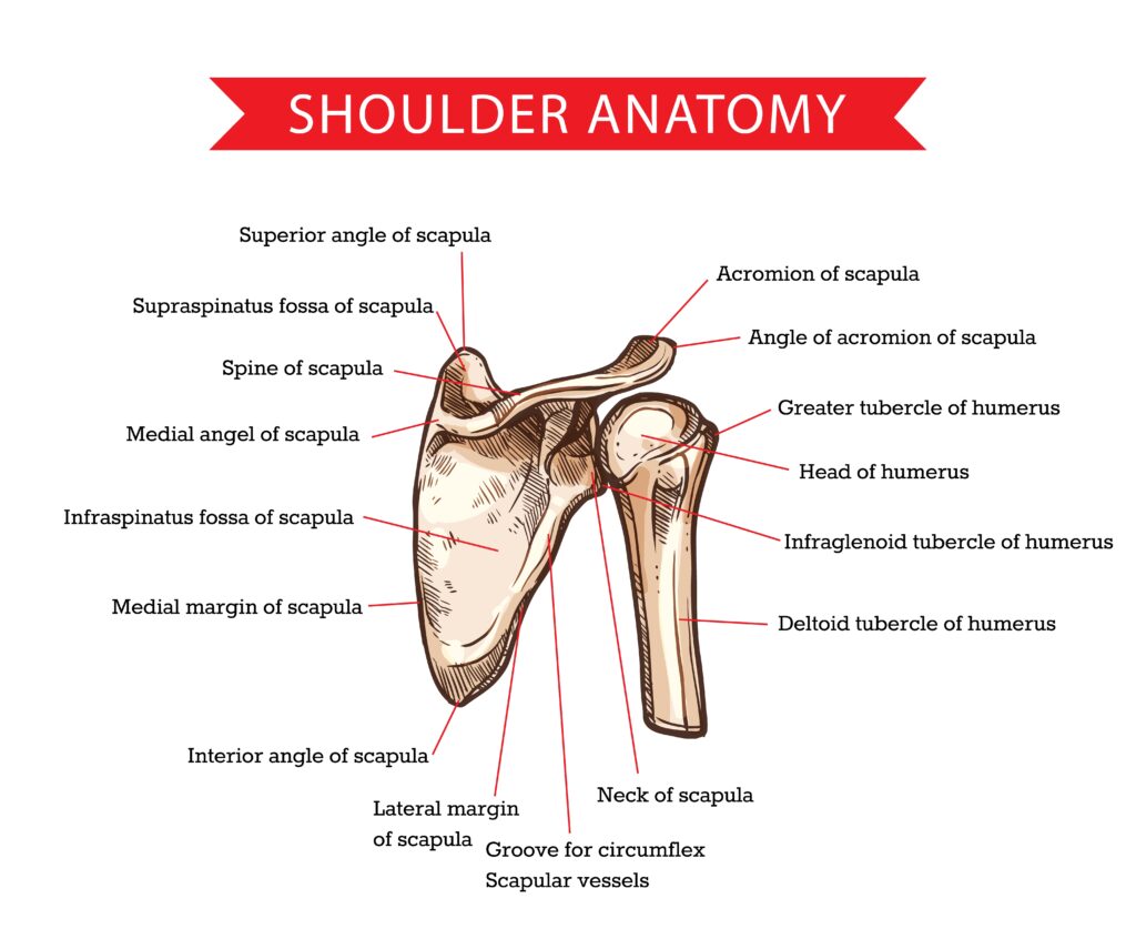
1.2 Scapula (Shoulder Blade)
The scapula, or shoulder blade, is a flat triangular bone that plays a critical role in shoulder movement and serves as an attachment point for several muscles. Key landmarks of the scapula include the acromion, which extends over the shoulder joint, and the glenoid fossa, which articulates with the humerus. These structures are vital for the stability and function of the shoulder joint.
The scapula provides a base for muscle attachment and plays a pivotal role in shoulder mobility. Its ability to move along the thoracic wall allows for a greater range of motion, enabling complex movements such as lifting, pushing, and pulling. Understanding the structure of the scapula helps in appreciating its importance in both the stability and mobility of the shoulder.
1.3 Humerus (Upper Arm Bone)
The humerus is a long bone located in the upper arm. It extends from the shoulder to the elbow. The proximal end of the humerus features the head, which fits into the glenoid cavity of the scapula to form the shoulder joint. This ball-and-socket arrangement allows for a wide range of motion, making the shoulder joint one of the most flexible joints in the human body.
The greater and lesser tubercles on the humerus provide attachment sites for key muscles involved in shoulder movement. These muscles facilitate various arm movements, including lifting, rotating, and stabilizing the shoulder.
The structure and articulations of the humerus are essential for understanding how the shoulder functions and why it is susceptible to certain injuries.
2. Joints of the Shoulder
The shoulder joint is a marvel of biomechanical engineering, consisting of four key joints: the glenohumeral, acromioclavicular, sternoclavicular, and scapulothoracic joints. These joints work together to provide the shoulder with its remarkable range of motion and stability. Each joint has a specific structure and function, contributing to the overall mechanics of shoulder movement.
Recognizing the structure and function of these joints helps appreciate the shoulder’s complexity. Each joint plays a unique role, from facilitating arm movements to providing stability and support. Let’s delve deeper into each of these joints to understand their significance.
2.1 Glenohumeral Joint
The glenohumeral joint is a highly mobile ball-and-socket joint, facilitating a wide range of motion due to its anatomical structure. This joint is formed by the articulation of the humeral head with the glenoid cavity of the scapula, allowing for extensive movement in multiple directions. However, this mobility comes at the cost of stability, making the glenohumeral joint prone to dislocation.
The stability of the glenohumeral joint is largely maintained by the surrounding rotator cuff muscles and the joint capsule. These structures work together to keep the humeral head securely in the glenoid cavity during movement, preventing dislocations and ensuring smooth, coordinated actions.
2.2 Acromioclavicular Joint (AC Joint)
The acromioclavicular joint is also known as the AC joint. It stabilizes the shoulder by linking the clavicle to the acromion of the scapula. This joint functions as a connection point between the acromion of the scapula and the clavicle, playing a crucial role in shoulder movement and stability.
The AC joint is supported by ligaments such as the acromioclavicular and coracoclavicular ligaments. These ligaments prevent dislocation and maintain the structural integrity of the joint, ensuring that the shoulder can perform a wide range of movements without compromising stability.
2.3 Sternoclavicular Joint (SC Joint)
The sternoclavicular joint is the only direct connection between the upper limb and the axial skeleton, facilitating arm movement. This joint connects the sternum to the clavicle, acting as the only bony link between the arm and the axial skeleton.
The SC joint is stabilized by several ligaments, including the anterior and posterior sternoclavicular ligaments. These ligaments enhance the stability of the SC joint while allowing for a wide range of movements, making it a vital component of the shoulder complex.
2.4 Scapulothoracic Joint
The scapulothoracic joint is not a true anatomical joint but rather a functional articulation between the scapula and the thoracic wall. This joint plays a crucial role in shoulder movement by allowing the scapula to glide over the thoracic rib cage.
The scapulothoracic joint is essential for shoulder mobility and overall upper limb function. It enables the scapula to move smoothly along the thoracic wall, facilitating a greater range of motion and contributing to the dynamic stability of the shoulder.
3. Muscles of the Shoulder
The shoulder region includes approximately eight muscles that work together to provide stability and movement for the shoulder joint. These muscles are essential for a wide range of movements, including lifting, rotating, and stabilizing the arm. Understanding the function of these muscles helps to appreciate the complexity and versatility of shoulder movements.
The muscles of the shoulder can be categorized into different groups, each with specific roles. The most notable groups include the rotator cuff muscles, the deltoid muscle, and other key muscles such as the trapezius and pectoralis major.
3.1 Rotator Cuff Muscles
The rotator cuff is primarily made up of four muscles that support the shoulder joint by allowing arm rotation and elevation. These muscles include:
Supraspinatus, which plays a key role in initiating arm elevation
Infraspinatus, which assists in the external rotation of the arm
Teres minor, which contributes to the rotation and adduction of the shoulder
Subscapularis, which is responsible for internal rotation of the arm
This structure is essential for maintaining shoulder stability and function.
These muscles work together to stabilize the shoulder joint and enable a wide range of arm movements. The rotator cuff tendons also play a crucial role in maintaining the stability of the glenohumeral joint, preventing dislocations and ensuring smooth, coordinated movements.
3.2 Deltoid Muscle
The deltoid muscle is divided into three distinct parts:
Anterior fibers, which are crucial for shoulder flexion, moving the arm forward
Middle fibers, which primarily facilitate shoulder abduction, lifting the arm away from the body
Posterior fibers, which assist with shoulder extension and external rotation, moving the arm backward and outward
The deltoid muscle plays a vital role in shoulder movement and stability. Its unique structure allows for a wide range of motions, making it essential for tasks that involve lifting, pushing, and pulling.
3.3 Other Key Muscles
In addition to the rotator cuff and deltoid muscles, several other key muscles contribute to shoulder movement and stability. The trapezius muscle aids in the elevation and rotation of the shoulder blade, playing a crucial role in shoulder movement. The trapezius helps in the movement and stabilization of the shoulder blade and neck.
The pectoralis major is involved in arm adduction and flexion, while the latissimus dorsi aids in the extension, adduction, and internal rotation of the shoulder. The serratus anterior is important for the upward rotation of the shoulder blade.
These muscles work together to ensure smooth and coordinated shoulder movements, contributing to the overall function and stability of the shoulder joint.
4. Ligaments of the Shoulder
Shoulder ligaments serve to connect bones, providing stability and support to the shoulder complex. These ligaments play a crucial role in maintaining joint stability and preventing dislocations. The ligaments of the shoulder include the glenohumeral ligaments, coracoclavicular ligament, and coracoacromial ligament, each contributing to the overall stability and function of the shoulder joint.
Recognizing the structure and function of these ligaments highlights their role in maintaining shoulder stability and preventing injuries. Let’s delve into the specifics of each ligament group.
4.1 Glenohumeral Ligaments
The glenohumeral ligaments include superior, middle, and inferior ligaments, each contributing to the stabilization of the glenohumeral joint. The inferior glenohumeral ligament complex is the primary structure preventing anteroinferior dislocation of the shoulder.
These ligaments provide dynamic stability to the shoulder joint by tightening during specific arm movements. They play a crucial role in maintaining the integrity of the glenohumeral joint, preventing dislocations, and ensuring smooth, coordinated movements.
4.2 Coracoclavicular Ligament
The coracoclavicular ligament consists of two parts: the conoid and trapezoid ligaments, which are crucial for maintaining stability at the acromioclavicular joint. These ligaments help stabilize the clavicle position in relation to the scapula, ensuring that the shoulder joint remains secure during movement.
The coracoclavicular ligament plays a vital role in shoulder stability, preventing dislocations, and maintaining the structural integrity of the acromioclavicular joint. Understanding this ligament’s function helps to appreciate its importance in overall shoulder health.
4.3 Coracoacromial Ligament
The coracoacromial ligament functions to limit upward displacement of the humeral head, which helps prevent impingement in the shoulder. It helps prevent superior dislocation of the humerus and acts as a protective arch over the shoulder joint.
This ligament plays a crucial role in maintaining shoulder stability, preventing injuries, and ensuring smooth, coordinated movements. Understanding the structure and function of the coracoacromial ligament helps to appreciate its importance in shoulder health.
5. Nerves of the Shoulder
The nerves that control shoulder movement and sensation originate from the brachial plexus, which is located in the armpit region. These nerves are responsible for both motor and sensory functions in the upper limb. Recognizing these nerves’ roles highlights the complexity and functionality of the shoulder.
The primary nerves involved in shoulder innervation include the brachial plexus, axillary nerve, and suprascapular nerve. Each of these nerves plays a crucial role in controlling muscle movements and providing sensation to the shoulder and upper limb.
5.1 Brachial Plexus
The brachial plexus is composed of a network of nerves that extend from the spinal cord to supply the arm with motor and sensory functions. This plexus plays a crucial role in innervating various muscles around the shoulder, enabling arm movement.
The brachial plexus is formed by the merging of spinal nerves C5 to T1, grouping into trunks, divisions, cords, and terminal branches. It plays a crucial role in controlling muscle movements in the shoulder, arm, and hand, making it essential for the overall function of the upper limb.
5.2 Axillary Nerve
The axillary nerve is responsible for supplying the deltoid muscle, which is vital for shoulder abduction. Injuries to the axillary nerve can lead to significant weakness in shoulder movement, particularly with raising the arm.
The axillary nerve innervates the deltoid and teres minor muscles, facilitating shoulder abduction and external rotation. Injuries to this nerve can result in sensory loss over the lateral shoulder and weakness in shoulder movement, highlighting its importance in shoulder function.
5.3 Suprascapular Nerve
The suprascapular nerve innervates both the supraspinatus and infraspinatus muscles, which are essential for shoulder stability and movement. Clinical issues related to the suprascapular nerve may result in shoulder pain and impaired arm functions due to muscle weakness.
Damage to this nerve can lead to weakness in arm elevation and shoulder pain. Understanding the role of the suprascapular nerve helps to appreciate its importance in maintaining shoulder stability and function.
6. Blood Supply to the Shoulder
The subclavian artery is responsible for delivering oxygen-rich blood to the shoulder area. The shoulder receives its arterial supply primarily from branches of the subclavian and axillary arteries.
Recognizing the shoulder’s blood supply highlights how nutrients and oxygen are delivered, ensuring health and function. Let’s delve into the specifics of the arterial and venous supply to the shoulder.
6.1 Arteries
The axillary artery, a continuation of the subclavian artery, supplies blood as it passes through the armpit. Branches from the axillary artery provide essential blood supply to various shoulder structures, including muscles and joints.
The subclavian artery transitions into the axillary artery, which is essential for supplying blood to the shoulder region. The posterior circumflex humeral artery, a branch of the axillary artery, plays a critical role in vascularizing the deltoid muscle. Understanding the arterial supply to the shoulder helps to appreciate how blood flow supports shoulder function and health.
6.2 Veins
The axillary vein is a major vessel that collects de-oxygenated blood from the shoulder and drains into the subclavian vein. Additional veins, such as the cephalic and basilic veins, also contribute to the venous drainage of the shoulder region.
The axillary vein is responsible for draining blood from the shoulder and is a continuation of the brachial veins. Venous drainage of the shoulder area is facilitated by the subclavian vein, which collects blood from the axillary vein.
Understanding the venous drainage of the shoulder helps to appreciate how blood is returned to the heart for reoxygenation.
7. Bursae of the Shoulder
Bursae are fluid-filled sacs that reduce friction between moving parts of the shoulder. These sacs are located in various parts of the body, including the shoulder, and play a crucial role in facilitating smooth movements and reducing wear and tear on the tissues.
Recognizing the function and location of bursae highlights their importance in maintaining shoulder health and preventing injuries. Let’s explore the specifics of the subacromial and subscapular bursae.
7.1 Subacromial Bursa
The subacromial bursa is positioned between the rotator cuff muscles and the outer shoulder muscles. Subacromial bursitis occurs when the bursa becomes inflamed, often associated with shoulder impingement.
The subacromial bursa is situated beneath the acromion and above the supraspinatus muscle, serving to protect the muscle from wear. Subacromial bursitis, often linked to shoulder impingement, arises from inflammation of the bursa due to repetitive overhead activities. Understanding the role of the subacromial bursa helps to appreciate its importance in maintaining shoulder health.
7.2 Subscapular Bursa
The subscapular bursa is located beneath the subscapularis muscle, providing cushioning for shoulder movements. Injuries to the subscapular bursa can lead to pain and restricted movement in the shoulder joint.
The subscapular bursa is found between the subscapularis muscle and the shoulder joint, aiding in reducing friction in this area. Injury or inflammation of the subscapular bursa can lead to pain and restrict movement in the shoulder, impacting daily activities. Understanding the role of the subscapular bursa helps to appreciate its importance in maintaining shoulder health.
8. Biomechanics of the Shoulder
The shoulder complex consists of multiple joints working continuously together for effective movement. The shoulder exhibits a high degree of mobility due to the structural design of the glenohumeral joint, making it the most flexible joint in the human body.
Recognizing the biomechanics of the shoulder reveals how its components work together for smooth, coordinated movements. Let’s explore the range of motion and scapulohumeral rhythm in more detail.
8.1 Range of Motion
The shoulder joint allows significant movement, including flexion, extension, abduction, and adduction. Shoulder movement includes internal and external rotation, contributing to its versatility.
Shoulder flexion allows the arm to move forward and upward, achieving an average motion of up to 180 degrees. Internal rotation of the shoulder typically allows for 70 to 90 degrees of movement, primarily facilitated by muscles like the subscapularis. Recognizing the shoulder’s range of motion highlights its flexibility and versatility.
8.2 Scapulohumeral Rhythm
The scapulohumeral rhythm describes the coordinated movements of the scapula and humerus during arm elevation. This rhythm typically follows a 2:1 ratio of glenohumeral to scapulothoracic motion during shoulder elevation.
The coordination between the scapula and humerus is crucial for effective shoulder movement, facilitating a more extensive range of motion. Scapulohumeral rhythm is important for shoulder function as it helps distribute movement across the shoulder girdle and prevents injury.
Shoulder Diagram
Common Shoulder Injuries
The shoulder’s extensive range of motion increases its vulnerability to injuries and dislocations. Common shoulder injuries include rotator cuff tendon tears, shoulder dislocations, and impingement syndromes. Rotator cuff tears can result from acute injuries or gradual wear, leading to pain, weakness, and decreased mobility in the shoulder.
Repeated shoulder dislocations can increase the risk of damage to muscles, ligaments, and tendons around the joint. It’s important to be aware of these potential injuries and seek appropriate medical attention if you experience persistent shoulder pain or discomfort.
Functional Movements of the Shoulder
The glenohumeral joint can achieve a remarkable range of motion, making it the most mobile joint in the human body. Internal rotation of the shoulder allows movement toward the body’s midline, typically covering a range of 70 to 90 degrees. External rotation moves the shoulder away from the midline and usually has a normal range of about 90 degrees.
Abduction, which involves lifting the arm away from the body, typically allows for a range of motion of 150 degrees. The scapulothoracic joint, while not a true joint, refers to the movement of the scapula over the thoracic cage, allowing for additional shoulder mobility.
Role of the Brachial Plexus
The brachial plexus is a complex network of nerves that originates from the spinal nerves C5 to T1, responsible for motor and sensory control of the upper limb. It is structured into roots, trunks, divisions, cords, and terminal branches, each playing a role in innervating specific muscle groups in the shoulder and arm.
The terminal branches of the brachial plexus include the musculocutaneous, axillary, radial, median, and ulnar nerves, each facilitating distinct movements in the upper limbs.
Frequently Asked Questions
What are the main bones that make up the shoulder joint?
The shoulder joint primarily consists of the clavicle, scapula, and humerus. These bones work together to provide a wide range of movement.
What is the role of the rotator cuff muscles in the shoulder?
The rotator cuff muscles are crucial for stabilizing your shoulder joint while allowing you to rotate your arm and lift it in various directions. They play a key role in keeping your shoulder functioning smoothly during everyday activities.
How does the glenohumeral joint contribute to shoulder mobility?
The glenohumeral joint is super flexible, acting like a ball-and-socket to let your arm move freely in multiple directions. This unique structure is key to shoulder mobility!
What is the function of the brachial plexus in the shoulder?
The brachial plexus is crucial for your shoulder because it controls both the movement and feeling in your upper limb by connecting nerves from the neck to the arm. So, when you move your arm or feel sensations in your shoulder area, you have the brachial plexus to thank!
How can common shoulder injuries like rotator cuff tears be prevented?
To prevent common shoulder injuries like rotator cuff tears, focus on strengthening your shoulder muscles and boosting flexibility with regular exercises, while also staying aware of any potential injury risks. Taking these simple steps can make a huge difference in keeping your shoulders healthy.

