A spine diagram helps you understand the structure of your spine and pinpoint where issues like pain, stiffness, or discomfort may be coming from. By identifying the specific region affected cervical (neck), thoracic (upper back), lumbar (lower back), sacral, or coccygeal you can better understand what might be causing the problem.
Common spine-related issues include herniated discs, spinal stenosis, muscle strain, and nerve compression. For instance, lower back pain affects nearly 80% of people at some point in their lives and knowing where it hurts is the first step to knowing why it hurts.
Visual tools like this have been shown to improve understanding and communication between patients and healthcare providers by up to 60%.
This article will break down each region of the spine, delve into the anatomy of vertebrae and intervertebral discs, and discuss common spinal issues.
Overview of Spine Anatomy
The spine is a bony structure that serves as the main support for the body, connecting various parts of the musculoskeletal system. The spine is a complex structure that enables various movements such as sitting, standing, and twisting, while also protecting the spinal cord and associated nerves.
This intricate system is composed of several bones and soft tissues, including vertebrae, ligaments, and muscles that work together to maintain our position and facilitate movement. The primary roles of the spine include providing structural support, allowing flexibility and movement, and protecting the spinal cord, which is crucial for transmitting signals between the brain and the rest of the body.
Understanding the spine’s anatomy helps us appreciate its functions and the need to maintain its health. This knowledge helps in recognizing and addressing potential issues before they escalate.
Regions of the Spine
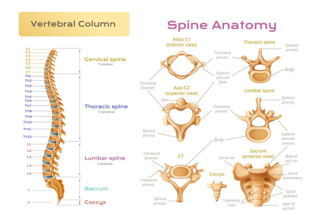
The spine is divided into five distinct regions, each with its own unique characteristics and functions. The cervical, thoracic, lumbar, sacral, and coccygeal sections each play a vital role in body support, movement facilitation, and spinal cord protection.
Each region has distinct characteristics and functions that are worth understanding.
Cervical Spine
The cervical spine consists of seven vertebrae that support the weight of the head and enable a wide range of head movements, such as turning, tilting, and nodding. This region maintains neck flexibility and mobility, enabling ease in everyday tasks.
Thoracic Spine
Located in the middle section of the spine, the thoracic spine connects the cervical spine above and the lumbar spine below. It has a kyphotic curve that helps accommodate the rib cage, which protects vital organs like the heart and lungs.
The thoracic spine’s alignment and curve are crucial for body balance and optimal weight distribution.
Lumbar Spine
The lumbar spine is responsible for bearing most of the body’s weight, making it a crucial region for activities involving lifting and carrying. It consists of five lumbar vertebrae that provide the necessary support and flexibility for the lower back.
Injuries here can impact mobility, yet those with L-1 to L-5 injuries may still experience increased motor movement in hips and knees and might walk with assistive devices.
Sacrum and Coccyx
The sacrum is formed by five fused vertebrae that connect the spine to the hip bones, forming a key part of the pelvic girdle. Below the sacrum is the coccyx, composed of four fused vertebrae, which provides attachment for ligaments and muscles of the pelvic floor.
The sacrum and coccyx together contribute to structural support, stability, and balance for the lower body.
Vertebrae Structure
Each vertebra in the spine consists of a weight-bearing anterior body and a posterior arch that encloses the spinal canal.
The vertebral bodies are lined with hyaline cartilage at their superior and inferior surfaces, allowing for smooth movement between adjacent vertebrae. This structure maintains a healthy spine and facilitates various movements without damaging the vertebrae.
Pedicles connect the vertebral body to the transverse processes, enhancing the overall stability of the vertebra. The vertebral arch features bony projections known as spinous process and transverse processes, which serve as muscle and ligament attachment sites. These processes support the muscles and ligaments that stabilize the spine and enable motion.
Facet joints are located on each side of the spine at every vertebral level, providing both flexibility and stability for spinal movement.
The articular processes at the junction of the laminae and pedicles form joints with adjacent vertebrae, further contributing to the spine’s flexibility and stability. The size and shape of vertebral bodies increase in the lower spine to better support the body’s weight.
Intervertebral Discs
Intervertebral discs enable movement and act as shock absorbers to protect the vertebrae. These discs consist of three key parts:
The nucleus pulposus, which is primarily composed of water, making up 66% to 86% of its structure. This provides the disc with its gel-like consistency and ability to absorb shocks.
The annulus fibrosus.
The cartilaginous endplates.
This structure is essential for maintaining spinal health and mobility.
The annulus fibrosus is structured with 15 to 25 layers of collagen fibers, providing both strength and flexibility. The outer layer of the annulus fibrosus is vascularized, allowing some nutrient supply to the avascular nucleus pulposus.
Intervertebral discs play a crucial role in distributing hydraulic pressure to prevent localized stress on vertebrae, ensuring the intervertebral disc can handle various physical activities without damage.
Spinal Cord and Nerves
The spinal cord is a cylindrical structure that extends from the brainstem to the lower back, relaying messages between the brain and the body. It facilitates voluntary movements and regulates automatic bodily functions through the signals that travel along its length. This essential communication network is protected by the vertebral column, ensuring its delicate structure remains intact.
At the end of the spinal cord is the cauda equina, a collection of nerve roots that serve the lower body. The cauda equina is crucial for transmitting sensory and motor signals to and from the lower limbs, playing a significant role in maintaining mobility and function as the spinal cord ends.
Facet Joints
Facet joints, located on each side of the spine at every vertebral level, facilitate back motion by providing both flexibility and stability. These joints are essential for allowing the spine to bend and twist while maintaining structural integrity, contributing to the overall mobility and functionality of the spinal column.
Muscles and Ligaments
Two main muscle groups provide support for the spine: the extensors and the flexors. These muscles work together to maintain spinal stability and movement.
The Psoas Major muscle, for example, plays a key role in flexing the thigh at the hip and supporting the backbone. The Quadratus Lumborum muscle facilitates lateral flexion of the vertebral column, while the Longissimus Thoracis muscle aids in extending and laterally flexing the vertebral column.
Ligaments provide stability to joints and help prevent injuries from excessive movements. Tendons, which connect muscles to bones, are crucial for transferring forces during movement, ensuring that the spine remains stable and aligned during various activities.
Poor muscle tone can lead to body misalignment and strain on the spine, emphasizing the importance of maintaining strong and flexible muscles.
Spine Diagram Labeled
Natural Curves of the Spine
A healthy spine exhibits three natural curves that resemble an ‘S’ shape, functioning as shock absorbers.
These natural spinal curves help distribute mechanical forces and reduce strain on vertebrae and surrounding tissues, ensuring the normal spine can handle various physical activities without damage.
The cervical lordosis is the inward curve of the neck, typically measuring between 20 to 40 degrees. Thoracic kyphosis refers to the outward curve of the upper and mid-back, which is also normal between 20 to 40 degrees. Lumbar lordosis is the inward curve of the lower back, with normal measurements ranging from 20 to 35 degrees.
These natural curves are essential for maintaining balance and reducing the risk of injury during movement.
Spinal Curves and Their Importance
The spinal curves play a vital role in maintaining balance and posture, absorbing shocks, and facilitating flexibility and mobility.
These curves are detailed further in the following subsections.
Natural Curves of the Spine
The spine features four primary curves: cervical, thoracic, lumbar, and sacral. The cervical curve supports the head and allows for a range of motion in the neck. The thoracic curve, which is convex, protects the heart and lungs. The lumbar curve helps to bear the weight of the upper body and provides stability.
The spine features four primary curves:
Cervical: supports the head and allows for a range of motion in the neck
Thoracic: protects the heart and lungs
Lumbar: helps to bear the weight of the upper body and provides stability
Sacral: provides support and stability to the pelvis
The sacral curve connects the spine to the pelvis and supports weight during sitting.
Importance of Spinal Curves
Spinal curves maintain balance and posture, absorb impact from physical activities, distribute stress during movement, and significantly contribute to flexibility and mobility.
Common Spine Disorders
Spine disorders can lead to pain and mobility issues due to damage in the vertebrae. Several types of spine disorders can affect the curvature and function of the spine, leading to discomfort and mobility issues.
Some common spine disorders are discussed in detail below.
Scoliosis
Scoliosis is characterized by an abnormal lateral curvature of the spine, which can form a ‘C’ or ‘S’ shape. Symptoms may include back pain, uneven shoulders, and difficulty standing upright.
Treatment for scoliosis may include physical therapy, bracing, and surgery in severe cases to correct alignment.
Lordosis
Lordosis, often referred to as swayback, is the exaggerated inward curvature of the spine, particularly in the lumbar region. Common causes of lordosis include obesity, osteoporosis, and certain neuromuscular conditions.
Managing lordosis may involve weight loss, physical therapy, and core-strengthening exercises.
Kyphosis
Kyphosis is a condition characterized by an excessive outward curvature of the spine, leading to a hunched posture. This condition can arise from developmental issues, degenerative diseases, or trauma and may cause back pain and stiffness.
Treatment for kyphosis includes physical therapy, pain management, and surgery in severe cases.
Herniated Disc
A herniated disc occurs when the inner gel-like core of a spinal disc bulges out through a tear in the outer layer. Symptoms can include pain, numbness, and weakness in the areas supplied by the affected nerves.
Preventing herniated discs involves maintaining a healthy weight, practicing good posture, and avoiding heavy lifting.
Keeping Your Spine Healthy
Maintaining a healthy spine is crucial for overall well-being and quality of life. Regular exercise enhances flexibility and strengthens the muscles that support the spine.
Incorporating back-strengthening and stretching exercises, such as planks, into your routine at least twice a week can significantly benefit spinal health. Good posture involves keeping the spine aligned while sitting, standing, or lifting to avoid strain.
Using correct lifting techniques, such as bending your knees and keeping the object close to your body, can help prevent back injuries. Frequent breaks and changing positions during prolonged activities minimize spine strain.
When to Seek Medical Attention
Certain symptoms indicate the need for immediate medical attention, as they could signify severe spinal issues.
Symptoms such as limited head and neck movement, paralysis below the injury site, and difficulty talking require prompt medical evaluation. Injuries in the cervical area may lead to tetraplegia, while injuries in the thoracic area could result in paraplegia, highlighting the severity of spinal injuries.
Persistent back pain, loss of sensation in the limbs, or any sudden changes in nerve function are also signs that you should consult a healthcare provider.
Bottom line
The spine is comprised of five distinct regions (cervical, thoracic, lumbar, sacral, and coccygeal) that each serve unique functions, including support, flexibility, and spinal cord protection.
The spine is a complex structure composed of bones, ligaments, muscles, and intervertebral discs that work together to provide support, facilitate movement, and protect the spinal cord. Each region of the spine, from the cervical to the coccygeal, plays a vital role in our daily activities.