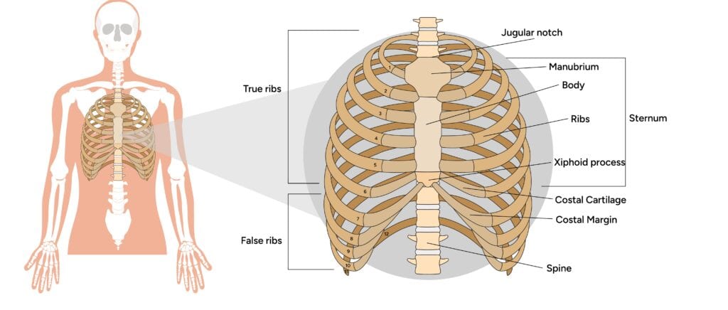
A sternum diagram is a useful guide that helps you understand the anatomy of your chest and the location of pain or discomfort around the sternum. By knowing the specific area of your sternum that hurts (upper, middle, or lower), you can identify whether the pain might be due to muscle strain, rib issues, or more serious conditions like heart-related problems or fractures.
Common causes of sternum pain include costochondritis (inflammation of the cartilage connecting the ribs to the sternum), trauma, or even heartburn. For instance, chest injuries or excessive coughing can often lead to sharp pain around the sternum.
Moreover, using a sternum diagram can assist in identifying the exact area of concern, leading to a more accurate self-diagnosis. Studies have shown that visual aids, such as diagrams, can improve diagnostic accuracy by up to 40%. Sounds pretty helpful, right?
That’s why we’re here to provide you with a detailed sternum diagram to help you diagnose your symptoms effectively and explore treatment options.
Detailed Anatomy of the Sternum
The sternum, commonly known as the breastbone, is a flat bone located at the middle of the chest. Serving a pivotal role in protecting vital organs such as the heart and lungs, the sternum is divided into three main parts: the manubrium, the body, and the xiphoid process. Each component has its unique structure and function, contributing to the sternum’s overall role in the human body.
Examining each part helps to grasp the sternum’s overall function.
Manubrium
The manubrium is the uppermost part of the sternum, characterized by its large quadrangular shape. At the top of the sternum, it meets the medial ends of the clavicles, forming sternoclavicular joints that ensure a sturdy link between the sternum and collarbones.
This joint aids in the mobility and stability of the upper chest. The manubrium also articulates with the first two ribs, ensuring a robust connection to the thoracic cage.
At its lower border, the manubrium meets the body of the sternum at the sternal angle, a significant anatomical landmark. The anterior surface of the manubrium attaches muscles like the pectoralis major and sternocleidomastoid, which are essential for chest and head movement.
The manubrium forms from a single primary ossification center, contributing to the sternum’s structural integrity.
Body of the Sternum
The sternal body is flat and elongated, forming the middle portion of the sternum. It articulates with the third to seventh ribs, as well as part of the second costal cartilage, through multiple articular facets. These links form the anterior chest wall and provide structural support to the thoracic cage. The lateral borders of the sternal body attach the costal cartilages of ribs 3 to 6, securing the ribcage.
Besides rib attachment, the sternal body also serves as a site for muscle connections. The pectoralis major muscle attaches to its anterior surface, playing a role in the movement of the upper arm. The transversus thoracis muscle originates from its posterior surface, aiding in chest wall movement and stability.
Inferiorly, the body of the sternum articulates with the xiphoid process, completing the structure of the sternum.
Xiphoid Process
The xiphoid process is a thin bony projection inferiorly located at the inferior end of the sternum. This small part can vary greatly in shape and size among individuals. Despite its variability, it serves as an attachment point for muscles like the rectus abdominis, internal oblique, and external oblique. These attachments are essential for trunk and abdomen movements.
Primarily, the xiphoid process functions as a muscular attachment point, playing a key role in the movement and stability of the anterior chest wall. Its location and structure make it an important landmark in both anatomy and surgical procedures. Recognizing the xiphoid process’s role highlights the sternum’s complexities and contributions to human anatomy.
Functions of the Sternum
The sternum protects vital thoracic organs, such as the heart and lungs, from external impacts and injuries. This protection is vital, as sternal fractures can cause severe damage to these organs. Its connection to the ribs and collarbones forms the ribcage, providing structural support to the thoracic cavity.
Visual aids often demonstrate the sternum’s connections to the clavicles and the true and false ribs, illustrating its role in the rib cage structure.
Moreover, the cartilage linking the sternum to the ribs allows for slight movements, aiding in the breathing process. This flexibility allows the lungs to expand and contract during respiration. These functions highlight the sternum’s importance in maintaining the thoracic cage’s structural integrity and functional capacity.
Common Conditions Affecting the Sternum
Physical strain, trauma, or congenital issues can lead to conditions affecting the sternum. Since the sternum protects vital organs, any condition compromising its integrity significantly impacts overall health. Treatments often include rest, medications, and sometimes surgery.
We will explore specific conditions such as sternal fractures, costochondritis, and congenital deformities like pectus excavatum and pectus carinatum.
Sternal Fractures
A sternal fracture is a break in the sternum, often resulting from auto accidents, falls, or blunt force trauma. The most common sternal fracture is a comminuted fracture, usually at the manubriosternal joint. This severe fracture often requires X-rays, CT scans, and ultrasound for detailed assessment of sternum sternal fractures.
Treatment varies with the severity of the fracture. Mild fractures typically need rest and pain medication, whereas severe cases might require surgery to correct the sternum’s position.
Costochondritis
Costochondritis involves inflammation of the rib cartilage connected to the sternum, causing localized sternum pain or tenderness.
This condition can result from physical strain, trauma, or underlying infections and arthritis. Symptoms often include chest pain that worsens with physical activity, coughing, stretching, and deep breathing.
Rest is the primary treatment, allowing inflammation to subside. Over-the-counter pain medications, such as NSAIDs, are recommended for pain management. Corticosteroid injections may be used in some cases to reduce inflammation and provide relief. Knowing these treatment options helps manage costochondritis effectively.
Pectus Excavatum and Pectus Carinatum
Pectus excavatum, or funnel chest, is a congenital condition where the sternum appears sunken, creating a concave chest. It can be associated with genetic disorders such as Marfan syndrome and Ehler’s Danlos syndrome.
Conversely, pectus carinatum involves the sternum protruding outward, resulting in a bird-like chest shape. Both conditions can impact thoracic organs and cause chest pain, particularly in certain positions or activities.
Treatment options for these conditions differ. Pectus carinatum is often treated with bracing, involving wearing a chest brace for an extended period to correct the shape. Severe cases may require surgery to remove cartilage and reposition the breastbone.
Blood Supply and Nerve Innervation
The internal thoracic artery primarily supplies blood to the sternum, providing multiple branches to the xiphoid process and other parts.
The internal thoracic veins facilitate venous drainage, directing blood into the brachiocephalic vein. This intricate blood supply is vital for the sternum’s health and function.
Intercostal nerves, originating from thoracic spinal nerves, innervate the sternum. Nerves from T1-T11 supply the xiphoid process and other parts, ensuring proper sensory and motor function.
Surgical Considerations
Sternotomy and other surgical procedures involving the sternum are vital for open cardiothoracic surgery, where the sternum is divided to access the heart and lungs. The xiphoid process serves as a midline landmark during these surgeries and may be removed (xiphoidectomy) for better visualization. Post-surgery, fixation techniques like titanium rib plating are used for sternal stability.
Innovative approaches like 3D-printed custom prosthetics are used for sternum reconstruction, enhancing recovery outcomes and stability.
Diagnostic Imaging of the Sternum
Diagnostic imaging is essential for evaluating the sternum and diagnosing related conditions. X-rays are commonly the first imaging technique to identify sternum issues. They quickly and effectively visualize fractures and other abnormalities. CT scans provide detailed images to evaluate chest injuries and conditions, offering a comprehensive sternum view.
MRI is useful for assessing soft tissue abnormalities related to the sternum. These noninvasive techniques provide vital information for diagnosing sternum-related conditions, ensuring proper treatment and management.
Sternum Diagram and Visual Aids
Diagrams often show the three main parts of the sternum: the manubrium, body, and xiphoid process, along with their relationships. These aids can demonstrate the sternum’s orientation relative to other thoracic features. Detailed aids, including 3D models, are used in education to clarify the sternum’s spatial orientation within the thorax.
These diagrams often highlight articulations with structures like the costal cartilages, illustrating the sternum’s role in the ribcage.
Key Takeaways
- The sternum has three main parts: the manubrium, body, and xiphoid process, each serving unique roles in protecting vital organs and aiding movement.
- Common conditions affecting the sternum include fractures, costochondritis, and congenital deformities, which require various treatments like rest or surgery.
- Diagnostic imaging, such as X-rays and CT scans, is crucial for identifying sternum-related issues, ensuring proper diagnosis and treatment.