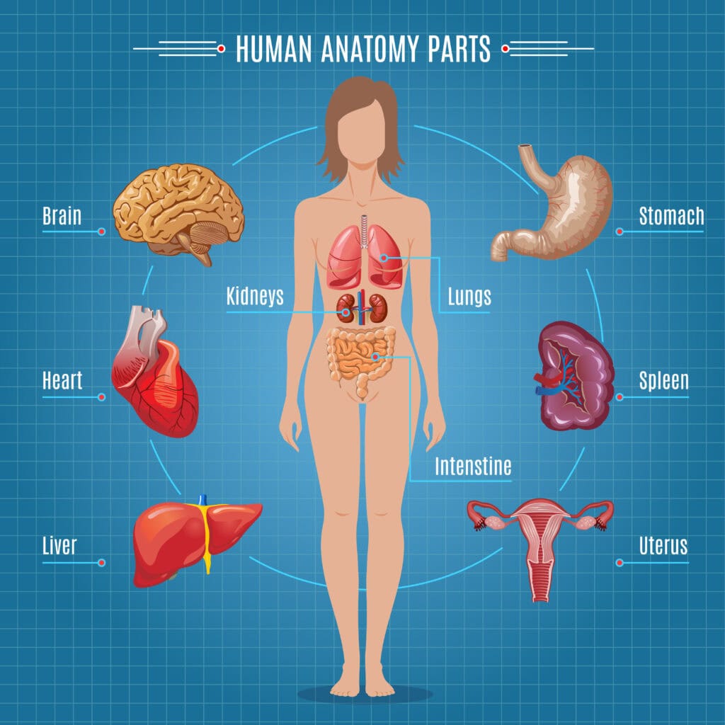
A female anatomy diagram provides a detailed visual representation of the key structures and organs in the female body.
By familiarizing yourself with the various organs, muscles, and systems, you can better identify areas of discomfort, potential health concerns, or just gain a deeper appreciation for the body’s incredible design.
Common topics covered in a female anatomy diagram include the reproductive system, the circulatory system, the skeletal system, and the muscular system. For example, recognizing changes in the reproductive system can help detect conditions like polycystic ovary syndrome (PCOS) or fibroids early on.
Moreover, studies have shown that visual aids, like detailed anatomy diagrams, significantly enhance our understanding and retention of information improving communication between patients and healthcare providers by up to 50%.
Looking for a female anatomy diagram? This guide will help you understand the key parts of the female reproductive system, including external and internal organs, and their functions.
Overview of Female Reproductive Anatomy
The female reproductive system is a marvel of biological engineering, comprising both external and internal organs that work in harmony to perform several critical functions. The external genitalia, such as the vulva, protect the internal reproductive organs and play a significant role in sexual pleasure.
Internally, the uterus, ovaries, and fallopian tubes are essential for reproduction, hormone regulation, and nurturing new life. Additionally, understanding the external female anatomy is crucial for comprehensive knowledge of the female reproductive system.
This system’s primary functions include hormone production, facilitating sexual intercourse, enabling pregnancy, and supporting breastfeeding. Each part, from the external genitalia to the ovaries, serves a specific purpose in maintaining reproductive health and ensuring the continuity of life.
External Female Genitalia
The term “vulva” refers collectively to the external female genitalia, which include:
the clitoris
vulvar vestibule
labia majora
labia minora
external urethral orifice
vaginal opening
hymen
These structures are not just anatomical features; they are guardians of the internal reproductive organs, shielding them from infections and injuries.
The labia majora are prominent, protective folds of skin that cover more delicate structures like the labia minora, clitoris, and vaginal opening. The labia minora are smaller, inner folds that encircle the clitoris and form the borders of the vulvar vestibule. Below the urethral opening lies the vaginal opening, a crucial component of the reproductive system.
Clitoris: A highly sensitive organ with numerous nerve endings that contribute to sexual pleasure.
Labia Majora: Protective outer folds that cover the inner structures.
Labia Minora: Inner folds that encircle the clitoris and form the vulvar vestibule.
Vaginal Opening: Located below the urethral opening, crucial for sexual intercourse and childbirth.
Each of these structures plays a role not only in protection but also in sexual arousal and pleasure, making them integral to the female reproductive anatomy.
Functions of External Genitalia
The external genitalia are multifunctional, serving as protectors, facilitators of sexual pleasure, and contributors to sexual arousal.
The mons pubis cushions the pubic bone during sexual intercourse and secretes pheromones, which may enhance attraction. The labia majora and labia minora protect the internal reproductive organs from infections and injuries, including the external genitals and pubic hair.
During sexual arousal, the vestibular bulbs and labia majora become engorged with blood, providing additional stimulation and enhancing pleasure. These functions highlight the importance of the external genitalia in both protective and sensory capacities, making them essential components of the female reproductive system.
Internal Female Reproductive Organs
The internal reproductive organs consist of the vagina, cervix, uterus, fallopian tubes, and ovaries, all of which are essential for reproduction. These organs work together to facilitate sexual arousal, intercourse, conception, pregnancy, and childbirth within the female reproductive tract.
Each organ has a specific role: the vagina serves as a conduit for menstrual fluid and a passage for childbirth, the cervix separates the vagina and uterus, and the uterus houses the fetus during pregnancy. The fallopian tubes transport eggs from the ovaries to the uterus, while the ovaries produce eggs and hormones.
The Vagina
The vagina is a muscular canal that plays a versatile role in the female reproductive system. It serves as a conduit for menstrual fluid, a passage for childbirth, and a receptacle for the penis during sexual intercourse. During childbirth, the vagina expands to allow the passage of the baby, showcasing its remarkable elasticity and strength.
Apart from its reproductive functions, the vagina also contributes to sexual pleasure, making it a crucial component of the female reproductive anatomy.
The Cervix
The cervix is located at the lower section of the uterus. It serves as a connection to the vagina. It serves as a gateway, allowing menstrual blood to pass from the uterus to the vagina and enabling sperm to enter the uterus during sexual intercourse.
The Uterus
The uterus is a pear-shaped muscular organ that plays a pivotal role in reproduction. It develops a lining, known as the endometrium, which is essential for supporting a fertilized egg. If fertilization occurs, the zygote implants in the uterine lining and develops into an embryo.
If the egg is not fertilized, the uterus sheds its lining during the menstrual cycle. Conditions like endometriosis and uterine fibroids can affect the uterus, causing pain and other complications.
Fallopian Tubes
The fallopian tubes are bilateral structures that connect the ovaries to the uterus. Their primary function is to transport the ovum from the ovaries towards the uterus.
These tubes also provide a site for fertilization, where sperm can meet and fertilize the egg.
Ovaries
The ovaries are small, almond-shaped organs that store eggs and produce hormones like estrogen and progesterone. At birth, a female has approximately 1 million to 2 million eggs in her ovaries, but this number decreases to around 300,000 to 500,000 by the time of the first period.
At the age of 37, a woman typically has around 27,000 eggs left. This number reflects the natural decline in egg quantity over time.
Blood Supply and Nerve Endings
The primary blood supply for the external female genitalia comes from the internal pudendal artery, with additional supply provided by its branches. Venous drainage is primarily through the external and internal pudendal veins, ensuring efficient circulation of blood vessels.
The pudendal nerve, originating from the second to fourth sacral spinal roots, is crucial for the sensory and motor innervation of the external genitalia. The genitofemoral nerve provides additional sensory innervation to the mons pubis.
Lymphatic drainage primarily leads to the superficial inguinal lymph nodes, with lymph from the clitoris draining into the deep inguinal lymph nodes.
Hormonal Regulation and Menstrual Cycle
The female reproductive system’s primary role includes facilitating sexual intercourse, hormone production, and enabling reproduction. Ovaries not only produce eggs but also secrete hormones that regulate the menstrual cycle and reproductive processes.
Key phases of the menstrual cycle include:
Ovulation: Typically occurs around day 14 of a standard 28-day menstrual cycle.
Luteal Phase: Characterized by the dominance of progesterone and averages about 14 days.
Menstruation: Triggered by a sharp decline in progesterone and estradiol levels.
The menstrual cycle is divided into two main cycles: the ovarian cycle and the endometrial cycle. The uterine lining thickens to prepare for the potential implantation of a fertilized egg, and if fertilization does not occur, the uterine lining sheds during menstruation.
Female Breast Anatomy
The female breast consists of glandular tissue organized into lobes, with each lobe containing smaller lobules connected to milk ducts. This glandular tissue is responsible for milk production, crucial for breastfeeding.
Breasts are primarily composed of adipose (fat) tissue, which contributes to their size and shape and provides cushioning. Montgomery’s glands, located in the areola, secrete an oily substance that protects the nipple and prevents chafing during breastfeeding.
Female Anatomy Diagram Labeled
Health Conditions Affecting Female Anatomy
Pelvic inflammatory disease (PID) can lead to infertility if left untreated, as it affects the reproductive organs. Symptoms of PID can include pelvic pain, fever, and unusual discharge; if untreated, it may result in long-term complications such as infertility.
Vulvar Cancer: A rare but serious condition often presenting with symptoms like persistent itching and changes in the vulvar skin.
Signs of Vulvar Cancer: Persistent itching, changes in vulvar skin color, or the appearance of lumps or sores.
These health conditions underscore the importance of regular medical check-ups and early diagnosis to ensure reproductive health and well-being.
Sexual Arousal and Pleasure
The clitoris, with its high density of nerve endings (around 8,000), is a primary contributor to sexual pleasure. Clitoral stimulation is often necessary for achieving orgasm among individuals with vaginas. The dorsal nerve of the clitoris is responsible for sensory input related to clitoral erection.
The G-spot is part of the clitoral complex, contributing to sexual pleasure. Anatomical features sensitive to touch, such as the clitoris, vulva, vagina, and breasts, play crucial roles in sexual arousal.
Conception and Pregnancy
During the ovulation phase of the menstrual cycle, an ovary releases an egg. Fertilization typically occurs in the fallopian tubes when sperm fertilizes an egg, creating a zygote. The zygote then travels to the uterus and divides into a blastocyst.
Implantation involves the blastocyst attaching to the uterine lining, and successful implantation triggers hormone release to maintain the uterine lining. This process underscores the intricate coordination within the female reproductive system.