

The supinator muscle is in the forearm and helps rotate your palm upward in a movement known as supination. Essential for actions like turning doorknobs or using tools, this muscle’s anatomy and function are crucial for diagnosing and treating forearm issues.
In this article, we delve into the supinator’s structure, Diagram, function, clinical relevance, and ways to keep it healthy and strong.
Key Takeaways
- The supinator muscle, located in the posterior forearm, is essential for forearm supination and interacts closely with the radial nerve, impacting forearm movements.
- Injuries or conditions such as supinator entrapment syndrome can occur due to repetitive motions, leading to radial nerve compression and affecting forearm functionality.
- Effective strengthening and rehabilitation protocols, including targeted exercises and stretching, are critical for maintaining the supinator muscle’s health and preventing injuries.
Supinator Muscle Anatomy
The supinator muscle, located in the posterior compartment of the forearm, is integral to forearm movement. This muscle’s anatomy is a testament to its complex and essential role in rotating the forearm.
Grasping its structure helps in understanding its function and clinical relevance.
Origin
The supinator muscle originates from several anatomical landmarks. The radial collateral ligament begins at the lateral epicondyle of humerus and connects to the annular ligament and the supinator crest of the ulna. Additionally, it attaches to the posterior surface of the ulna, providing a robust foundation for its function.
These origins are key to understanding the supinator muscle’s role in forearm movement and its anatomical relationships.
Insertion
The supinator muscle inserts on the proximal third of the radius. It covers both the anterior surfaces and posterior surfaces of this region, ensuring efficient transmission of force during supination movements.
This insertion enables the muscle to rotate the radius efficiently, ensuring smooth and controlled forearm supination.
Layers
The supinator muscle is composed of two distinct layers: a superficial layer and a deeper layer. The superficial layer lies over the deep layer, and both layers contribute to the muscle’s overall function and efficiency.
The deep layer encircles the neck of the radius above the radial tuberosity, significantly contributing to forearm movements.
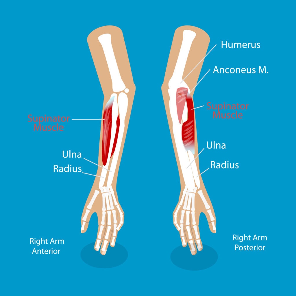
Location of the Supinator Muscle
The supinator muscle resides in the posterior compartment of the forearm, above the radial tuberosity, and curves around the upper third of the radius. This positioning is strategic for its primary function of forearm supination.
The muscle’s two layers of fibers encapsulate the radial shaft, providing stability and control during movement.
The deep branch of the radial nerve passes between these two layers, highlighting the muscle’s crucial anatomical relationship with the nerve.
This proximity is significant in conditions like supinator entrapment syndrome, where repetitive supination movements can cause nerve compression and associated symptoms.
Supinator Muscle Diagram
Anatomical Relationships
The supinator muscle’s anatomical relationships are vital for understanding its function and potential clinical issues.
Positioned in the deep compartment of the posterior forearm, this muscle wraps around the upper third of the radius, establishing connections with various structures. Its proximity to the radial nerve is particularly important, as this relationship influences both mobility and stability in the forearm.
Repetitive forearm movements, especially supination and pronation, increase the risk of developing conditions like supinator entrapment syndrome. Compression of the deep branch of the radial nerve near the supinator muscle can result in paralysis of associated muscles, emphasizing the clinical importance of these anatomical relationships.
Cubital Fossa
The cubital fossa, located in the anterior aspect of the elbow, serves as a crucial anatomical landmark for clinical examinations. The floor of this fossa is formed by the brachialis and supinator muscles, contributing to its structural integrity. The supinator muscle forms the distal part of the floor, crucial for the proper function of the elbow joint.
Radial Nerve
The radial nerve’s interaction with the supinator muscle is critical for forearm function. The deep branch of the radial nerve travels through the supinator muscle. It then transforms into the posterior interosseous nerve. This nerve is crucial for innervating various forearm muscles, including the supinator.
Innervation and Blood Supply
Innervation and blood supply are vital for the supinator muscle’s function and health. The posterior interosseous nerve, originating from spinal nerves C7 and C8, is responsible for its innervation.
The pathway of this nerve through the supinator muscle is crucial for its function.
Innervation
The posterior interosseous nerve primarily innervates the supinator muscle. This nerve is a branch of the radial nerve. This deep branch travels through the supinator muscle and emerges as the posterior interosseous nerve.
The precise innervation ensures the supinator muscle functions efficiently.
Blood Supply
The blood supply to the supinator muscle is complex, with different sources nourishing its superficial and deep layers. The superficial layer receives blood from the radial artery via its radial recurrent branch, and the deep layer is supplied by the ulnar artery through the posterior interosseous and posterior interosseous recurrent arteries.
The dual arterial supply ensures the muscle receives adequate oxygen and nutrients for optimal function.
Function of the Supinator Muscle
The supinator muscle plays a pivotal role in forearm movements, primarily responsible for supinating the forearm. This action involves rotating the radius laterally, placing the radius parallel to the ulna.
Its cylindrical shape and strategic positioning on the posterior side of the forearm enable efficient performance of this function.
Forearm Supination
The supinator muscle primarily drives slow and unopposed supination movements. It laterally rotates the radius at the proximal radioulnar joint, facilitating forearm supination.
During rapid or heavy supination efforts, the biceps brachii enhances the supinator muscle’s action, ensuring smooth and powerful rotation.
Synergistic Action
The supinator muscle also collaborates with other muscles during forearm supination. In slow, unopposed movements, it acts as the prime mover.
In more dynamic movements, it works with the biceps brachii and other forearm muscles to achieve fluid and controlled supination.
Strengthening the Supinator Muscle
Strengthening the supinator muscle enhances forearm stability and improves grip strength. Focusing on specific exercises can significantly enhance the muscle’s functionality and prevent conditions like supinator entrapment syndrome.
Exercises for Supination
Several exercises effectively target the supinator muscle. Supination with dumbbells and resistance bands is particularly beneficial. Reverse wrist curls and towel wrist curls also engage the supinator muscle and improve its strength.
Tips for Effective Strengthening
Using lighter weights, especially for beginners, helps focus on proper form and prevent injuries when strengthening the supinator muscle. Consistent practice and a variety of exercises prevent plateaus and enhance muscle development.
Exercises like the overhead press can also strengthen the supinator and other upper body muscles simultaneously.
Stretching and Rehabilitation for the Supinator Muscle
Stretching and rehabilitation maintain the flexibility and functionality of the supinator muscle. Injuries from overuse or trauma often lead to symptoms like swelling and limited mobility.
Stretches can enhance flexibility and aid in correcting supination.
Stretches to Improve Supinator Flexibility
Stretches like the supinator stretch technique and wrist rotation stretches can significantly improve the flexibility of the supinator muscle. A forearm supination stretch, rotating the palm upwards while the forearm is supported on a flat surface, is particularly effective.
Wrist flexion and extension stretches relieve tension in the forearm, contributing to overall muscle health.
Rehabilitation Protocols After Injury
Rehabilitation after a supinator muscle injury involves several steps. Resting the affected area and applying ice initially helps reduce inflammation. Gentle exercises like wrist curls and controlled supination movements become essential once initial inflammation decreases.
Consulting a physical therapist helps create a tailored rehabilitation program that ensures a gradual increase in activity levels to prevent re-injury.
Clinical Relevance
The supinator muscle’s clinical relevance extends to various forearm conditions. Rapid supination, especially during quick, unopposed movements, depends heavily on this muscle.
Compression of the radial nerve at the arcade of Frohse can result in symptoms similar to lateral epicondylalgia, highlighting the muscle’s clinical importance.
Supinator Entrapment Syndrome
Repetitive forearm movements often cause supinator entrapment syndrome, leading to compression of the radial nerve and potentially resulting in nerve syndrome. Symptoms, including pain and weakness in the forearm or wrist, can significantly impact daily activities.
Patients may experience deep aching pain in the lateral elbow, which worsens with activities involving forearm rotation.
Radial Nerve Pathologies
Radial nerve pathologies can cause motor dysfunction and sensory deficits due to its interaction with the supinator muscle. Recognizing these pathologies is crucial for diagnosing and treating conditions affecting forearm mobility and stability.
Assessment Techniques
Assessing the supinator muscle’s health and function is crucial for diagnosing forearm-related conditions and injuries. Physical examination and diagnostic imaging both play essential roles in this process.
Physical Examination
Practitioners often palpate the area around the proximal radius while testing for resistance during forearm supination to assess the supinator muscle. This involves extending the patient’s arm with the forearm in a neutral position before applying resistance.
Observing muscle contraction and palpating the proximal radius help evaluate the muscle’s function and detect abnormalities.
Diagnostic Imaging
Imaging techniques like MRI and ultrasound visualize the supinator muscle and adjacent structures for abnormalities. MRI is particularly useful for detailed visualization of the muscle, revealing structural abnormalities or injuries.
Ultrasound imaging offers real-time dynamic assessment, aiding in the accurate diagnosis and management of muscle pathologies.
Wrap Up
From its intricate origin and insertion points to its layered structure and vital innervation and blood supply, each aspect contributes to its efficient function.
Frequently Asked Questions
What is the primary function of the supinator muscle?
The primary function of the supinator muscle is to supinate the forearm by rotating the radius laterally, which turns the palm upwards. This action is crucial for various activities requiring palm orientation.
How can I strengthen my supinator muscle?
To strengthen your supinator muscle, focus on exercises like supination with dumbbells, resistance band supination, reverse wrist curls, and towel wrist curls. Consistently performing these exercises will enhance muscle strength effectively.
What are the symptoms of supinator entrapment syndrome?
The symptoms of supinator entrapment syndrome primarily manifest as deep aching pain in the lateral elbow, along with pain and weakness in the forearm or wrist, particularly exacerbated by activities that involve forearm rotation.
How is the supinator muscle innervated?
The supinator muscle is innervated by the posterior interosseous nerve, a branch of the radial nerve, which passes through the muscle. This innervation enables its function in supinating the forearm.


