

The thyroid cartilage is an essential structure in your neck. It protects the vocal cords and supports the larynx. In this article, learn about its anatomy, diagram, functions, and clinical importance.
Key Takeaways
- The thyroid cartilage is a vital component of the laryngeal skeleton, providing protection for the vocal cords and facilitating voice modulation through its structural features and muscle attachments.
- Developmental variations and pathologies of the thyroid cartilage, such as fractures, infections, and tumors, highlight the need for accurate clinical assessment using imaging techniques like ultrasound, MRI, and CT scans.
- The relationship between thyroid cartilage and other laryngeal cartilages, including the cricoid and arytenoid, is essential for maintaining laryngeal function during respiration and phonation.
Anatomy of the Thyroid Cartilage
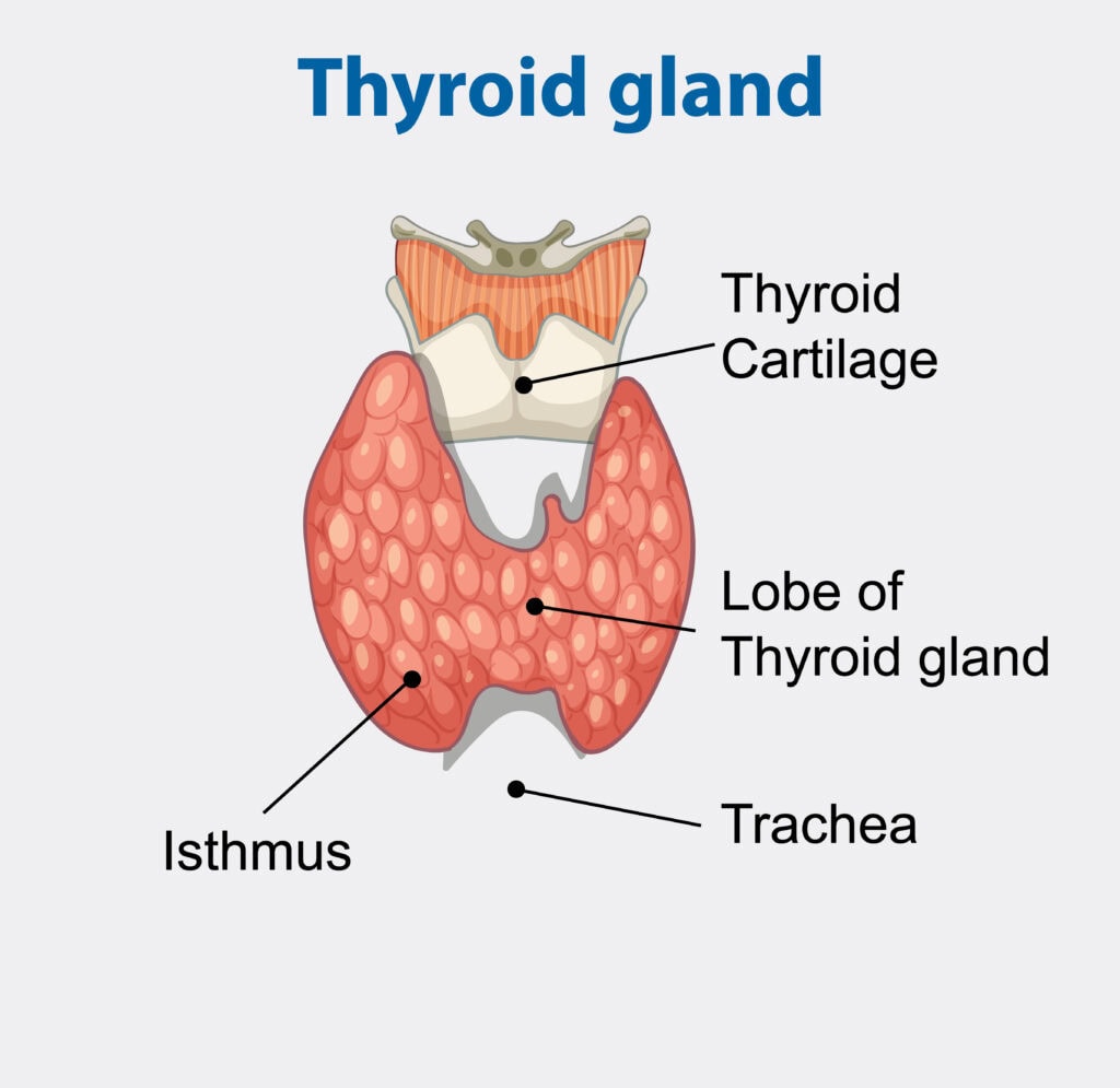
The thyroid cartilage is a tough, flexible tissue that forms the front part of the larynx, serving as a key component of the laryngeal skeleton. It consists of two thyroid laminae that meet at a peak called the laryngeal prominence, commonly known as the Adam’s apple.
This prominent feature is more noticeable in males due to the angle at which the laminae meet, creating a visible protrusion.
Located underneath the hyoid bone and above the thyroid gland, the thyroid cartilage is intricately tied to the hyoid bone by the thyrohyoid membrane. This positioning allows the thyroid cartilage to serve multiple functions, such as protecting laryngeal structures and providing muscle attachment points.
The thyroid cartilage forms a significant part of the larynx cartilage, contributing to the structure and function of the voice box. Features like the thyroid laminae and laryngeal prominence are key to understanding anatomy head and neck.
The cricoid cartilage, situated below the thyroid cartilage, forms a critical connection point, further highlighting the importance of the thyroid cartilage in the laryngeal skeleton.
Transitioning into the details, we will explore the specific features of the thyroid laminae and notches, the superior and inferior horns, and the oblique line and muscle attachments. These details deepen the understanding of thyroid cartilage anatomy and its components.
Thyroid Laminae and Notches
Each thyroid lamina is a flat plate that joins at the front, forming a prominent notch. This V-shaped notch is a distinctive feature, contributing to the overall structure of the thyroid cartilage. The thyroid cartilage features both a superior thyroid notch and an inferior thyroid notch, each playing a role in the anatomy of the laryngeal cartilages.
The superior notch notably forms the visible protrusion called the adam’s apple or laryngeal prominence.
These notches and the laminae they form are crucial for the structural integrity of the thyroid cartilage, aiding in its primary functions such as protecting the laryngeal cavity and providing attachment points for ligaments and muscles.
Superior and Inferior Horns
The superior and inferior horns of the thyroid horns cartilage are essential projections that contribute to its overall structure. The superior horn extends towards the hyoid bone, and the inferior horn connects with the cricoid cartilage at the cricothyroid joint.
These horns provide critical attachment points for ligaments and play a vital role in the movement and stability of the laryngeal skeleton.
Oblique Line and Muscle Attachments
The oblique line is a prominent feature located on the lateral surface of the thyroid lamina. This anatomical landmark is crucial as it delineates areas for muscle attachment, influencing laryngeal function.
Several important muscles, including the sternothyroid and thyrohyoid muscles, attach to the oblique line, playing a significant role in regulating the position and tension of the thyroid cartilage.
These muscle attachments are vital for various functions such as voice production and airway dynamics. These laryngeal muscles regulate the thyroid cartilage’s movement and tension, ensuring optimal laryngeal function for sound production and vocal cord protection, involving several muscles.
Function of the Thyroid Cartilage
The thyroid cartilage serves multiple crucial functions, making it an indispensable part of the laryngeal skeleton.
One of its primary functions is to protect and support the vocal cords, safeguarding the delicate structures of the larynx from external trauma. Additionally, it plays a vital role in voice modulation by providing a structural foundation for the associated laryngeal musculature involved in sound generation.
Developmental variations in the thyroid cartilage can significantly influence respiratory and vocal functions, highlighting its importance in sound production.
The thyroid cartilage, being one of the nine cartilages in the laryngeal skeleton, collaborates with other cartilages to maintain airway structure and facilitate sound production.
The following subsections delve deeper into its specific functions.
Protecting Laryngeal Structures
The thyroid cartilage acts as a shield for the larynx, which protects it from external trauma. This protective role is paramount, especially in safeguarding the vocal cords against injury during activities such as speaking and swallowing.
Its superficial position makes it the primary shield for delicate laryngeal structures, ensuring that the vocal cords remain unharmed during various actions.
Modulating Voice Pitch
The thyroid cartilage plays a crucial role in modulating voice pitch during vocalization. Its position and shape adjustments influence vocal cord tension, thereby affecting pitch and voice quality.
This adjustment is facilitated through its movement at the cricothyroid joint, allowing for precise control over voice modulation.
When the thyroid cartilage tilts forward, it increases the tension on the vocal cords, resulting in a higher pitch.
Conversely, when it tilts backward, the tension decreases, producing a lower pitch. This dynamic adjustment is crucial for various vocal expressions, enabling us to produce a range of sounds and tones.
Muscle Attachment Points
The oblique line on the thyroid lamina serves as an attachment point for several key muscles involved in phonation and respiration.
These muscles, including the sternothyroid and thyrohyoid, are crucial for moving the larynx and controlling vocalization. The thyroid cartilage anchors these muscles, facilitating complex vocal functions and protecting the airway.
Thyroid Cartilage Diagram
Developmental Variants and Clinical Significance
Developmental variations in the thyroid cartilage are common and often benign, meaning they typically do not have clinical implications. However, understanding these variations is essential, especially in clinical settings where accurate diagnosis and treatment are paramount.
Laryngeal ultrasound is a valuable tool for identifying human anatomy variations, aiding in the detection of anomalies that might otherwise go unnoticed.
Neck sonography is a prevalent imaging approach for assessing variations in the thyroid cartilage. This technique allows for detailed visualization, helping clinicians distinguish between normal anatomical variations and potential pathologies.
The following subsections explore specific Developmental variant and their clinical significance.
Ossification and Calcification
Ossification and calcification are natural processes that occur in the thyroid cartilage as we age. The process typically begins in the lower part of the thyroid lamina and the inferior horn, gradually spreading over time.
This slow calcification process doesn’t usually have clinical significance but is an important consideration in imaging studies and clinical assessments.
Cyst-like Changes
Cyst like change in the thyroid cartilage are generally benign and have no clinical significance. These cyst-like lesions are not regarded as pathologies and are often discovered incidentally during imaging studies.
Understanding these changes is crucial for clinicians to avoid unnecessary interventions and to reassure patients about the benign nature of these findings.
Clinical Cases and Findings
A clinical case report involved a 32-year-old woman who was referred for neck sonography and MRI. The imaging studies revealed no contrast enhancement on the T1-weighted postcontrast image, indicating normal findings.
The final clinical outcome was considered normal, underscoring the benign nature of the observed clinically oriented anatomy variations.
Chondrosarcomas and chondromas are conditions that are included in the differential diagnosis of thyroid cartilage lesions. They are important to consider when evaluating such lesions.
However, the benign nature of most findings highlights the importance of accurate diagnosis and careful evaluation to avoid misdiagnosis and unnecessary treatments.
Related Laryngeal Cartilages
The thyroid cartilage is the largest and one of the most significant components of the laryngeal skeleton, but it’s not alone in its function.
The larynx is composed of nine cartilages, including the cricoid, arytenoid, and epiglottis, which all play crucial roles in maintaining the structure and function of the voice box. Understanding the relationship between these cartilages can provide a more comprehensive view of laryngeal anatomy and physiology.
The cricoid cartilage, for instance, articulates with the thyroid cartilage below, forming a key connection in the laryngeal structure.
Similarly, the arytenoid cartilages and the epiglottis have their unique roles and interactions with the thyroid cartilage, ensuring the smooth operation of the larynx during respiration and phonation.
Let’s delve into each of these related laryngeal cartilages in the following subsections.
Cricoid Cartilage
The cricoid cartilage is a complete ring of hyaline cartilage that provides structural support to the larynx and connects to the thyroid cartilage at the cricothyroid joint, forming the cricoid ring. This connection is crucial as it allows for the movement and adjustment necessary for voice modulation and airway protection.
The superior horn of the thyroid cartilage extends upward towards the hyoid bone, while the inferior horn connects to the cricoid cartilage, highlighting the integral relationship within the laryngeal skeleton.
Arytenoid Cartilages
The arytenoid cartilages, shaped like pyramids, are crucial for adjusting the tension of the vocal cords during sound production. These cartilages play a key role in the movement of the vocal cords, allowing them to abduct and adduct as needed for phonation.
This movement occurs at the cricoarytenoid joint, enabling precise control over voice pitch and quality.
Epiglottis
The epiglottis is a leaf-shaped structure composed of elastic cartilage, which gives it the flexibility needed to cover the larynx. Acting as a protective flap, the epiglottis covers the airway during swallowing, preventing food and liquids from entering the respiratory tract.
This critical function ensures that the airway remains clear, allowing for safe and efficient breathing and swallowing.
Thyroid Cartilage and Imaging Techniques
Imaging techniques such as ultrasound, MRI, and CT scans are invaluable tools for visualizing the thyroid cartilage and identifying any abnormalities.
These techniques provide unique perspectives on the thyroid cartilage, aiding in the detection of normal variations and potential pathologies. Cyst-like structures in the thyroid cartilage, for example, are often discovered incidentally during imaging studies and are usually benign.
Such imaging studies are crucial in clinical settings, where accurate diagnosis can prevent misinterpretations and unnecessary treatments. The ability to visualize the thyroid cartilage in detail helps clinicians distinguish between benign variations and more serious conditions that may require intervention.
Let’s explore the specific imaging techniques used for studying the thyroid cartilage in the following subsections.
Laryngeal Ultrasound
Laryngeal ultrasound is a key imaging technique for visualizing the thyroid cartilage. This non-invasive method allows clinicians to detect anatomical variants and assess the thyroid cartilage in detail.
Neck sonography is commonly used for this purpose, providing high-resolution images that aid in the evaluation of both normal and abnormal findings.
MRI and CT Scans
MRI offers superior soft tissue contrast compared to CT, making it highly effective for assessing the thyroid cartilage and surrounding structures. However, MRI is often limited by cost and availability.
CT scans, on the other hand, are commonly employed to evaluate the extent of thyroid cartilage involvement in various conditions, including incidental thyroid nodules, which are frequently detected and often benign.
Both MRI and CT scans play crucial roles in the comprehensive assessment of the thyroid cartilage.
Pathologies Involving the Thyroid Cartilage
The thyroid cartilage is vital for protecting the larynx and vocal cords by providing a sturdy anterior wall. However, it is not immune to various pathologies, including fractures, infections, and tumors.
Understanding these conditions is crucial for effective diagnosis and treatment. Imaging methods such as CT and ultrasound are essential for assessing thyroid cartilage in cancer cases, offering high diagnostic accuracy.
MRI is generally more sensitive than CT for detecting middle thyroid cartilage invasion, with sensitivity rates reaching up to 96%. These imaging techniques help clinicians identify the extent of cartilage involvement, guiding appropriate treatment strategies.
Fractures and Trauma
Fractures of the thyroid cartilage can occur due to incidents such as motor vehicle accidents and sports injuries. Management of these fractures may range from rest and non-invasive treatments to surgical intervention, depending on the severity of the injury.
Infections and Inflammation
Thyroiditis is an inflammatory condition affecting the thyroid cartilage, which may lead to symptoms like pain and swelling. This condition can occur in various forms, including autoimmune thyroiditis like Hashimoto’s and subacute thyroiditis.
Acute infectious thyroiditis is a rare form caused by an infectious organism and typically requires antibiotic treatment.
Tumors and Malignancies
Tumors in the thyroid cartilage can vary from benign growths to malignancy cancers, significantly impacting treatment approaches and prognosis.
Symptoms may include hoarseness or difficulty breathing, necessitating prompt medical attention. Malignancies of the thyroid cartilage are rare but can occur, requiring surgical intervention and possibly additional therapies.
The prognosis for thyroid cancer is generally favorable, especially for common types like papillary thyroid cancer.
Risk factors for thyroid cancer include female sex, exposure to radiation, and genetic predispositions. Treatment options may involve surgery, radioactive iodine therapy, and in some cases, chemotherapy. Early diagnosis and treatment are crucial for improving outcomes.
Wrap Up
The thyroid cartilage is a fundamental component of the laryngeal skeleton, playing crucial roles in protecting laryngeal structures, modulating voice pitch, and serving as an attachment point for various muscles.
Frequently Asked Questions
What is the primary function of the thyroid cartilage?
The primary function of the thyroid cartilage is to protect and support the vocal cords, thus safeguarding the delicate structures of the larynx from external trauma.
How does the thyroid cartilage modulate voice pitch?
The thyroid cartilage modulates voice pitch by adjusting its position and shape, thereby affecting the tension of the vocal cords. This dynamic alteration is crucial for producing a range of vocal sounds.
What are some common developmental variants of the thyroid cartilage?
Common developmental variants of the thyroid cartilage consist of ossification and calcification, usually initiating in the lower sections of the lamina and inferior horn. These variations can affect the structure and function of the thyroid cartilage.
How are cyst-like changes in the thyroid cartilage typically discovered?
Cyst-like changes in the thyroid cartilage are typically discovered incidentally during imaging studies, such as ultrasound or CT scans. Therefore, regular imaging may reveal these alterations even in the absence of symptoms.
What imaging techniques are used to assess the thyroid cartilage?
Laryngeal ultrasound, MRI, and CT scans are effective imaging techniques for assessing the thyroid cartilage, each providing distinct advantages in evaluation.


