
What is the tibial nerve and why is it important? The tibial nerve is crucial for movement and sensation in the lower leg and foot. This article covers its anatomy, functions, and related disorders to provide a clear and comprehensive understanding.
Key Takeaways
The tibial nerve branches from the sciatic nerve and plays a key role in innervating muscles and providing sensation in the lower leg and foot.
It has multiple branches for motor and sensory functions, including significant roles in plantarflexion and sensation in the foot and heel.
Common disorders related to the tibial nerve include tarsal tunnel syndrome and injuries, which can significantly impact mobility and may require surgical intervention.
Anatomical Pathway of the Tibial Nerve
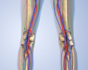
The tibial nerve is a major player in the complex orchestra of our nervous system, branching off from the sciatic nerve, which itself is a powerhouse of lower limb innervation. As it descends through the popliteal fossa, located behind the knee, it continues its journey alongside the popliteal artery, ensuring a close-knit relationship with crucial vascular structures.
From the popliteal fossa, the tibial nerve travels deep into the calf, nestled among the muscles. This pathway is not just a straight road; it’s a corridor bustling with anatomical interactions. The nerve’s route under the gastrocnemius and soleus muscles showcases its strategic positioning, allowing it to innervate key muscles and provide sensory feedback from different parts of the lower leg and foot.
Tibial Nerve Diagram
Branches of the Tibial Nerve
The tibial nerve is a master of multitasking, giving rise to numerous branches that ensure both motor and sensory functions throughout the leg and foot. These branches are categorized into muscular, cutaneous, articular, and terminal types, each with specific roles.
Understanding these branches helps us appreciate the nerve’s comprehensive role in lower limb functionality.
3.1 Muscular Branches
The muscular branches of the tibial nerve are primarily responsible for motor innervation. They supply 21 muscles in the posterior leg, including the powerful gastrocnemius, soleus, and plantaris muscles. These muscles are essential for movements such as plantarflexion-key for walking, running, and maintaining balance.
3.2 Cutaneous Branches
On the sensory side, the tibial nerve gives rise to several cutaneous branches. Notable among these is the sural nerve, which provides sensation to the skin over the lower leg and lateral aspect of the foot. Additionally, the medial calcaneal nerve supplies the skin on the heel, ensuring we can feel the ground beneath us.
3.3 Articular Branches
Articular branches of the tibial nerve are pivotal for joint sensation. They innervate the knee and ankle joints, playing a crucial role in joint movement and stability. This sensory feedback is essential for coordinated movement and avoiding injuries.
3.4 Terminal Branches
As the tibial nerve reaches the foot, it divides into its terminal branches: the medial and lateral plantar nerves. These branches are responsible for both sensory and motor functions in the foot, including the intricate control of the toes and the sensation across the plantar surface.
Tarsal Tunnel
The tarsal tunnel is a narrow passageway located behind the medial malleolus, formed by the flexor retinaculum. Within this tunnel, the tibial nerve runs alongside the posterior tibial vessels, protected yet vulnerable to compression. Compression of the tibial nerve in this area can lead to tarsal tunnel syndrome, characterized by pain, tingling, and numbness in the foot.
Tarsal tunnel syndrome is often caused by swelling, injuries, or anatomical variations that put pressure on the nerve. Understanding the anatomy of the tarsal tunnel is crucial for diagnosing and treating this condition, which can significantly impact mobility and quality of life.
Course of the Tibial Nerve
The tibial nerve’s journey from the thigh to the foot is a testament to its critical role in lower limb function. As it descends from the sciatic nerve, it navigates through various anatomical landmarks, including the popliteal fossa and the tarsal tunnel, ensuring it can deliver motor and sensory innervation effectively.
In the Thigh
In the thigh, the tibial nerve travels deep to the gastrocnemius muscle and along the distal border of the popliteus muscle. Its journey through the popliteal fossa, accompanied by the popliteal artery, highlights its strategic placement for innervating the lower leg.
This course ensures it is well-protected yet accessible for its numerous branches.
In the Leg
Once in the leg, the tibial nerve descends beneath the soleus muscle, accompanied by the posterior tibial vessels. It travels through the tarsal tunnel, a critical passage that protects the posterior tibial nerve but can also be a site of compression.
This pathway ensures the nerve can reach and innervate all necessary structures in the lower leg and foot.
In the Foot
In the foot, the tibial nerve bifurcates into the medial and lateral plantar nerves. These terminal branches provide motor and sensory innervation to the foot, playing essential roles in foot movement and sensation. This division ensures precise control and feedback for all areas of the foot.
Motor Functions of the Tibial Nerve
The tibial nerve is a powerhouse of motor control, overseeing the movement of 21 different muscles in the leg and foot. Its primary targets include the gastrocnemius, soleus, and plantaris muscles, crucial for actions like plantarflexion, which are vital for walking, running, and maintaining balance.
Posterior Leg Muscles
The posterior leg muscles, including the gastrocnemius, soleus, and tibialis posterior muscle, rely heavily on the tibial nerve for their function. The gastrocnemius, known as the calf muscle, is essential for plantarflexing the foot at the ankle joint.
The soleus muscle works alongside it, providing stability during standing and walking.
Intrinsic Foot Muscles
The intrinsic foot muscles receive their innervation from the medial and lateral plantar nerves, branches of the tibial nerve. These muscles are essential for fine motor control of the toes and maintaining the arch of the foot, ensuring stability and balance during movement.
Sensory Functions of the Tibial Nerve
Sensory functions of the sensory nerve are vital for transmitting sensations from the leg and foot back to the brain. This nerve allows us to feel textures, temperature, and pain, making it crucial for protective reflexes and coordinated movements.
Cutaneous Innervation
Cutaneous innervation by the tibial nerve includes branches like the medial sural cutaneous nerve and sural nerve, which provide sensation to the back of the leg and the lateral aspect of the foot. The medial calcaneal nerve, another sensory branch, supplies the skin on the posterior aspect of the heel, ensuring comprehensive sensory coverage.
Medial and Lateral Plantar Nerves
The medial and lateral plantar nerves are key players in foot sensation. The medial plantar nerve supplies the skin on the medial aspect of the foot’s sole and the plantar surface corresponding to three and a half toes.
The lateral plantar nerve covers the lateral aspect of the foot, including the fourth and fifth toes, ensuring detailed sensory input across the lateral plantar branches.
Common Disorders Related to the Tibial Nerve
Disorders related to the tibial nerve can lead to significant mobility issues and chronic pain if not addressed promptly. Conditions like tarsal tunnel syndrome and tibial nerve injuries are common, each presenting unique challenges and requiring specific treatments.
Tarsal Tunnel Syndrome
Tarsal tunnel syndrome occurs due to compression of the tibial nerve within the tarsal tunnel, often caused by swelling, injuries, or anatomical variations. Symptoms include pain, tingling, and numbness in the foot.
Treatment ranges from conservative methods like rest and anti-inflammatory medications to surgical interventions for severe cases.
Tibial Nerve Injury
Tibial nerve injuries are usually the result of trauma, such as fractures or deep cuts, leading to sensory and motor deficits. Such injuries can cause significant functional impairment, and treatment often involves addressing the underlying cause and, in some cases, surgical repair for tibial nerve damage and tibial nerve dysfunction.
Surgical Considerations
Surgical interventions for tibial nerve issues are critical for restoring function and alleviating pain. These procedures include nerve decompression and repair, each requiring careful assessment and timely execution for optimal recovery.
5.1 Tibial Nerve Decompression
Decompression surgery is indicated for patients with significant nerve compression symptoms that do not improve with conservative treatment. The procedure involves relieving pressure on the tibial nerve, thereby restoring function and alleviating pain.
Proper surgical technique is crucial for successful outcomes.
5.2 Nerve Repair and Grafting
Nerve repair and grafting techniques are essential for severe tibial nerve injuries. These procedures may involve direct suturing of nerve ends or using grafts to bridge gaps. Early intervention typically leads to better recovery outcomes, emphasizing the importance of timely surgical care.
6. Anatomical Variations
Anatomical variations in the tibial nerve can significantly influence clinical outcomes. Differences in branching patterns and cross-sectional areas are often linked to conditions like foot neuropathy.
Recognizing these variations is crucial for accurate diagnosis and effective treatment.
6.1 Common Variations
Common variations in the tibial nerve include different bifurcation patterns and the presence of anomalous branches. These variations can affect the medial and lateral plantar nerves, influencing the innervation of the foot and potentially leading to unique clinical presentations.
Such variations are important considerations during surgical procedures and when diagnosing nerve-related conditions.


