
Visualize uncovering a firm, whitish-yellow bump on your skin that, initially, appears harmless. This perplexing condition, termed calcinosis cutis, can unfold a realm of concerns. Whether it’s tied to an autoimmune disease or a prior injury, comprehending why these calcium deposits emerge is vital for handling the scenario. Suppose treatments could assist in alleviating your discomfort? We should delve into the causes and management options for calcinosis cutis jointly.
What Is Calcinosis Cutis?
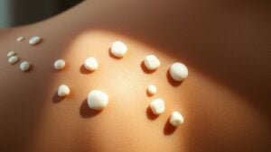
Calcinosis cutis could sound intimidating, but don’t worry; many others are also facing this unusual skin condition. Fundamentally, it involves abnormal calcium deposits in your skin and foundational tissues, leading to firm, whitish-yellow lumps called papules, plaques, or nodules.
You may experience these deposits due to various reasons. For instance, dystrophic calcinosis often occurs in damaged skin from autoimmune diseases like scleroderma.
Metastatic calcinosis is linked to systemic mineral imbalances, usually from chronic conditions like kidney disease. On the other hand, idiopathic calcinosis comes with no clear cause and appears in diverse forms.
Grasping these aspects can help you traverse your path with calcinosis cutis more confidently. Others share this experience!
Types of Calcinosis Cutis
Upon considering calcinosis cutis, you may find it unexpected to discover that multiple forms exist, each with distinct traits.
Dystrophic calcification arises in injured regions, whereas metastatic calcification connects to wider systemic concerns.
Next, there’s idiopathic calcinosis, where no obvious reason emerges-each variation contributes uniquely to grasping your situation.
Dystrophic Calcification Characteristics
Although it could seem concerning, dystrophic calcification frequently develops in regions where your tissue has been harmed or irritated, without any problems in your total calcium or phosphate levels. This type of calcification is the most common in conditions like systemic sclerosis and dermatomyositis.
You could notice firm, whitish or yellowish lesions appearing as papules or nodules, particularly on your fingers, elbows, or knees. These calcium deposits are linked to local tissue damage in collagen, elastin, or fat.
Sometimes, you could even experience ulceration, leading to a chalky discharge of calcium phosphate or carbonate. Grasping these characteristics can assist you in recognizing and addressing any concerns regarding your skin health effectively.
Metastatic Calcification Causes
Metastatic calcification can occur once your blood levels of calcium or phosphate become higher, often due to various root health issues. It’s commonly linked to conditions like chronic kidney disease or hyperparathyroidism, where abnormal calcium metabolism leads to skin deposits. other causes include hypervitaminosis d and milk-alkali syndrome.
you should know that this type of calcification occurs in normal tissues, unlike dystrophic calcification, especially as serum calcium-phosphate product exceeds 70 mg²/dl². common sites for these deposits include blood vessels, kidneys, and lungs.
keep an eye out for calciphylaxis, a severe condition that affects small blood vessels and can lead to necrosis, particularly in dialysis patients. it’s essential to monitor your health to manage these risks effectively.
Idiopathic Calcinosis Presentation
Idiopathic calcinosis cutis presents a unique set of challenges, as it arises without any prior tissue injuries or fundamental health issues related to calcium metabolism.
This condition often manifests as subcutaneous nodules that can appear either as solitary or multiple firm, white or yellow growths on your skin.
For instance, scrotal calcinosis leads to yellowish nodules on the scrotum, while children could develop hard, white papules on their face or extremities.
Additionally, milia-like idiopathic calcinosis, particularly in individuals with Down syndrome, shows tiny white facial papules that can clear up over time.
The rare tumoral calcinosis features large, painless calcified material around joints, causing little bother, but raises questions about its origins.
Causes of Calcinosis Cutis
When it comes to calcinosis cutis, you may ponder what leads to those calcium deposits in your skin.
There are several causes, ranging from fundamental health conditions like autoimmune diseases to iatrogenic factors tied to medical procedures.
Grasping these different causes can help you recognize the signs and know at what point to seek help.
Types of Calcinosis Cutis
Calcinosis cutis can take various forms, each with distinct causes that can impact your skin. You could encounter dystrophic calcinosis cutis, often linked to inflamed or damaged skin, especially in conditions like scleroderma.
Metastatic calcinosis cutis develops from abnormal calcium metabolism, frequently seen in kidney failure or hyperparathyroidism.
Then there’s idiopathic calcinosis cutis, which has no known cause and can include rare types like scrotal calcinosis.
Iatrogenic calcinosis cutis arises from medical interventions, such as calcium-rich IV treatments or multiple heel pricks in infants.
Grasping these types can help you talk to your doctor more effectively about your skin health and any changes you notice. Don’t hesitate to reach out should you have concerns.
Underlying Medical Conditions
Recognizing the circumstances that can trigger calcinosis cutis assists you in better associating the links between your skin changes and possible concealed health issues. Autoimmune diseases, such as systemic lupus erythematosus and systemic sclerosis, frequently result in these calcium deposits.
In case you have chronic kidney disease, especially in case you’re on dialysis, be mindful that you’re at a greater risk. Hyperparathyroidism can also raise calcium levels, leading to calcinosis cutis.
Additionally, trauma, infections, or inflammation can prompt localized deposits even without fundamental conditions. Genetic factors, like pseudoxanthoma elasticum, could disrupt your connective tissue, resulting in further complications.
Comprehending these potential causes can enable you to discuss your health with a doctor more effectively and seek personalized guidance for your skin concerns.
Iatrogenic Causes and Factors
Comprehending the potential for iatrogenic causes of calcinosis cutis is crucial, particularly for those who could undergo frequent medical interventions. Here are some key factors that you should be aware of:
- Repeated heel sticks in neonates can lead to calcium deposition at the puncture sites.
- Extravasation of calcium-containing IV solutions, like calcium gluconate, can cause localized calcification.
- Prolonged use of electrode paste during EEG or EMG procedures can trigger iatrogenic calcinosis.
- Injections of phosphate-containing medications, such as calcium polystyrene sulfonate, can result in calcification.
- Premature infants receiving parenteral nutrition are at risk due to subcutaneous calcium and phosphate administration.
Understanding these factors helps you recognize and address potential risks in medical settings, ensuring better care for yourself or your loved ones.
Idiopathic Calcinosis Cutis
While you may not have heard of idiopathic calcinosis cutis before, it’s a condition that could catch many parents and teens off guard. This skin issue shows up as small, solitary deposits of calcium, often appearing as painless calcified nodules on the scalp, face, or extremities in children and adolescents.
Some could even experience scrotal calcinosis, with multiple firm skin lesions on the scrotal area. These cutaneous calcification occurrences typically don’t indicate any fundamental tissue damage or metabolic problems, making them somewhat mysterious.
In most cases, treatment isn’t necessary unless the lesions become uncomfortable, in which case surgical removal can be an option. So, there’s no need to worry unduly; support and comprehension are key in managing this condition.
Iatrogenic Calcinosis Cutis
Iatrogenic calcinosis cutis can surprise you because it stems from medical treatments rather than a primary condition.
You could observe calcium deposits forming near injection sites or even in more widespread areas after certain interventions.
Grasping the causes and how to address this issue is key to managing your health effectively.
Causes of Iatrogenic Calcinosis
While you ponder how medical treatments can occasionally have unintended consequences, iatrogenic calcinosis cutis mightn’t initially cross your mind. This condition arises from various medical procedures that inadvertently lead to calcium deposits in the skin.
Here are some common causes:
- Repeated heel sticks in neonates
- Extravasation of calcium gluconate during IV therapy
- Use of electrode paste in EEG or EMG procedures
- Intramuscular injections of calcium-containing medications
- Trauma from repeated venipuncture or subcutaneous heparin injections
These scenarios highlight the fact that well-intentioned medical interventions can sometimes result in troublesome side effects like iatrogenic calcinosis.
Grasping these causes can help you better manage medical treatments.
Symptoms and Presentation
Upon reflecting on the aftermath of medical treatments, unforeseen skin changes like iatrogenic calcinosis cutis can be unsettling. These lesions usually appear as firm, white or yellowish papules, plaques, or nodules, often showing up at sites of previous interventions.
You could observe them months after procedures like intravenous calcium infusions or repeated heel sticks in newborns. The overlying skin can feel tense or hardened, while soft tissues beneath might also be affected.
In some cases, ulceration could even occur. It’s crucial to differentiate these changes from other calcifications, as imaging, such as X-rays or ultrasounds, typically reveals dense, punctate calcium deposits confined to the affected area.
Identifying these signs promptly is key to managing your skin’s health.
Treatment and Management Strategies
Addressing iatrogenic calcinosis cutis doesn’t have to be a daunting journey, especially once you grasp the appropriate steps to take. You’ll want to investigate these treatment options:
- Discontinue calcium chloride or other causative agents.
- Use topical corticosteroids or intralesional steroids to control inflammation.
- Consider nonsteroidal anti-inflammatory medications for pain relief.
- Should lesions persist or cause discomfort, surgical excision or laser therapy could be required.
- For stubborn cases, CO2 laser ablation or dermabrasion can be beneficial.
While some lesions might heal on their own, preventive actions, like ensuring correct IV catheter placement, can aid in stopping the condition from arising initially.
Your collaboration with healthcare providers is vital for customized support during this process.
Signs and Symptoms of Calcinosis Cutis
Whenever you observe abnormal bumps on your skin, it’s natural to want to grasp what they could be, particularly should they’re causing discomfort.
In calcinosis cutis, you might notice firm, whitish or yellowish papules, plaques, or nodules, often appearing on your fingertips, elbows, or knees. While many lesions are painless, ulcerated spots could discharge chalk-like material from calcium deposits, possibly leading to secondary infections.
Should you’re managing painful fingertip deposits, it can really hurt, while larger plaques can limit your mobility. Severe cases could even cause deep tissue calcification, leading to functional disability.
Keeping an eye on these signs is essential for comprehending your skin and seeking any needed medical advice.
Diagnosis of Calcinosis Cutis
To precisely identify calcinosis cutis, your healthcare provider will initially assess the abnormal skin lesions you could be encountering. They’ll look for firm, whitish, or yellowish deposits in your skin and might perform a biopsy to confirm calcium deposits in the dermis or subcutaneous tissue.
Additionally, you could undergo laboratory tests to evaluate serum calcium levels, phosphate, or parathyroid hormone to rule out other issues. Imaging techniques could also be employed to check the extent of any calcification.
- Biopsy for confirmation
- Laboratory tests for calcium levels
- Imaging to assess location
- Histopathology for diagnosis
- Differential diagnosis to rule out infections or malignancies
These steps help secure an accurate diagnosis of calcinosis and guide your next steps.
Treatment Options for Calcinosis Cutis
At the time of managing calcinosis cutis, you have several options tailored to your specific situation. For localized or painful lesions, surgical excision or CO2 laser treatment could ease discomfort, even though recurrence can occur.
Pharmacological options include calcium channel blockers like diltiazem, which might reduce lesions in autoimmune cases. Bisphosphonates, such as pamidronate, are used to prevent calcium deposits in dystrophic calcinosis. Sodium thiosulfate is gaining attention as a treatment, particularly for severe types like calciphylaxis.
Additionally, some doctors could contemplate low-dose warfarin or off-label probenecid and colchicine to help dissolve deposits through targeting inflammation. Exploring these pathways can support you in your path toward relief and improved skin health.
Risks and Complications of Calcinosis Cutis
Existing with calcinosis cutis can feel like a never-ending uphill battle, particularly while unforeseen complications emerge. You could encounter serious health challenges that complicate your daily life. Here are some potential risks:
- Severe pain, especially near joints or pressure areas
- Increased likelihood of ulcerated lesions and skin breakdown, leading to infection risks
The severe subtype known as calciphylaxis can cause non-healing ulcers, with a high mortality rate. Large calcinosis deposits can compress nerves or blood vessels, resulting in numbness or poor circulation. A tripled risk of digital necrosis in systemic sclerosis patients.
Being aware of these risks lets you manage your path with more awareness, allowing you to seek appropriate help and support.
Management of Symptoms in Calcinosis Cutis
At times handling calcinosis cutis, it’s crucial to address the pain and discomfort these calcium deposits can cause in your daily life. For effective pain management, consider nonsteroidal anti-inflammatory drugs (NSAIDs) or colchicine, which can help reduce inflammation and discomfort.
In the event you have smaller calcified nodules, topical corticosteroids or intralesional steroid injections may ease localized swelling.
In cases where deposits are severely painful or ulcerated, surgical excision could be an option, though keep in mind that recurrence rates can be high.
Also, don’t overlook proper wound care-keeping ulcerated lesions clean with antiseptic dressings prevents infections and supports healing.
Together, these strategies can help improve your quality of life while managing symptoms effectively.
Prognosis for Patients With Calcinosis Cutis
While the outlook for individuals with calcinosis cutis can vary greatly, grasping your prognosis can be empowering. Comprehending what could lie ahead helps you manage your condition better, particularly when you have autoimmune diseases.
Here are some key points to ponder about your prognosis:
- Spontaneous resolution can happen in some cases.
- Dermatomyositis-associated calcinosis means a poorer prognosis with progressive lesions.
- Up to 30% of cases could ulcerate, raising the infection risk.
- Surgical excision might provide temporary relief, but recurrence rates can be high.
- Calciphylaxis carries a significant mortality rate, often due to complications like sepsis.
Being informed strengthens you in the management of calcinosis and allows for better planning along your path.
Preventive Measures for Calcinosis Cutis
If you’re looking to prevent calcinosis cutis, focusing on managing core health conditions is crucial. Start by keeping an eye on any root cause, like rheumatoid arthritis or scleroderma, which can lead to dystrophic calcification.
If you have chronic kidney disease, regularly monitor your calcium and phosphate levels to maintain normal serum calcium and prevent issues.
Plus, it’s essential to avoid skin trauma; be gentle with your skin to reduce localized calcinosis risk.
When using intravenous treatments involving calcium or phosphate, follow protocols diligently to prevent iatrogenic calcinosis.
Finally, protect your skin from UV damage with sunscreen and protective clothing to guard against idiopathic calcinosis. Taking these steps can help keep your skin healthy and minimize risks.
When to Seek Medical Attention for Calcinosis Cutis
As calcinosis cutis progresses, identifying the optimal moment to seek medical help is essential for your health. You should respond promptly in the event you observe any of the following alterations:
- Skin lesions turn painful or ulcerated.
- Lesions display indications of infection or blood leakage.
- You encounter challenges with joint movement or functionality.
- Widespread calcification arises, suggesting potential systemic concerns.
- You detect signs such as tiredness or extreme thirst, which could indicate hypercalcemia.
Do not delay in consulting a healthcare provider in the event you encounter any of these indicators.
Timely intervention can avert complications and result in successful treatment, ensuring your comfort and wellness. Your well-being is vital, so remain vigilant and proactive!


