Ponder those small, hard lumps on your face as little surprises that most people don’t want to find. They can be puzzling and sometimes a bit concerning. You may question what caused them and whether there’s a way to deal with it. Grasping calcium deposits, or calcinosis cutis, can help you feel more in control. We should examine what they are, why they show up, and what you can do about them, to arm you with knowledge and options for your skin health.
Calcium Deposits on the Face
Upon initially observing tiny bumps on your face, you might ponder whether they’re merely a skin concern or a more significant problem.
Those whitish or yellowish bumps could be calcinosis cutis, which occurs whenever calcium phosphate builds up under your skin. Often, these deposits stem from metabolic imbalances, autoimmune diseases like scleroderma, or tissue damage from acne or trauma.
Sometimes, there’s no clear reason, especially in kids and young adults. Should you be curious about treatment, recall that while many cases are harmless, they might need attention should they cause pain or cosmetic issues.
Doctors typically rely on blood tests or imaging to confirm their presence, guiding you toward the best option for your situation.
Symptoms of Calcium Deposits
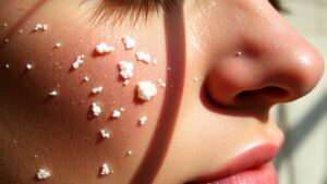
Should you observe small, hard, white or yellowish lumps forming on your face, it could be beneficial to acquaint yourself with the symptoms of calcium deposits. These lesions typically begin as firm, pimple-like bumps frequently emerging beneath the skin. Initially, they can display redness or itching but often stay asymptomatic. When punctured, they can release a chalky, paste-like substance, which may surprise you. In certain instances, you could encounter tenderness or pain, and while uncommon, complications such as skin ulcers or joint mobility limitations can occur.
| Symptoms | Description |
|---|---|
| Lumps Appearance | Small, hard, white or yellowish lumps |
| Initial Response | Can start with redness or itching, often asymptomatic |
| Puncturing Result | Leakage of chalky, paste-like substance |
Types of Calcinosis Cutis
Should you’ve encountered those firm, unusual bumps on your face, grasping the fundamental types of calcinosis cutis can offer clarity. There are several forms to be aware of.
Idiopathic calcinosis appears without any known cause, while Dystrophic calcinosis develops in damaged tissues, often linked to autoimmune conditions like scleroderma.
Metastatic calcinosis, on the other hand, arises from systemic mineral imbalances, commonly due to kidney issues. Finally, iatrogenic calcinosis stems from medical interventions, such as calcium IV fluids, which can result in localized deposits.
Comprehending these types can help you and your healthcare provider manage your situation more effectively, ensuring you receive the appropriate care and treatment customized to your needs.
Causes of Calcinosis Cutis
Grasping the causes of calcinosis cutis can feel overwhelming, particularly as you’re faced with those surprising bumps on your face. One major reason is autoimmune diseases, like scleroderma or dermatomyositis, where calcium deposits form in damaged or inflamed skin.
Additionally, kidney disease often leads to increased calcium and phosphate levels in your blood, which can trigger metastatic calcinosis cutis. Should you’ve had medical procedures involving calcium, you could experience iatrogenic calcinosis cutis from the treatments.
Dysmorphic Calcinosis Cutis
Identifying calcium deposits on your face can be unnerving, particularly when they emerge suddenly. Dysmorphic calcinosis cutis often forms in damaged or abnormal skin due to autoimmune disorders, like dermatomyositis or scleroderma.
You could notice firm, whitish-yellow nodules or plaques on your skin, especially in areas that have experienced trauma or inflammation. Even with normal calcium and phosphate levels, these spots can cause discomfort, sometimes leaking a chalky substance or limiting your joint movement.
Addressing the root condition triggering these deposits is crucial. Treatment could involve corticosteroids or calcium channel blockers. In severe cases, surgical excision might be necessary. Grasping these aspects can help you feel more in control of your situation.
Iatrogenic Calcinosis Cutis
Iatrogenic calcinosis cutis can sneak up on you after certain medical treatments, leaving behind firm, white or yellowish nodules at injection sites.
It’s usually caused by things like calcium gluconate injections or using calcium-containing pastes during tests like EEGs and EMGs.
Grasping the causes is key, so let’s delve into how these medical interventions can lead to unwanted calcium deposits on your skin.
Causes of Iatrogenic Calcinosis
Grasping the causes of iatrogenic calcinosis cutis can aid you in identifying and avoiding potential problems linked to specific medical treatments. This condition often arises from medical procedures, especially in instances calcium-containing solutions, like calcium chloride paste, extravasate during intravenous therapy.
Such incidents can cause localized tissue calcification, which could be surprising. Additionally, certain injectable medications, particularly those containing phosphate, can lead to calcium deposition at injection sites. It’s essential to monitor for any skin injury during these processes.
Through comprehending these causes, you can better discuss any concerns with your healthcare provider and take proactive steps, ensuring careful administration techniques are followed to minimize risks. Your health deserves the best care!
Treatment and Management Options
Finding the right treatment for calcinosis cutis, particularly as soon as it’s linked to medical procedures, can feel a bit overwhelming, but don’t worry-you’re not alone in this. Initially, it’s essential to stop the source, like any calcium-containing IV solutions.
Your healthcare provider could also run blood tests or take a tissue sample to better understand your condition. Medications, such as aluminum hydroxide, can help bind phosphate and reduce calcium deposits.
For stubborn lesions, options like surgical excision or laser therapy can be effective. In some cases, your doctor could suggest calcium channel blockers to help manage the situation.
Keep in mind, prompt intervention helps prevent complications, so don’t hesitate to reach out for help as soon as you notice these calcium deposits.
Metastatic Calcinosis Cutis
Metastatic calcinosis cutis can feel alarming as soon as you spot calcium deposits appearing unexpectedly on your skin.
It usually happens due to higher calcium or phosphate levels in your blood, often because of root health issues like chronic kidney disease.
Comprehending how this condition develops can help you grasp its causes and available treatment options, putting you in a better position to manage it.
Causes and Risk Factors
When considering the root causes and risk factors of metastatic calcinosis cutis, grasping how our body’s mineral equilibrium can influence our skin is crucial. Increased calcium or phosphate levels in your blood frequently provoke this condition.
Long-term kidney disease or hyperparathyroidism can result in calcinosis cutis, as they disturb your calcium balance. Situations like renal failure or excessive vitamin D consumption can also lead to calcium accumulations in your skin.
It’s vital to observe that these metastatic deposits form in healthy tissue, without prior injury or inflammation. Comprehending these fundamental causes aids you in identifying potential risks, directing you toward seeking appropriate care when necessary.
Diagnosis and Treatment Options
Understanding the diagnosis and treatment of metastatic calcinosis cutis is vital for effectively managing this condition. Your path begins with blood tests that check for increased calcium and phosphate levels, typically over 10.5 mg/dL and 4.5 mg/dL, respectively. Chronic kidney disease or hyperparathyroidism often fuels these abnormalities.
Imaging studies like CT scans reveal diffuse calcium deposits beneath your skin, especially near joints. In terms of treatment, you’ll find it focuses on tackling the root metabolic issues, such as using phosphate binders or medications like cinacalcet.
In severe cases of hypercalcemia, bisphosphonates could assist in reducing calcium buildup. Surgery is rarely recommended, reserved for truly troublesome lesions due to risks of recurrence and ulceration.
Idiopathic Calcinosis Cutis
While the exact cause of idiopathic calcinosis cutis remains a mystery, it’s a condition that tends to catch people off guard, especially since it pops up without any warning signals. Known as calcinosis cutis, this condition typically appears as small, firm nodules, white or yellowish, mainly affecting children and young adults.
You could notice these hardened deposits on your face or extremities. What’s interesting is that this type of calcinosis isn’t associated with any fundamental tissue damage or metabolic issues. Although the lesions are usually painless, they’ll sometimes become uncomfortable in case injured.
In case these deposits bother you, treatments like surgical excision or laser therapy could be options despite most cases needing no intervention.
Diagnosis of Calcium Deposits on the Skin
At times you notice calcium deposits on your skin, getting a proper diagnosis is key.
Dermatologists often start with a physical exam and might suggest blood tests to check your calcium levels or use imaging studies to see deeper deposits.
Each method plays a role in helping you find the right treatment and understand what’s happening beneath the surface.
Physical Examination Techniques
Evaluating calcium deposits on the skin can be a bit of a puzzle, but don’t worry; dermatologists have a toolkit full of techniques to help them make sense of those mysterious bumps on your face.
They start with a visual inspection, noting the size, color, and distribution of lesions to differentiate between calcium deposits and other conditions like milia.
Palpating the area reveals firmness and mobility-calcium deposits usually feel hard. Using a dermatoscope, doctors can spot white or yellowish clumps beneath the skin’s surface.
Should it be necessary, a skin biopsy can confirm the presence of hydroxyapatite crystals. Sometimes, they could also check serum calcium levels to rule out any foundational systemic issues, helping guarantee you receive the right care.
Laboratory Testing Methods
Grasping the fundamental causes of calcium deposits on your skin often starts with a few key laboratory tests that can uncover essential details about your body’s chemistry. Blood tests measure your levels of calcium, phosphate, vitamin D, and parathyroid hormone (PTH), helping identify any metabolic imbalances.
A skin biopsy utilizing von Kossa or Alizarin red staining can confirm the presence of calcium deposits through highlighting mineralized tissue under a microscope. Additionally, urinalysis can assess calcium excretion to rule out issues like hypercalciuria.
Should autoimmune disorders be suspected, tests for antinuclear antibodies can help in diagnosis. Together, these methods provide clarity on the root reasons for those pesky calcium deposits.
Imaging Studies Utilization
While grasping the fundamental causes of calcium deposits on your skin often includes laboratory tests, imaging studies play a vital role in visualizing these deposits directly. These techniques help in precisely diagnosing your condition, ensuring you get the right treatment.
| Imaging Study | Purpose |
|---|---|
| X-rays | Detect calcium deposits as white, dense areas on the skin or foundational tissues. |
| Ultrasound | Differentiate deposits from cysts or tumors by examining texture and location. |
| CT scans | Provide detailed cross-sectional images to evaluate the extent and depth of calcifications. |
| MRI | Identify soft tissue involvement around calcium deposits without radiation exposure. |
Each imaging method offers unique perspectives, helping your doctor create a customized management plan. Don’t hesitate to ask questions about these options during your consultation!
Treatment Options for Calcium Deposits
Should you’ve found yourself handling calcium deposits on your face, many others share your experience, and there’s a variety of treatment options that can help. For larger or painful deposits, surgical removal is often recommended. Dermatologists might use a scalpel or laser to guarantee precise extraction.
In case you’re managing milder cases, topical treatments like corticosteroids or calcium channel blockers can reduce inflammation and slow deposit formation. For smaller deposits, intralesional steroid injections can effectively shrink them.
Laser therapy, especially carbon dioxide lasers, works well for superficial deposits, minimizing scarring compared to surgery. Also, tackling any root conditions could help prevent future deposits, so be sure to discuss all your options with your dermatologist.
Home Remedies and Lifestyle Changes
After exploring various treatment options for calcium deposits, you could be looking for ways to manage the condition at home. Here are some effective home remedies and lifestyle changes that might help:
- Apply warm compresses twice daily to soften small calcium deposits.
- Reduce dietary calcium intake to under 1,000 mg daily to slow new deposits.
- Massage affected areas with olive oil for about 5 minutes to enhance skin elasticity.
- Stay hydrated through drinking 2-3 liters of water each day for better skin health.
- Use fragrance-free moisturizers with urea or lactic acid to prevent dryness.
These simple adjustments can make a noticeable difference and help you feel more comfortable with your skin. You’re taking positive steps towards better skin health, and that’s commendable!
Medications for Calcium Deposits
Provided you’re handling calcium deposits and seeking relief, medications can be a vital part of your path. Your doctor could suggest calcium channel blockers like diltiazem to enhance blood flow and slow down further buildup. Should you’re experiencing inflammation, corticosteroids like prednisone can aid with autoimmune-related cases.
For painful deposits, colchicine is often prescribed for its anti-inflammatory effects. In certain situations, warfarin can assist through limiting calcium deposition in small blood vessels. Should hypercalcemia be a concern, bisphosphonates like alendronate could regulate your calcium metabolism effectively.
It’s important to discuss these options with your healthcare provider to find a customized approach since your calcium intake and general health play critical roles in managing calcium deposits.
Surgical Procedures for Severe Cases
At times you’re managing severe calcium deposits on your face, surgery can often provide the relief you need. Several surgical procedures are available, depending on the nature of your deposits:
- Surgical excision for large or painful deposits
- Carbon dioxide laser ablation for precision with minimal scarring
- Curettage, scraping out smaller lesions
- Arthroscopic debridement for those affecting facial joints
Post-surgical care is crucial due to slow healing and risk of infection
Each method carries its unique benefits and considerations, so it’s essential to discuss them with your healthcare provider. You deserve to feel comfortable and confident in your skin, and these procedures can help pave the way for a brighter, clearer future.