You might suppose that blunting of the costophrenic angle isn’t a major concern, but it can actually indicate quite a bit about your health. This change on chest X-rays often signals issues like fluid buildup or even fundamental lung problems. Whether you’re curious about what it means or concerned about your own results, grasping the causes and implications is key. Let’s examine what those details reveal and what steps could come next.
Definition of Costophrenic Angle Blunting
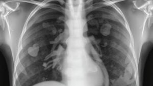
Costophrenic angle blunting could sound complex, but it’s simply a sign that something’s a little off in your respiratory system. This occurs as the sharp angle created by your diaphragm and chest wall loses its definition on a chest radiograph.
Typically, this angle should be about 30 degrees, and blunting here could suggest an issue like pleural effusion, where fluid accumulates in the pleural space, or other abnormalities like thickening or atelectasis.
You may observe blunting in one angle (unilateral) or both (bilateral), depending on what’s happening. Identifying this sign is crucial, as it can be the key to grasping deeper respiratory concerns, helping you and your healthcare provider address any fundamental issues effectively.
Common Causes of Blunting
At the time it comes to comprehending costophrenic angle blunting, it aids to recognize the various reasons why it could occur. One of the most common culprits is pleural effusion, which is the buildup of fluid in the pleural space, noticeable on imaging provided there’s over 200 mL.
Additionally, pleural thickening from conditions like tuberculosis can also obscure the angle, even without fluid. Should you have COPD, lung hyperexpansion could flatten your diaphragm, creating a false blunting effect.
Subpulmonic effusions can mimic blunting through raising the hemidiaphragm without showing clear fluid on frontal views.
Diagnostic Implications of Blunting
Upon spotting blunting of the costophrenic angle in imaging, it frequently indicates the necessity for a thorough plunge into your health. This finding can suggest the presence of pleural fluid, leading you to ponder conditions like heart failure or infections.
Your healthcare provider could recommend further tests, such as fluid analysis provided that drainage is necessary. Grasping whether your pleural effusion is transudative or exudative can help pinpoint the cause.
Symptoms like fever or dyspnea, along with distinct imaging features, guide treatment choices such as antibiotics or procedures like thoracentesis.
Follow-up imaging is vital; should blunting persist, it might signal more serious issues, including fibrothorax or malignant thickening, necessitating continued evaluation to safeguard your well-being.
Associated Conditions Leading to Blunting
At times, blunting of the costophrenic angle pops up on imaging; it’s often a signal to look closer at what’s happening inside your body. Congestive heart failure (CHF) commonly causes this blunting due to pleural fluid accumulation, appearing in 60-90% of decompensated cases.
Tuberculosis also plays a role, leading to exudative pleural effusion in 80% of instances, often with specific enzyme levels. Pneumonia can result in blunting if it’s paired with parapneumonic effusions, and malignancies like lung or breast cancer contribute through similar means, showing a significant likelihood of detecting cancer cells.
Rheumatoid arthritis might lead to sterile exudative effusions, affecting glucose and pH levels. A Chest X-ray offers crucial understanding into these associated conditions.
Imaging Techniques for Evaluation
At the time you’re evaluating costophrenic angle blunting, imaging techniques play a vital role in obtaining accurate results. To begin with, chest radiography is often your primary step, but advanced methods like ultrasound and CT scans provide more detailed understanding.
Each technique has its strengths, helping you identify pleural effusions and any foundational problems with clarity.
Initial Imaging Modality
Should you suspect costophrenic angle blunting, you might consider about the ideal approach to tackle this issue. The primary-line imaging method is chest radiography, particularly erect PA/lateral views. These views are essential to evaluate pleural fluid accumulation effectively.
Here’s a quick summary of various imaging modalities:
| Imaging Modality | Usefulness | Sensitivity/Specificity |
|---|---|---|
| Chest Radiographs | Initial identification of blunting | Variable |
| Ultrasound | Quick bedside confirmation | 93% |
| CT Scans | Superior evaluation of blunting | 98% |
| MRI | Complex cases evaluation | Indeterminate findings |
These approaches help you understand the causes of blunting in the chest, guiding treatment decisions along the way.
Advanced Imaging Techniques
While exploring advanced imaging techniques for evaluating costophrenic angle blunting, you’ll find that each method offers unique advantages in diagnosing pleural fluid-related issues. For instance, CT scans can catch as little as 2-5 mL of fluid accumulation and accurately identify the causes of blunting with a striking 99% accuracy. Should you be seeking sensitivity, chest ultrasound excels with a 93% sensitivity rate.
Lateral decubitus radiographs improve detection sensitivity to 83%, markedly up from standard upright views. MRI is crucial for examining malignant pleural disease, boasting a 97% accuracy rate where other methods fall short.
Plus, digital tomosynthesis provides a detailed 3D view, detecting 85% of subtle pleural abnormalities that routine X-rays could overlook. Each option arms you with essential information for effective diagnosis.
Management Strategies and Next Steps
Managing costophrenic angle blunting effectively involves comprehending the root causes and tailoring your approach based on those specifics.
Should you be handling pleural effusion, start with thoracentesis for fluid analysis to determine the cause.
For tuberculosis-related issues, you’ll need an antibiotic regimen, like RIPE therapy, plus regular check-ins via imaging for 6-9 months.
In case malignancy is at play, therapeutic thoracentesis is key, and you could consider pleurodesis to prevent recurrence.
Should blunting continue despite treatment, a computed tomography (CT) scan can pinpoint trapped lung areas.
Also, using ultrasound-guided procedures can help minimize the risk of iatrogenic pneumothorax to keep you safe and sound.
Each step brings you closer to better management and comfort.