FLAIR hyperintensities appear as bright spots on MRI scans and frequently indicate something is amiss in the brain or surrounding tissues. These spots can originate from bleeding around the brain, infections like meningitis, or even cancer spreading to the membranes. Impeded blood flow from a blocked artery may also create these signals. Whereas FLAIR aids in identifying serious conditions like strokes or multiple sclerosis, occasionally what seems worrying turns out to be nothing more than an imaging anomaly. Determining which is which necessitates a discerning eye.
Overview of FLAIR MRI Technology
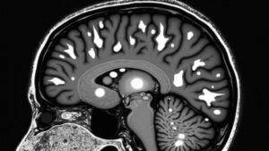
Though MRI scans can be complex, the FLAIR sequence makes certain brain conditions easier to locate. Fluid-attenuated inversion recovery (FLAIR) is an advanced MRI imaging technique that suppresses the signal from cerebrospinal fluid (CSF), helping highlight abnormalities near brain surfaces.
Unlike T1-weighted imaging, FLAIR uses a specific inversion pulse to darken CSF, improving contrast for lesions that might blend in with fluid. This sequence excels in detecting subtle changes, like those in acute stroke or meningitis, where traditional methods could fall short.
While T2-weighted images can miss subtle details, FLAIR enhances sensitivity, especially when combined with contrast-enhanced scans. By reducing CSF interference, radiologists gain clearer views of abnormalities, making FLAIR a critical tool in neuroimaging. Its precision aids in timely diagnosis, guiding better treatment decisions.
Pathologic Causes of FLAIR Hyperintensities
FLAIR hyperintensities can signal several fundamental brain conditions, each with distinct causes that appear bright on MRI scans. Subarachnoid hemorrhage increases protein and blood products in cerebrospinal fluid (CSF), shortening T1 and prolonging T2 signals.
Meningitis, due to inflamed meninges, raises CSF protein and cell counts with similar effects. Meningeal carcinomatosis often shows FLAIR hyperintensity from tumor cells disrupting CSF, sometimes clearer than contrast-enhanced T1 scans. Leptomeningeal melanosis, rare but notable, darkens CSF with melanin, altering relaxation times.
Contrast-related FLAIR hyperintensity occurs when gadolinium leaks into CSF, mimicking pathology in kidney-impaired patients. Each condition’s unique features-like protein shifts, melanin presence, or contrast leakage-warrant careful evaluation to distinguish true illness from mimics. Identifying these causes helps pinpoint accurate diagnoses without unnecessary alarm. Comprehending MRI patterns eases patient concerns while guiding precise clinical decisions.
Vascular and Flow-Related Anomalies on FLAIR
Slow or disrupted blood flow in the brain can create unusual bright spots on FLAIR MRI scans, often pointing to vascular issues that need attention. These FLAIR hyperintensities may appear as hyperintense vessels on FLAIR, signaling hemodynamic impairment due to arterial stenosis or arterial occlusion.
When blood moves too slowly (sluggish flow), it can cause abnormal signals, especially near the middle cerebral artery. Vascular anomalies, like narrowed or blocked vessels, disrupt normal circulation, leading to these bright spots. Sometimes, stagnant blood mixes with cerebrospinal fluid (CSF), making the vessels stand out more on FLAIR imaging.
While not always hazardous, these findings help doctors spot hidden circulation problems before they worsen. Identifying these signs promptly can guide treatment and prevent complications.
Imaging Artifacts vs. True Pathologic Findings
Distinguishing between real brain abnormalities and misleading signals on FLAIR scans can be tricky, even for experienced doctors. Artifacts often mimic true FLAIR hyperintensity, requiring careful analysis to avoid misdiagnosis.
Magnetic susceptibility artifacts occur near tissue interfaces with metal, distorting signals and creating false bright spots. CSF pulsation artifacts appear as hyperintensity in basal cisterns, mimicking disease due to fluid movement. Vascular pulsation causes phase-encoding ghosting, where blood flow creates duplicate ghost images.
Other factors like head motion or supplemental oxygen (via paramagnetic effect shortening T1 relaxation) can also alter scans. Identifying these artifacts helps radiologists separate them from true pathology, ensuring accurate diagnoses. Comprehending these nuances prevents unnecessary worry and guides proper follow-up when real abnormalities exist.
Clinical Applications and Diagnostic Interpretation
Comprehending whenever FLAIR hyperintensities signify genuine neurological concerns-rather than imaging peculiarities-assists doctors in making better decisions for patients. The FLAIR sequence shows bilateral white matter abnormalities on magnetic resonance imaging, helping diagnose conditions like multiple sclerosis or ischemic strokes. These hyperintense areas stand out against suppressed cerebrospinal fluid (CSF) signals, making lesions easier to spot.
Clinicians depend on these images to distinguish between normal aging changes and serious issues, ensuring patients receive timely care. Monitoring disease progression becomes clearer with repeated scans, tracking how lesions evolve across time. Symptoms like memory loss or weakness often prompt further evaluation, but clinical context is key-not all bright spots mean danger.
Pairing imaging with patient history, doctors can pinpoint causes and tailor treatments effectively.
Conclusion
FLAIR hyperintensities serve as silent narrators of the brain’s concealed struggles, revealing conditions from strokes to infections. While they guide diagnosis, differentiating genuine disease from imaging deceptions remains crucial. As the old saying goes, “Not all that glitters is gold”-some bright spots deceive, while others demand swift action. Comprehending their causes and meanings guarantees improved care, transforming uncertainty into clarity for those traversing neurological obstacles.