

Liver anatomy involves understanding the liver’s structure, location, and functions within the human body. In this article, we will explore the liver’s lobes, surfaces, blood supply, and its role in vital functions such as metabolism and detoxification.
Key Takeaways
The liver is a vital organ located in the upper right abdomen, divided into four lobes, and secured by ligaments for stability.
It plays a crucial role in metabolism, detoxification, and bile production, supporting overall health with functions like nutrient processing and blood protein synthesis.
Liver diseases such as hepatitis, cirrhosis, and liver cancer highlight the need for early diagnosis and treatment, often involving procedures like liver biopsies and transplants.
Liver Structure and Location
The liver is a large, reddish-brown organ located in the right upper abdomen, just beneath the rib cage. Weighing approximately 3 to 3.5 pounds, it is positioned behind the ribs, well-protected from external injury. This strategic placement not only safeguards it but also enables it to efficiently perform its numerous functions.
Lobes of the Liver
The liver is divided into four anatomical lobes:
The right lobe, which is the largest
The left lobe, which is significantly smaller
The caudate lobe, located between the left and right lobes, situated anteriorly and superiorly
The quadrate lobe
The quadrate lobe, on the other hand, is positioned between the gallbladder and the fissure for the ligamentum teres hepatis. The left and right lobes are divided by the falciform ligament, and a central fissure known as the porta hepatis separates the caudate lobe from the quadrate lobe.
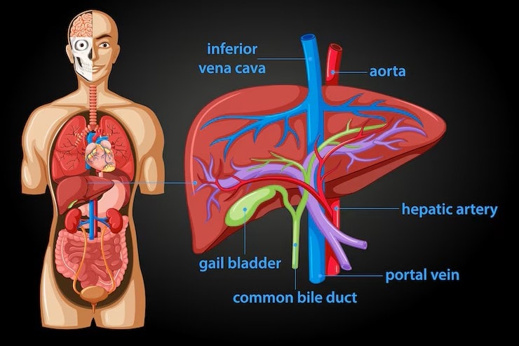
Liver Anatomy Diagram
Surfaces of the Liver
The liver has two primary surfaces. These are the diaphragmatic surface and the visceral surface of the liver. The diaphragmatic surface is smooth and convex, conforming to the shape of the diaphragm.
The visceral surface, covered by the visceral peritoneum, helps protect and support the liver.
Ligaments of the Liver
The liver is anchored in place by several key ligaments. The falciform ligament splits the liver into the left and right lobes and also divides the subphrenic recess. The coronary ligament attaches the liver to the diaphragm, consisting of peritoneal folds.
These ligaments collectively provide stability and maintain the liver’s position within the abdominal cavity.
Liver Location and Gross Structure
The liver is primarily located in the upper right quadrant of the abdomen, predominantly situated on the right side beneath the ribs. This strategic positioning ensures it remains well-protected and optimally functional.
Position and Size of the Liver
In adults, the liver usually weighs about three pounds and is comparable in size to a football. Weighing approximately 1.8 kg in males and 1.3 kg in females, it is the largest solid organ in the human body. Situated beneath the diaphragm and above the stomach, the liver spans an area that can be approximated by placing a hand over the right side under the ribs.
Its top surface is convex, matching the diaphragm’s shape, while its inferior surface is uneven and concave.
Lobes of the Liver
The liver is divided into two main lobes: the larger right lobe and the smaller left lobe. Additionally, the caudate and quadrate lobes are smaller subdivisions that play crucial roles in the liver’s overall function.
Fissures and Ligaments
The liver is anchored to the diaphragm and abdominal wall by several ligaments, including the falciform ligament, which helps attach the liver to the anterior abdominal wall. The coronary ligament consists of peritoneal folds that attach the liver to the diaphragm, and the triangular ligaments provide additional support.
Liver Histology
Various organizational units like classic lobules, portal lobules, and hepatic acini help in examining the liver’s structure. These microscopic units are crucial for understanding the liver’s complex functions, including blood processing and bile production.
Liver Lobules
Classic liver lobules are hexagonal in shape, featuring a central vein surrounded by portal tracts. Hepatocytes within these lobules are arranged in plates that radiate from the central vein towards the portal tracts.
Comprising the hepatic artery, portal vein, and bile duct, the portal tracts facilitate efficient blood flow and bile production.
Biliary System
Bile produced by hepatocytes drains through bile canaliculi, leading to the bile duct system. These channels collect bile produced by hepatocytes and transport it through bile ductules to larger bile ducts, aiding digestion.
Blood Supply to the Liver
The liver receives blood from two main sources: the hepatic artery, which supplies oxygen-rich blood, and the portal vein, which brings nutrient-rich blood from the gastrointestinal tract.
Blood from the lobules is drained by the central vein into the central veins and hepatic veins, which then return it to the venous system.
Liver Blood Supply
The liver’s blood supply consists of a unique architecture featuring two major inlets: the hepatic artery and the portal vein. The dual blood supply is vital for delivering the oxygen and nutrients required for the liver’s metabolic functions.
Hepatic Artery
Delivering oxygen-rich blood, the hepatic artery ensures the liver maintains its metabolic functions. This artery carries oxygenated blood from the systemic circulation directly to the liver, supporting the organ’s high metabolic demands.
Portal Vein
The portal vein is responsible for transporting nutrient-rich blood from the gastrointestinal tract to the liver, facilitating the processing of absorbed nutrients. Carrying deoxygenated, nutrient-loaded blood from the small intestine, this vein allows the liver to metabolize and distribute these nutrients.
Hepatic Veins
Hepatic veins drain deoxygenated blood from the liver back to the heart, playing a crucial role in overall circulatory efficiency. These veins ensure processed blood is returned to the venous system for recirculation.
Liver Sinusoids
Liver sinusoids are specialized capillaries that allow for the mixing of oxygenated and nutrient-rich blood from the hepatic artery and portal vein. Kupffer cells in the sinusoids act as macrophages, filtering pathogens and debris from the blood.
Hepatic stellate cells in the sinusoids contribute to vitamin A storage and play a role in liver fibrosis.
Biliary System
The biliary system is essential for bile production, transport, and storage, aiding in the digestion of fats. This system includes the bile ducts, which transport bile from the liver to the gallbladder and intestines.
Bile Production and Function
Bile produced by the liver helps in the emulsification and absorption of dietary fats. This yellow or orange fluid is crucial for breaking down fats into smaller particles, making them easier to digest.
Bile Ducts and Their Pathways
Bile produced by hepatocytes drains through bile canaliculi into bile ductules, which then transport it to larger bile ducts. The common hepatic duct is a major pathway that carries bile from the liver to the gallbladder and intestines, facilitating digestion.
Gallbladder and Its Role in Bile Storage
The gallbladder is a small, pear-shaped organ that stores bile produced by the liver. When needed, the gallbladder releases bile into the small intestine to aid in the digestion of fats.
Liver Lobes and Segments
The liver has an anatomical division into two lobes. The larger right lobe and the smaller left lobe are separated by the falciform ligament. This division is crucial for understanding its structure and function.
Right Lobe of the Liver
The right lobe is subdivided into anterior and posterior segments by the right hepatic vein, each segment performing specific functions within the liver.
Left Lobe of the Liver
The left lobe contains the medial segment known as segment IV and the lateral segments designated as segments II and III, each contributing to the liver’s overall functionality.
Functional Segments of the Liver
The liver is classified into eight functional segments based on the Couinaud classification, which defines each segment’s vascular supply.
These segments highlight the liver’s structural and functional complexity.
Liver Functions
The liver plays a crucial role in processing nutrients from the digestive system and maintaining metabolic homeostasis. It manages the metabolism of carbohydrates, proteins, and lipids.
Metabolism of Carbohydrates
The liver converts excess glucose into glycogen for storage during periods of high blood sugar. During fasting, it converts glycogen back into glucose to maintain blood sugar levels, balancing glycogen synthesis and breakdown.
Protein Synthesis
Liver cells are responsible for producing most blood proteins essential for maintaining blood volume and pressure. It also synthesizes blood-clotting enzymes like fibrinogen and prothrombin.
Lipid Metabolism
The liver converts carbohydrates into fats for energy storage and produces lipoproteins, which transport fats in the bloodstream. It is also essential for synthesizing cholesterol, crucial for hormone production and cellular structure.
Detoxification
Detoxifying harmful substances, the liver breaks them down into less harmful compounds. Through enzymatic processes, it converts ammonia produced from protein metabolism into urea, facilitating its excretion.
Storage Functions
The liver stores significant amounts of vitamins A, D, E, K, and B12, as well as minerals like copper and iron. It also accumulates glycogen, which can be converted back to glucose as needed.
Bile Production and Secretion
Bile produced by the liver helps in the emulsification and absorption of dietary fats. This bile is secreted into bile ducts and stored in the gallbladder, ready to be released into the intestines for digestion.
Liver and Digestive System
Producing bile, the liver plays a critical role in breaking down fats for digestion. This process is essential for nutrient absorption from the gastrointestinal tract.
Role in Digestion
Bile produced by the liver contains bile salts that facilitate the emulsification process of fats, making them easier to digest. This emulsification facilitates fat absorption in the small intestine.
Liver and Intestinal Absorption
The liver processes nutrients absorbed from the intestine via the portal circulation, ensuring they are distributed throughout the body. This processing maintains nutrient balance and overall health.
Liver Regeneration
The liver has remarkable regenerative capabilities, allowing it to recover from significant injuries and maintain function. This unique ability allows it to regrow to its original size even after losing up to 90% of its tissue.
Ability of the Liver to Regenerate
Normal liver cells called hepatocytes are primarily responsible for liver maintenance and repair. These cells proliferate to restore liver mass and functionality, showcasing its regenerative mechanisms.
Factors Affecting Liver Regeneration
Age and certain health conditions can significantly influence the liver’s ability to regenerate. Conditions like fibrosis and steatosis can impair the liver’s regenerative capacity.
Liver Diseases and Disorders
Liver disease encompasses conditions that progressively damage the liver over time, often without early symptoms.
Understanding these diseases aids in early diagnosis and treatment.
Hepatitis
Hepatitis is characterized by inflammation of the liver, which can be caused by viral infections, toxins, or autoimmune issues. Symptoms often include abdominal pain, jaundice, fatigue, and dark urine.
Cirrhosis
Cirrhosis results from severe liver scarring, which can arise from chronic liver diseases and significantly impairs liver function. The progression of cirrhosis occurs in stages, leading to increasing liver damage and associated complications.
Fatty Liver Disease (NAFLD/NASH)
Non-Alcoholic Fatty Liver Disease (NAFLD) is characterized by excess fat accumulation in the liver not due to alcohol consumption. It can progress to Non-Alcoholic Steatohepatitis (NASH), a more severe form that can lead to cirrhosis.
Liver Cancer (Hepatocellular Carcinoma)
Hepatocellular Carcinoma is the most common type of liver cancer and typically arises in the setting of chronic liver disease. Common risk factors include chronic viral hepatitis and cirrhosis.
Diagnosis typically involves imaging tests, blood tests, and occasionally a biopsy.
Liver Failure
Liver failure is classified into acute and chronic types, with chronic liver failure being a gradual process leading to severe health complications. Treatment focuses on the underlying cause and may involve liver transplantation.
Clinical Relevance
Knowledge of liver anatomy is essential for diagnosing and treating liver conditions.
Biopsies, imaging, and enzyme tests are critical tools for this purpose.
Liver Biopsy and Imaging
A liver biopsy assesses liver tissue to identify damage or disease. Techniques like ultrasound, CT, and MRI often assess liver conditions before biopsy, ensuring accurate diagnosis.
Liver Transplantation
Liver transplants are indicated for severe liver diseases such as cirrhosis and acute liver failure.
Post-operative care involves monitoring for organ rejection and managing immunosuppressive therapy.
Liver Enzyme Tests
Liver function tests, such as ALT and AST levels, help assess liver health and detect abnormalities. Interpreting these tests aids in diagnosing liver conditions and monitoring treatment efficacy.
Blood Supply and Circulation
The liver’s blood supply consists of two primary sources: the hepatic artery for oxygenated blood and the portal vein for nutrient-rich blood. This dual blood supply is vital for liver functions.
Hepatic Artery and Portal Vein
The hepatic artery carries oxygenated blood directly from the systemic circulation to the liver. The hepatic portal vein transports nutrient-laden, deoxygenated blood from the small intestine to the liver for processing.
These blood sources are crucial for the liver’s metabolic activities.
Hepatic Veins and Inferior Vena Cava
The hepatic veins are responsible for draining deoxygenated blood from the liver into the inferior vena cava. This process ensures processed blood returns to the heart for recirculation.
Lymphatic Drainage
The lymphatic drainage of the liver consists of pathways that facilitate the removal of excess fluids and waste products. Draining into the hepatic lymph nodes at the porta hepatis, the superficial lymphatic system filters lymphatic fluids and contributes to the immune response.
Microscopic Anatomy
The liver’s microscopic structure consists of hexagonal units known as lobules, which contain rows of liver cells that radiate from a central point. These lobules are crucial for understanding the liver’s intricate functions.
Liver Lobules
Liver lobules are hexagonal structures defined by a central vein surrounded by portal tracts containing blood vessels and bile ducts. Each lobule features a central vein surrounded by hepatocytes, with the portal triad at each corner.
Liver Cells
Hepatocytes are the primary cells in the liver, positioned next to blood-filled sinusoids and canaliculi where bile is secreted. Stellate cells store vitamin A and regulate blood flow within the sinusoids, while Kupffer cells, a type of macrophage, play a critical role in the liver’s immune response.
Functions of the Liver
The liver performs primary functions including metabolism, immunity, digestion, detoxification, vitamin storage, and bile production. It is responsible for over 500 vital functions, showcasing its complexity and necessity for overall health.
Metabolism and Detoxification
Hepatocytes, the liver’s primary cells, handle metabolic processes like detoxification and protein synthesis. Facilitating exchange with blood, these cells play a crucial role in metabolism.
The liver also plays a significant role in regulating cholesterol levels and breaking down medications and toxins.
Bile Production
Bile produced by the liver is typically clear yellow or orange and helps digest fats. This bile breaks down fats into smaller particles, making them easier to absorb.
Synthesis of Plasma Proteins
The liver synthesizes albumin and clotting factors, which are critical for maintaining osmotic pressure and facilitating blood coagulation. These blood plasma proteins are crucial for various bodily functions and overall health.
Clinical Aspects and Diseases
Understanding clinical aspects and common liver diseases aids in early diagnosis and effective treatment. Hepatocellular carcinoma, cirrhosis, and portal hypertension are among the significant conditions affecting the liver.
Hepatocellular Carcinoma
Hepatocellular carcinoma is the most common type of liver cancer, typically arising in the setting of chronic liver disease. Early symptoms may be vague but can include abdominal fullness, jaundice, and unexplained weight loss.
Treatment options encompass surgery, tumor ablation, embolization, radiation therapy, and immunotherapy.
Cirrhosis and Portal Hypertension
Cirrhosis can cause severe complications like portal hypertension, marked by increased blood pressure in the portal vein. Common signs of portal hypertension include esophageal varices, rectal hemorrhoids, and caput medusae.
The TIPS procedure can significantly lower portal pressure and help manage complications from portal hypertension, such as variceal bleeding.
Liver Transplantation
Liver transplantation is a critical treatment for patients with severe liver diseases, offering a chance for prolonged life and improved quality of life.
Post-operative care is crucial and includes monitoring for organ rejection and managing immunosuppressive medications to prevent rejection.
Surgical Considerations
Surgical considerations related to the liver are essential due to its complex structure and critical functions. Careful preoperative evaluation minimizes risks and ensures successful outcomes.
Percutaneous Liver Biopsy
A percutaneous liver biopsy is often performed using ultrasound guidance to minimize risk and ensure accuracy. This procedure, which involves obtaining tissue samples with a needle inserted through the skin, primarily diagnoses liver diseases.
Risks include bleeding, infection, and pain at the biopsy site, though complications are rare.
TIPS Procedure
The TIPS procedure is used to treat portal hypertension related to liver damage or portal vein thrombosis. This procedure creates a shunt between the portal vein and the inferior vena cava to reduce portal pressure.


