
Ear anatomy involves studying the three main parts of the ear: the outer ear, middle ear, and inner ear. Each part plays a crucial role in hearing and balance. The outer ear captures sound waves. The middle ear amplifies vibrations. The inner ear converts these vibrations into signals for the brain. This article explores each part in detail, explaining their structures and functions.
Key Takeaways
The ear is divided into three main parts: outer ear, middle ear, and inner ear, each with specific functions for hearing and balance.
Understanding ear anatomy helps recognize symptoms of common ear disorders like hearing loss and infections, which can significantly impact quality of life.
Maintaining ear health involves good hygiene, avoiding loud noise exposure, and seeking timely medical care to prevent issues.
What is Ear Anatomy ?
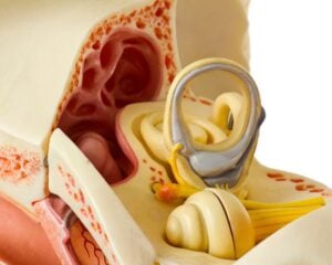
Ear anatomy is the study of the structural components that make up the ear and their respective functions. The ear serves two primary roles: facilitating hearing and maintaining balance. Anatomically, the ear is divided into three main parts: the outer ear, middle ear, and inner ear, each playing a distinct role in auditory and balance processes.
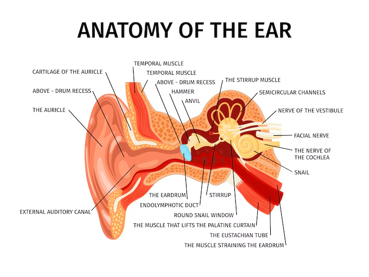
The outer ear is visible externally and consists of the auricle (also known as the pinna) and the external auditory canal, which directs sound waves towards the tympanic membrane (eardrum).
The middle ear houses three small bones called ossicles, which are crucial in conducting sound vibrations. Finally, the inner ear contains the cochlea and semicircular canals, essential for processing sound and balance. Understanding these components provides a foundational insight into how our ears function.
Ear Diagram
Outer Ear Anatomy
The outer ear plays a significant role in capturing sound waves and directing them deeper into the ear. Its structure varies in shape and size among individuals, contributing to unique ear appearances. The outer ear comprises three main components: the auricle (pinna), external auditory canal (ear canal), and the tympanic membrane (eardrum).
Each part of the outer ear has a specific function, from collecting sound waves to protecting the inner structures. The auricle helps in sound localization, while the ear canal directs sound waves towards the tympanic membrane. The tympanic membrane then vibrates in response to these sound waves, initiating the hearing process.
Examining these components more closely.
Auricle (Pinna)
The auricle, or pinna, is the visible part of the ear made up of cartilage and skin. Its primary function is to collect sound waves from the environment and funnel them into the ear canal. The shape and size of the auricle vary among individuals, giving each person a unique ear appearance.
This structure not only aids in hearing by directing sound waves but also assists in determining the direction from which sounds originate.
External Auditory Canal (Ear Canal)
The external auditory canal, commonly known as the ear canal, is a tube-like structure that directs sound waves towards the eardrum. Measuring about one inch long, the canal is divided into cartilaginous and bony sections.
It contains ceruminous glands that produce earwax, which protects the ear by trapping debris and preventing infections.
Tympanic Membrane (Eardrum)
The tympanic membrane, or eardrum, is a thin, flexible membrane that vibrates when sound waves hit it. It separates the outer ear from the middle ear and plays a crucial role in transmitting sound vibrations to the ossicles.
Remarkably, the tympanic membrane remains the same size from infancy to adulthood, resembling a dime in diameter.
Middle Ear Anatomy
The middle ear acts as a cavity that amplifies sound waves before they reach the inner ear. It is situated between the tympanic membrane and the oval window and contains vital structures that enhance sound transmission.
The middle ear’s main components include the ossicles, the Eustachian tube, and the oval window. These structures work together to ensure efficient sound transmission and pressure equalization. Let’s explore these components in detail.
Ossicles (Small Bones)
The ossicles are the three smallest bones in the human body: the malleus (hammer), incus (anvil), and stapes (stirrup). These bones transmit sound vibrations from the eardrum to the inner ear. The ossicles amplify sound vibrations, enhancing the overall hearing process.
The stapes, the smallest ossicle, plays a crucial role in transmitting these vibrations to the cochlea through the oval window.
Eustachian Tube
The Eustachian tube is a canal that connects the middle ear to the back of the nose and throat. Its primary function is to equalize pressure between the middle ear and the external environment, preventing damage to the eardrum.
Additionally, the Eustachian tube facilitates the drainage of fluid from the middle ear, helping to prevent infections.
Oval Window
The oval window is a membrane-covered opening between the middle ear and the inner ear. It receives vibrations from the stapes, which are then transferred into the cochlea. This process initiates the conversion of sound vibrations into neural signals, leading to the perception of sound.
Inner Ear Anatomy
The inner ear is situated adjacent to the middle ear and is responsible for both auditory and balance functions. It contains intricate structures that transform sound vibrations into electrical signals and help maintain balance.
Key components of the inner ear include the cochlea, vestibular system, and auditory nerve. Each of these structures plays a vital role in processing sound and providing balance information. Let’s delve deeper into these components.
Cochlea
The cochlea is a spiral-shaped, fluid-filled structure that transforms sound vibrations into electrical signals for the brain. It contains hair cells that detect sound frequencies and intensities.
The cochlea’s unique snail-like structure is divided into chambers filled with fluid that reacts to sound vibrations, making it essential for hearing.
Vestibular System
The vestibular system includes the semicircular canals, utricle, and saccule, and is responsible for maintaining balance and spatial orientation by detecting head movements. The semicircular canals are oriented perpendicularly, allowing for accurate detection of head movement.
This system is crucial for detecting rotational movements and facilitating balance.
Auditory Nerve
The auditory nerve, also known as the eighth cranial nerve, transmits electrical impulses from the cochlea to the brain. It is responsible for the sensation of sound and conveys both auditory and balance information to the brain.
Common Ear Disorders and Conditions
Ear disorders can significantly impact hearing and balance, affecting overall quality of life. Common ear disorders include hearing loss, ear infections, and balance disorders. Understanding these conditions and their symptoms is crucial for timely intervention and treatment.
Hearing loss can result from damage to any part of the ear, including the eardrum, ossicles, cochlea, or auditory nerve. Ear infections, such as otitis externa and otitis media, can cause pain and hearing difficulties. Balance disorders, often related to inner ear issues, can lead to dizziness and vertigo.
Hearing Loss
Hearing loss can occur due to various factors, including damage to the eardrum, ossicles, cochlea, or auditory nerve. There are different types of hearing loss: conductive, sensorineural, and mixed. Age-related hearing impairment is a common type that occurs gradually over time.
Recognizing early signs of hearing loss, such as needing to raise the volume or frequently asking for repetition, is important for timely intervention.
Ear Infections
Ear infections are common conditions that affect different parts of the ear. Otitis externa, also known as swimmer’s ear, is an infection of the outer ear canal often caused by water remaining in the ear after swimming, creating a moist environment that aids bacterial growth. Otitis media is a middle ear infection that is particularly common in children and can cause ear pain, fever, and hearing difficulties.
Chronic ear infections can lead to persistent fluid in the middle ear, affecting hearing. Symptoms of ear infections include ear pain, fluid discharge, and a sense of fullness in the ear. Factors like exposure to secondhand smoke and frequent colds can increase the risk of ear infections, especially in young children.
Balance Disorders
Balance disorders are often caused by issues within the vestibular system of the inner ear. Conditions like Meniere’s disease can lead to severe episodes of dizziness, ringing in the ears, and even hearing loss. Disruptions in the vestibular system can affect balance and spatial orientation, leading to symptoms such as dizziness, vertigo, and balance instability.
How Ear Anatomy Affects Hearing and Balance
The structure of the ear is integral to its functions in hearing and maintaining balance. Each part of the ear plays a specific role in processing sound and sensory information:
The outer ear captures sound waves
The middle ear amplifies and transmits these waves
The inner ear converts them into electrical signals while also providing balance information
Grasping how each part of the ear contributes to hearing and balance helps in understanding our auditory and equilibrium systems. Delving into the roles of the outer, middle, and inner ear provides more detail.
The Role of the Outer Ear in Sound Collection
The outer ear, comprising the auricle and ear canal, is optimized to capture sound waves and direct them towards the eardrum. The shape of the auricle helps in sound localization by introducing time delays for incoming sound waves reach.
Earwax produced in the ear canal protects the ear by trapping debris and preventing infections.
The Middle Ear’s Role in Sound Transmission
The middle ear contains the ossicles, which amplify and transmit sound vibrations from the eardrum to the inner ear. The Eustachian tube helps maintain equal air pressure on both sides of the eardrum, ensuring proper sound transmission and preventing damage.
This design allows for efficient conduction of sound waves from the outer to the inner ear.
The Inner Ear’s Role in Sound Processing and Balance
The inner ear’s cochlea converts sound vibrations into electrical signals that the brain interprets as sound. The vestibular system, comprising the semicircular canals, utricle, and saccule, works with the brain to maintain balance and spatial orientation.
Together, these structures ensure accurate sound perception and balance maintenance.
External Ear Anatomy
The external ear, also known as the auricle or pinna, includes the visible part of the ear and the external auditory canal. The auricle aids in capturing sound waves and directing them toward the eardrum.
The external auditory canal, approximately 4 cm long, has a curved S shape that protects the tympanic membrane from external elements.
Middle Ear Structure
The middle ear, also known as the tympanic cavity, contains the tympanic membrane and the ossicular chain. The tympanic membrane, or eardrum, is a thin oval membrane that serves as a barrier between the outer ear and the middle ear. It vibrates in response to sound waves, converting them into mechanical vibrations that are transmitted to the ossicles.
The ossicular chain consists of three bones: malleus, incus, and stapes, each playing a distinct role in sound transmission. The malleus connects directly to the tympanic membrane and transmits sound vibrations to the incus. The stapes interfaces with the oval window to convey sound energy into the inner ear.
Inner Ear Components
The inner ear comprises the cochlea and the vestibular system, both essential for hearing and balance. The cochlea, a snail-shaped structure, contains fluid and tiny hair cells that detect sound frequencies and intensities. Movement of the stereocilia in the cochlea generates electrical signals sent to the brain.
The vestibular system includes the otolith organs (saccule and utricle) and the semicircular canals. These structures detect linear accelerations, gravitational forces, and rotational movements of the head, helping maintain balance.
Ear Canal and Tympanic Membrane
The ear canal, or external acoustic meatus, connects the outer ear to the tympanic membrane. It helps direct sound waves towards the tympanic membrane, where they are converted into mechanical vibrations. Ceruminous glands in the ear canal produce earwax, which has antibacterial properties and helps trap debris.
The tympanic membrane, commonly known as the eardrum, serves as the boundary between the outer ear and the middle ear. Its vibrations in response to sound waves initiate the process of hearing by transferring these vibrations to the ossicles in the middle ear.
Nerve Supply to the Ear
The nerve supply to the ear is crucial for its function. Motor control for the external ear muscles is primarily provided by branches of the facial nerve. Sensory and motor innervation are essential for the ear’s overall function, as they enable both movement and sensation.
Blood Supply and Lymphatics
The auricle receives arterial blood mainly from branches of the superficial temporal artery (STA) and the posterior auricular artery (PAA). An arterial arcade known as the ‘helical rim arcade’ connects branches of the STA and PAA, ensuring blood flow across the helical rim.
This blood supply is vital for maintaining the health and function of the auricle.
Muscles of the Ear
The ear consists of both intrinsic and extrinsic muscles. Intrinsic muscles originate and insert within the ear itself, contributing to its shape and movement. Extrinsic muscles, such as the superior auricular, anterior auricular, and posterior auricular muscles, attach to the ear from other parts of the body and are primarily responsible for positioning the ear.
Muscles like the tensor tympani and stapedius regulate the movement of the ossicles to protect the ear from loud sounds. While many extrinsic muscles are vestigial, some individuals can still move their ears slightly, thanks to these muscles.
Embryological Development of the Ear
The ear begins to develop from the otic placode, a thickening of ectoderm around the fourth week of gestation. By the sixth week, the structures of the external ear, including the auricle, become recognizable. The formation of the external auditory canal occurs from the first pharyngeal groove, starting in the fifth week.
By the tenth week of development, the external ear structures are largely formed and positioned. Cartilage starts to replace the mesenchyme in the ear structures around the sixth to seventh week.
Symptoms of Ear Conditions
Recognizing the symptoms of ear conditions is crucial for timely intervention. Common signs of an ear infection include ear pain, fever, difficulty hearing, and a sense of fullness in the ear. In young children, additional symptoms like irritability, loss of balance, and pulling at the ear can indicate an ear infection.
Persistent earache lasting more than three days or recurring infections warrant a consultation with a healthcare provider. Fluid drainage from the ear can indicate an infection, especially if it is accompanied by pain. Adults experiencing ear pain and fluid discharge may also be suffering from an ear infection.
A high fever can be a symptom of ear infections, especially in younger children. If fluid is leaking from the ear or there’s noticeable swelling, these are urgent symptoms that require immediate medical attention.
Diagnostic Tests for Ear Health
Various diagnostic tests help assess ear health and identify hearing issues. Hearing assessments, known as audiometry tests, determine the presence and severity of hearing loss. Pure-tone testing measures the softest sounds a person can detect at various pitches. Bone conduction tests assess the function of the inner ear by bypassing the outer and middle ear.
Auditory brainstem response (ABR) tests check the pathways from the inner ear to the brain, while speech testing evaluates how well individuals understand spoken words. Tympanometry evaluates the movement of the eardrum and can identify issues such as fluid in the middle ear.
Care and Maintenance of Ear Health
Maintaining ear health is similar to taking care of the rest of your body, requiring good hygiene, healthy lifestyle choices, and timely medical consultations. Protecting ears from excessive noise exposure is crucial, as prolonged loud noises can lead to hearing loss.
Maintaining ear health involves several key practices:
Good hygiene
Healthy lifestyle choices
Timely medical consultations
Protecting ears from excessive noise exposure, as prolonged loud noises can lead to hearing loss.
The ear canals are generally self-cleaning, and there is usually no need for manual cleaning unless there are specific issues. Using Q-tips inside the ears can push wax deeper and potentially block the ear canal, rather than cleaning it effectively.
Hydrogen peroxide can be safely used for ear cleaning if there are no previous ear surgeries or perforations in the eardrum.


