A Large Intestine Diagram helps you understand the structure and function of one of the most important parts of your digestive system. By clearly identifying sections like the cecum, colon, rectum, and anus, you can better understand how your body processes waste and absorbs water.
The large intestine has five main segments: cecum, ascending colon, transverse colon, descending colon, and sigmoid colon, all crucial for water absorption and feces formation. Common issues associated with the large intestine include constipation, IBS (Irritable Bowel Syndrome), inflammation (like colitis), and even colon cancer. Did you know that over 10% of the global population suffers from chronic digestive conditions involving the large intestine?
Research shows that anatomical diagrams improve comprehension and health outcomes by up to 50% in some cases! So, Need a large intestine diagram? This article provides a detailed, labeled diagram along with descriptions of each segment’s functions.
Anatomy of the Large Intestine
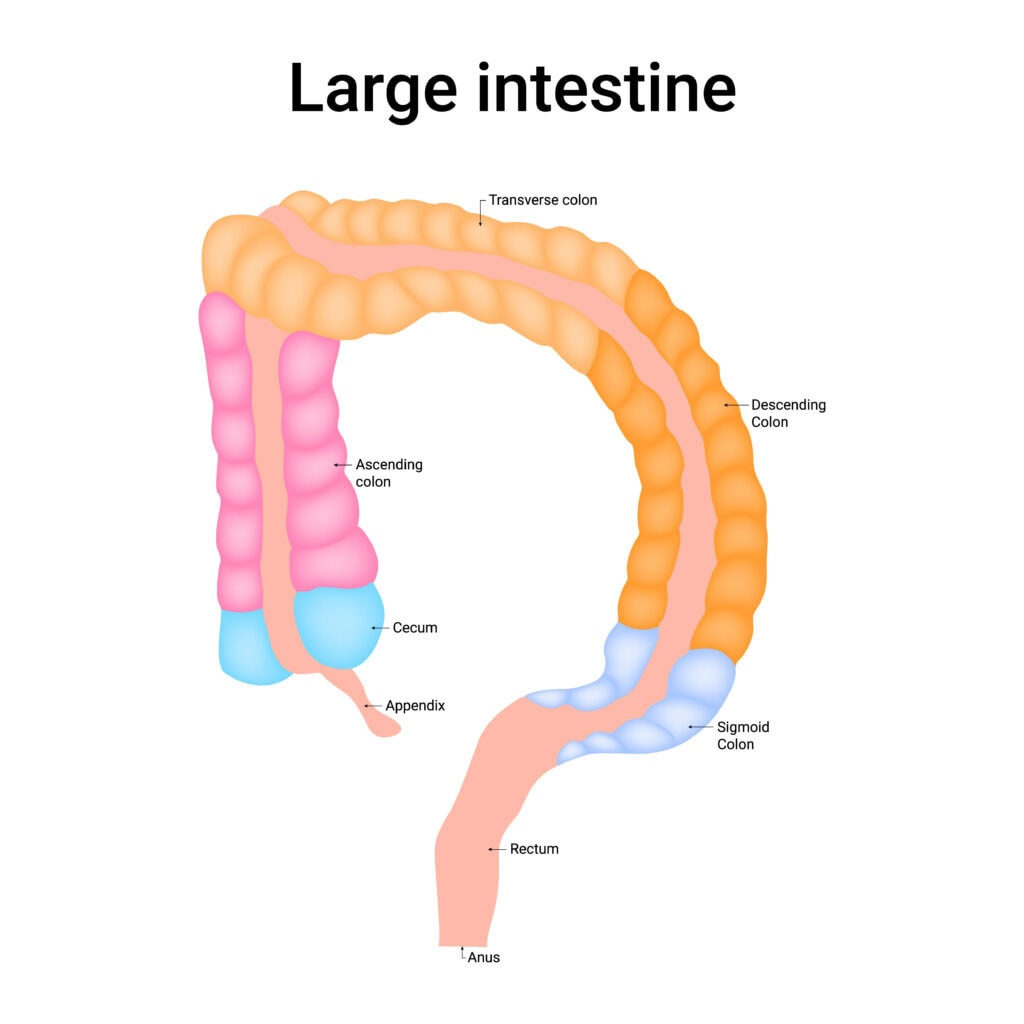
The large intestine consists of five primary segments: the cecum, ascending colon, transverse colon, descending colon, and sigmoid colon.
These segments work together to absorb water and electrolytes and to form and store feces.
The large intestine also features unique structures such as omental appendices, haustra, and teniae coli, which aid in its functions.
Cecum and Appendix
The journey through the large intestine begins at the cecum, an 8-centimeter-long pouch that receives waste from the small intestine.
The cecum acts as a storage site for chyme, regulating its movement into the colon. This process is crucial as it ensures that the material is moved at a pace that allows for maximum absorption of water and electrolytes.
Attached to the cecum is the appendix, a small tube-like structure.
Although once considered a vestigial organ, the appendix is now believed to play a role in maintaining gut flora and supporting the immune system. Positioned in the abdominal cavity and connected to the cecum, the appendix plays a significant role in digestion.
Colon Segments
The colon is divided into four segments, each with its specific function. The ascending colon, about 20 centimeters long, is retroperitoneal and absorbs water and electrolytes. Moving waste along the digestive tract, it leads into the transverse colon.
The transverse and descending colon continue this process, but it is the sigmoid colon, with its S-shaped structure and 35 to 40 centimeters in length, that plays a critical role in converting waste into stool.
The segmented appearance of the colon, due to haustra, allows for efficient absorption and movement of waste material.
Rectum and Anal Canal
The rectum, typically 12 to 15 centimeters long, stores feces before elimination. It holds the waste until the body is ready to expel it, ensuring that defecation occurs at an appropriate time.
The anal canal, about 5 centimeters long, contains structures like anal valves and sphincters to control feces expulsion. The smooth muscle internal anal sphincter and the skeletal muscle external anal sphincter collaborate to provide both involuntary and voluntary control.
Blood Supply to the Large Intestine
The large intestine’s blood supply is vital for its function, with arterial supply primarily provided by the superior and inferior mesenteric arteries. This system ensures all sections receive adequate blood flow, facilitated by the marginal artery of Drummond.
Venous drainage is managed by the superior and inferior mesenteric veins and the inferior rectal veins, efficiently removing deoxygenated blood.
Arterial Supply
The superior mesenteric artery supplies the midgut portions of the large intestine through branches like the ileocolic, right colic, and middle colic arteries. The inferior mesenteric artery, including the left colic artery, serves the hindgut structures such as the descending colon and rectum.
This arterial network ensures the large intestine receives the blood needed for effective function.
Venous Drainage
Venous drainage from the large intestine is primarily handled by the superior and inferior mesenteric veins. Midgut derivatives drain into the superior mesenteric vein via colic veins, while hindgut derivatives drain directly into the inferior mesenteric vein.
The rectum’s drainage is managed by the middle and inferior rectal veins drain, completing the efficient venous return system.
Innervation of the Large Intestine
Innervation by the enteric and autonomic nervous systems (ENS and ANS) is critical for coordinating the large intestine’s activities. These systems work together to regulate bowel movements and secretion, ensuring the smooth operation of the large intestine.
Enteric Nervous System
The enteric nervous system (ENS) consists of about 200 to 600 million neurons organized into myenteric and submucosal ganglia. This system operates independently from the central nervous system, regulating local reflexes and gut motility, thus playing a crucial role in the digestive process.
Autonomic Nervous System
The autonomic nervous system (ANS) includes the sympathetic and parasympathetic systems, which balance each other to regulate large intestine functions.
The sympathetic system inhibits digestive activities by reducing motility and constricting sphincters, while the parasympathetic system enhances digestion by promoting peristalsis and increasing mucus secretion.
Functions of the Large Intestine
The large intestine absorbs water and electrolytes, forms and eliminates feces, and supports the immune system through its microflora. These functions are crucial for maintaining overall health and ensuring the digestive process is efficient.
Absorption
One of the primary roles of the large intestine is the reabsorption of fluids and electrolytes. This occurs through osmosis, influenced by the absorption of electrolytes like sodium, actively transported through various channels. Beneficial bacteria in the large intestine also produce essential vitamins such as vitamin K and various B vitamins, contributing to overall health.
Absorbing water, electrolytes, and vitamins is vital for maintaining the body’s homeostasis. This process ensures that the body retains essential fluids and nutrients while forming solid waste to be eliminated.
Feces Formation and Elimination
Forming and eliminating feces are critical functions of the large intestine. Feces consist of indigestible food, bacteria, inorganic salts, unabsorbed substances, epithelial cells, and water.
The sigmoid colon significantly contributes by contracting to increase pressure and move stool into the rectum. The descending colon stores feces. Eventually, this waste will be emptied into the rectum for elimination.
The nervous system regulates this: sympathetic innervation slows motility, while parasympathetic innervation increases it to induce defecation and relax the internal anal sphincter. This coordination ensures that waste is efficiently and effectively expelled from the body.
Microflora and Immunity
The large intestine houses extensive microflora critical for digestion and immunity. These commensal bacteria assist in the absorption of vitamins and help break down fiber through fermentation, producing short-chain fatty acids and gas.
The large intestine produces colonic bacteria that also produce essential vitamins, including Vitamin B, K, and biotin, contributing to overall health.
Additionally, the large intestine supports the immune system by assisting in antibody creation to combat bacteria and prevent infections. The appendix aids in maintaining gut flora and mucosal immunity, highlighting the interconnected functions of the large intestine.
Large Intestine Diagram with Labels
Common Disorders of the Large Intestine
Issues such as diverticular disease, inflammatory bowel disease (IBD), and colorectal cancer are prevalent and can significantly impact one’s health.
Conditions often present with symptoms like constipation, diarrhea, abdominal cramps, and dehydration, highlighting the need for timely medical intervention.
Diverticular Disease
Diverticular disease includes both diverticulosis and diverticulitis, characterized by sac-like protrusions called diverticula in the colon.
Diverticulosis often occurs in the sigmoid colon and can progress to diverticulitis if these pouches become inflamed or infected. Symptoms of diverticulitis include abdominal pain, nausea, vomiting, and low-grade fever, which can be quite distressing. Treatment for uncomplicated diverticulitis typically involves oral antibiotics to manage the infection.
Diverticula can lead to complications if not managed properly. Regular check-ups and a fiber-rich diet can help prevent the formation and inflammation of diverticula, ensuring the large intestine remains healthy and functional.
Inflammatory Bowel Disease
Inflammatory bowel disease (IBD), including Crohn’s disease, causes chronic inflammation in the large intestine.
Crohn’s disease can affect any part of the gastrointestinal tract but often impacts the large intestine, leading to symptoms such as fever, abdominal pain, diarrhea, and weight loss.
Managing IBD requires a comprehensive approach, including medication, lifestyle changes, and sometimes surgery, to control inflammation and maintain quality of life.
Colorectal Cancer
Colorectal cancer involves malignant growths developing in the large intestine. Risk factors include age, family history, and lifestyle factors such as diet.
Symptoms often include changes in bowel habits, blood in the stool, and unexplained weight loss.
Colonoscopy is the gold standard for detecting colorectal cancers and polyps, allowing for immediate biopsy if necessary. This procedure enables direct visualization of the colon, facilitating the removal of polyps and other abnormalities during the examination.
Diagnostic Imaging and Diagrams
Diagnostic imaging techniques are essential for visualizing the large intestine and diagnosing various conditions. X-rays, CT scans, and colonoscopy provide detailed images and direct visualization, aiding in accurate diagnosis and treatment of large intestine disorders.
Detailed labeled diagrams further enhance understanding and serve educational purposes, offering clear visual guidance on the large intestine’s structure and function.
X-rays and CT Scans
X-rays and CT scans are critical diagnostic tools that help identify conditions affecting the large intestine, such as tumors and blockages. These imaging techniques provide detailed images, allowing healthcare professionals to detect abnormalities and plan appropriate treatment strategies.
Colonoscopy
Colonoscopy involves an endoscope inserted into the rectum to visualize the entire colon for abnormalities. This procedure is essential for detecting colorectal cancer and polyps, allowing for immediate biopsy and removal of polyps during the examination. Regular colonoscopy screenings are vital for early detection and prevention of colorectal cancer, especially for those with risk factors.
Screening methods for colorectal cancer often include colonoscopy and stool tests, helping detect early signs of cancer and other abnormalities. These procedures are critical for maintaining colon health and preventing serious conditions.
Wrap Up
At the end of the day, the large intestine, though often overshadowed by other parts of the digestive system, is essential for our well-being.