

Leg bones like the femur, tibia, and fibula play crucial roles in movement and stability. This article delves into their anatomy, functions, and common issues you should know about.
Key Takeaways
Leg bones include the femur, tibia, and fibula, each playing crucial roles in supporting weight, facilitating movement, and ensuring stability.
The knee joint is a complex structure consisting of bones, menisci, and ligaments, which work together to provide flexibility and stability during movement.
Common leg bone issues include fractures, osteoporosis, and stress fractures, each requiring different treatment strategies including physical therapy, medications, and sometimes surgery.
Structure and Function of Leg Bones
The leg is a marvel of biological engineering, comprising major bones such as the femur, tibia, and fibula. Together, these bones form a complex structure that enables a wide range of movements while supporting the body’s weight.
The femur, or thigh bone, links the hip to the knee joint, facilitating mobility and weight-bearing. The tibia, commonly known as the shin bone, is the primary weight-bearing bone of the lower leg, while the fibula, a thinner bone running parallel to the tibia, provides lateral stability and muscle attachment.
Leg Bones Diagram
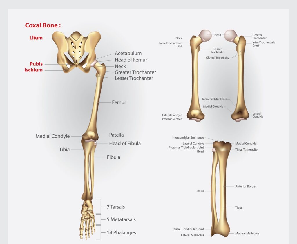
Femur
The femur, the longest bone in the human body, stretches from the hip joint to the knee joint, connecting the pelvic bone to the lower leg bones. This connection is essential for the range of movements we often take for granted, such as walking, running, and jumping. The femoral head, which fits into the hip joint, allows for a wide range of motion, while the distal femur interacts with the tibia to form the knee joint, facilitating flexion and extension.
Moreover, the femur’s robust structure supports significant muscular attachments, including the thigh muscles and the gastrocnemius muscle, which is vital for movements like standing on your toes.
The lateral and medial condyles of the femur articulate with the tibial condyles, ensuring the smooth operation of the knee joint. This intricate setup underscores the femur’s critical role in both mobility and stability, particularly at the medial condyle.
Tibia
The tibia, or shin bone, is the primary weight-bearing bone of the lower leg, supporting approximately 85% of the body’s weight. This robust bone plays a pivotal role in maintaining the structural integrity of the lower limb. The proximal tibia consists of the tibial condyles, which interact with the femur to form part of the knee joint, while the distal tibia connects to the ankle joint, facilitating foot movements.
The tibia’s ability to bear weight and withstand impact is crucial for activities such as walking, running, and jumping. Its anatomical design, including the tibial shaft and the tibial tuberosity, which serves as an attachment point for the patellar tendon, highlights its importance in both stability and mobility.
Without a healthy tibia, the basic functions of the lower leg would be severely compromised.
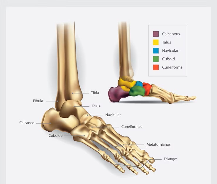
Foot Bone Anatomy Diagram
Fibula
The fibula, a thin bone located alongside the tibia, plays a key role in providing lateral stability to the lower leg and ankle joint. Although it does not bear significant weight, the fibula forms crucial connections with the tibia at both the proximal and distal ends, contributing to the structural integrity of the lower leg. Its primary function is to serve as an attachment point for muscles, including the flexor digitorum longus and the extensor digitorum longus, which are essential for foot movements.
Despite its slender appearance, the fibula is vital for maintaining balance and stability, particularly in the ankle joint. It helps to prevent excessive movements that could lead to injuries.
Common issues affecting the fibula include fractures, which can result from high-impact activities or direct trauma, underscoring the importance of this often-overlooked bone.
Knee Joint Anatomy
The knee joint is a complex and highly functional part of the human body, essential for various movements. It consists of three main bones: the femur, tibia, and patella, all working together to provide stability and flexibility.
Encased in a fibrous capsule and lined with a synovial membrane, the knee joint is well-protected and lubricated, allowing for smooth and efficient movements.
Menisci
The menisci are two semicircular cartilaginous structures located between the femur and tibia within the knee joint. There are two menisci in each knee: the medial meniscus and the lateral meniscus. These structures play a crucial role in stabilizing the knee joint, absorbing impact, and protecting the joint surfaces from wear and tear.
The menisci also act as shock absorbers, distributing the load across the knee joint and reducing stress on the articular cartilage. This function is vital for activities that involve significant impact, such as running and jumping. The menisci prevent bones from grinding together, preserving the knee joint’s integrity and longevity.
Ligaments
Four principal ligaments provide stability to the knee joint: the medial collateral ligament (MCL), lateral collateral ligament (LCL), anterior cruciate ligament (ACL), and posterior cruciate ligament (PCL). The MCL and LCL are located on the sides of the knee, controlling sideways movements and providing lateral stability.
The ACL and PCL are located inside the knee joint, forming an ‘X’ shape that controls the forward and backward motion of the knee. These ligaments are crucial for maintaining the stability and proper function of the knee joint, especially during dynamic activities that involve sudden changes in direction or speed. Injuries to these ligaments can significantly impact mobility and may require extensive rehabilitation.
Ankle Joint and Foot Bones
The ankle joint, also known as the talocrural joint, connects the lower leg bones (tibia and fibula) to the talus, allowing for various foot movements. This joint, along with the intricate structure of the foot bones, provides the necessary support and flexibility for daily activities.
Understanding the anatomy of the ankle and foot bones is essential for appreciating their roles in movement and stability.
Tarsal Bones
The tarsal bones comprise seven irregularly shaped bones in the hindfoot, including the calcaneus (heel bone) and talus. These bones provide the structural support needed for the foot’s complex movements and contribute to the foot’s natural curvature, which is essential for balance and shock absorption.
The calcaneus is the largest tarsal bone and serves as the primary weight-bearing bone during walking and standing. The talus, which articulates with the tibia and fibula at the ankle joint, plays a critical role in the foot’s mobility and stability. Together, the tarsal bones form the foundation of the foot’s structure and function.
Metatarsal Bones
The five metatarsal bones form the intermediate structure between the tarsal bones and the phalanges of the toes. Each metatarsal bone is aligned with the corresponding toe and plays a crucial role in weight distribution during movement. The first metatarsal, which supports the big toe, is the thickest and bears the most weight.
These bones connect to both the phalanges and tarsal bones, forming critical joints that facilitate movement and stability. The alignment and structure of the metatarsals are essential for efficient and balanced foot movements, particularly during activities like walking, running, and jumping.
Phalanges
The phalanges are the bones that make up the toes, with each toe containing three phalanges except for the big toe, which has two. These bones are divided into three segments: proximal, middle, and distal, allowing for the flexibility and dexterity required for various movements.
The phalanges work in conjunction with the metatarsals and tarsal bones to facilitate balance and propulsion during walking and running. Their structure and arrangement are crucial for maintaining stability and absorbing impact, making them essential components of the foot’s anatomy.
Blood Supply and Nerve Innervation
The blood supply and nerve innervation of the leg are vital for maintaining the health and functionality of the leg bones and muscles. The arteries, veins, and nerves work together to ensure that the leg receives adequate blood flow and sensory input, which is crucial for movement and overall well-being.
Arteries
The anterior tibial artery is responsible for supplying blood to the anterior compartment of the leg, ensuring that the muscles and tissues receive the oxygen and nutrients they need to function. The posterior tibial artery, on the other hand, provides blood to the posterior compartment of the leg and branches into the medial and lateral plantar arteries, which supply the foot.
The popliteal artery, located behind the knee, plays a crucial role in supplying blood to the lower leg. It bifurcates into the anterior and posterior tibial arteries, ensuring that both the front and back parts of the leg are well-nourished. This arterial network is essential for maintaining the health and functionality of the leg.
Veins
The deep veins of the leg, such as the anterior tibial vein and posterior tibial veins, accompany the arteries and help return deoxygenated blood to the heart. The posterior tibial vein is particularly important as it drains blood from the posterior compartment of the leg and works closely with the posterior tibial artery.
The great saphenous vein, the longest vein in the body, runs along the medial side of the leg and drains into the femoral vein. These veins play a crucial role in maintaining proper circulation and ensuring that the leg muscles and tissues receive an adequate supply of oxygen and nutrients.
Nerves
The sciatic nerve is the most important nerve for the lower extremity, dividing above the knee into the tibial nerve and the common peroneus nerve. The tibial nerve runs along the tibial artery and bifurcates below the medial malleolus into the medial and lateral plantar nerves, providing motor and sensory innervation to the posterior leg muscles and the foot.
Injury to the sciatic nerve can result in significant motor and sensory loss in the posterior thigh, leg, and foot, highlighting its importance. The common fibular nerve, which branches into the superficial fibular nerve and deep fibular nerve, also plays a vital role in innervating the muscles of the lower leg and foot.
Together, these nerves ensure that the leg functions properly and responds to sensory stimuli.
Types of Leg Bones
Leg bones are categorized into three main types: upper leg bones, lower leg bones, and foot and ankle bones. The femur is the largest bone in the leg, connecting to the tibia and fibula at the knee joint. Each type of bone has unique functions and characteristics that contribute to the overall structure and function of the leg.
Upper Leg Bones
The upper leg bones include the femur and the patella. The femur, or thigh bone, is the longest bone in the body and plays a vital role in supporting body weight and facilitating movement. It connects the hip joint to the knee joint, allowing for a wide range of motions such as walking, running, and jumping. The femur’s robust structure supports significant muscular attachments, including the thigh muscles and the gastrocnemius muscle, which are essential for various movements.
The patella, or kneecap, is a sesamoid bone that protects the knee joint and improves the leverage of the thigh muscles. It plays a crucial role in knee extension by acting as a fulcrum for the quadriceps tendon, which connects the quadriceps muscle to the patella. This setup enhances the efficiency of the knee joint and is essential for movements such as kicking and jumping.
Lower Leg Bones
The lower leg bones comprise the tibia and fibula. The tibia, commonly known as the shin bone, is the primary weight-bearing bone of the lower leg, supporting approximately 85% of the body’s weight. It connects to the femur at the knee joint and to the talus at the ankle joint, playing a critical role in maintaining the structural integrity of the lower limb. The tibia’s robust design, including the tibial shaft and tibial condyles, highlights its importance in both stability and mobility.
The fibula runs alongside the tibia and provides lateral stability to the lower leg and ankle joint, including the lateral malleolus. Although it does not bear significant weight, the fibula serves as an attachment point for several muscles, including the extensor digitorum longus and the flexor hallucis longus, which are essential for foot movements. This bone is crucial for maintaining balance and stability, particularly during dynamic activities.
Foot and Ankle Bones
The foot and ankle bones include the tarsals, metatarsals, and phalanges. The tarsal bones, a group of seven bones, form the rear part of the foot and contribute to its structure and movement. These bones, including the calcaneus and talus, provide the necessary support and flexibility for daily activities such as walking and running.
The metatarsal bones, five long bones in the foot, connect the tarsals to the toes and play a crucial role in weight distribution during movement. The phalanges are the bones that make up the toes, with each toe containing three phalanges except for the big toe, which has two.
Together, these bones form the intricate structure of the foot and ankle, enabling a wide range of movements and providing stability.
Functions of Leg Bones
Leg bones serve multiple essential functions, including providing support, facilitating movement, protecting organs, and enabling blood cell production.
These functions are crucial for maintaining overall health and mobility, making the leg bones indispensable components of the human body.
Support and Structure
Leg bones form a rigid framework that supports the weight of the body and maintains its shape. The femur, tibia, and fibula work together to provide structural support and stability to the leg, enabling various activities from standing to running. This structural integrity is essential for maintaining overall posture and alignment of the body, distributing weight across the lower limbs, and protecting surrounding tissues and organs from impact.
The dense cortical bone of the leg bones provides the strength and rigidity necessary for load-bearing. This arrangement allows for the distribution of weight during activities such as walking and running, ensuring that the body remains balanced and stable.
The leg bones are crucial for maintaining the integrity of the skeletal system and enabling posture.
Movement and Mobility
Leg bones act as levers for muscles, allowing for a wide range of movements at joints. The femur, tibia, and fibula work in conjunction with muscles and tendons to facilitate activities such as walking, running, and jumping. The upper leg muscles contribute to stabilizing the body and enabling movements at the hips and knees, while the lower leg muscles facilitate movements of the foot and ankle.
Anterior leg muscles are primarily responsible for lifting the foot and extending the toes, while lateral muscles stabilize the foot during walking or running and allow for side-to-side movements. Posterior leg muscles assist in flexing and pointing the toes, enabling activities like jumping, sprinting, and plantar flexion.
Muscle strains in the legs are often caused by overstretching or overuse during physical activities, highlighting the importance of proper conditioning and care.
Protection of Vital Organs
Leg bones play a crucial role in safeguarding internal organs from physical trauma and injury. The arrangement of leg bones provides a protective framework for vital organs located in the pelvic region, ensuring that they are shielded from impacts and injuries.
This function is vital for maintaining the overall health and well-being of the body.
Blood Cell Production (Bone Marrow)
Red bone marrow located in leg bones is crucial for producing blood cells, including red and white blood cells. This process, known as hematopoiesis, occurs within the bone marrow and is essential for maintaining the body’s blood supply and immune function. The stem cells within red bone marrow are responsible for generating the various components of blood, playing a critical role in overall health.
As individuals age, yellow bone marrow replaces some of the red bone marrow, primarily storing fat that can be utilized for energy as required. Bone marrow also serves as a storage site for fat, highlighting its multifunctional role within the body.
Common Conditions Affecting Leg Bones
Several common conditions can affect leg bones, including fractures, osteoporosis, and stress fractures. These conditions can significantly impact mobility and overall health, making it essential to understand their causes, symptoms, and treatment options.
Fractures
Fractures in the leg bones often result from high-impact activities or accidents, leading to severe pain, swelling, and possible deformity at the site of injury. Commonly affected bones include the femur, tibia, and fibula, with fibula fractures often caused by avulsion from muscle or ligament stress, or through hyperextended knees.
Symptoms of tibia fractures include severe pain and swelling, while fibula fractures can cause localized pain and swelling at the site of injury. Treatment typically involves immobilization, casting, and in some cases, surgical intervention to realign the bones and ensure proper healing.
Osteoporosis
Osteoporosis is a condition that weakens bones, increasing the risk of fractures, particularly in weight-bearing bones such as the distal femur and shin bone. This condition usually has no obvious symptoms until a fracture occurs, making early detection through bone density screening crucial.
Common risk factors for osteoporosis include being female and being an adult over the age of 50. Treatment options include exercise, vitamin and mineral supplements, and medications such as bisphosphonates, which help strengthen bones and reduce the risk of fractures.
Stress Fractures
Stress fractures are small cracks in the bone that often result from repetitive stress and overuse, commonly affecting athletes. These fractures are characterized by pain that worsens with physical activity and improves with rest. Commonly affected areas include the tibia and metatarsals in the foot.
Management of stress fractures involves rest, modifying activity, and possibly physical therapy to facilitate recovery. Proper conditioning and gradual increase in activity levels can help prevent stress fractures, ensuring the health and functionality of the leg bones.
Diagnostic Tests for Leg Bone Issues
Diagnostic tests are essential for identifying issues with leg bones, enabling timely and effective treatment. These tests include imaging tests and bone density tests, which help assess the condition of the bones and detect any anomalies.
Imaging Tests
Imaging tests such as X-rays, CT scans, and MRIs are crucial for diagnosing leg bone conditions. X-rays are effective for identifying bone fractures and assessing bone alignment, generally serving as the first imaging method used to detect bone fractures in the leg. If an X-ray does not reveal a suspected fracture, additional imaging techniques such as CT scans or MRIs may be employed to uncover hidden fractures.
CT scans provide detailed images of bone structures, revealing fractures and tumors not visible on standard X-rays. These imaging tests are essential for accurately diagnosing leg bone conditions and guiding appropriate treatment plans.
Bone Density Tests
Bone density tests, particularly using DEXA scans, are pivotal in evaluating bone density and diagnosing osteoporosis. These tests provide critical information regarding the mineral density of bones and help assess fracture risk.
DEXA scans utilize low radiation exposure, offering a safer alternative compared to conventional X-rays. Bone density testing is crucial for diagnosing conditions like osteopenia and osteoporosis, enabling early intervention and treatment to prevent fractures.
Treatment Options for Leg Bone Conditions
Treatment options for leg bone conditions vary based on the type and severity of the condition. These options include physical therapy, surgical interventions, and medications, each playing a crucial role in managing and treating leg bone issues.
Physical Therapy
Physical therapy plays a crucial role in the recovery process after leg bone injuries, focusing on enhancing flexibility, mobility, and strength through tailored exercise programs. Rehabilitation exercises are often essential after immobilization to regain flexibility and strength in the affected area.
In rehabilitation, physical therapy focuses on restoring mobility and strength in the affected leg after injury. This approach is vital for ensuring a full recovery and preventing future injuries, making it an indispensable part of the treatment process.
Surgical Interventions
Surgical interventions may be necessary for severe leg bone conditions, such as complex fractures or significant misalignments. These procedures often involve immobilization and surgery to realign bones, especially in the fibula and tibia. Surgery ensures proper healing and restores the normal function of the leg, making it a crucial option for severe cases.
Post-surgery rehabilitation, including physical therapy, is essential for regaining strength and mobility in the affected leg. The combination of surgical intervention and rehabilitation ensures a comprehensive approach to treating severe leg bone conditions.
Medication
Medications like bisphosphonates are commonly prescribed to help strengthen bones in patients with osteoporosis and reduce the risk of fractures. These medications are crucial for managing osteoporosis, a common condition that leads to weakened bones and an increased risk of fractures.
In addition to osteoporosis treatment, medications may be prescribed to manage pain resulting from bone conditions or fractures. Pain management is an essential aspect of treatment, ensuring that patients remain comfortable and are able to participate in rehabilitation activities.


